The nervous system and its significance in the body. Classification of the nervous system and the relationship of its parts.
The function of the nervous system
is to control the activities of various systems and apparatuses that make up the whole organism, to coordinate the processes occurring in it, to establish relationships between the body and the external environment.
Nerves
penetrate into all organs and tissues, form numerous branches with receptor (sensitive) and effector (motor, secretory) endings, and together with the central sections (brain and spinal cord) ensure the unification of all parts of the body into a single whole. The nervous system regulates the functions of movement, digestion, respiration, excretion, blood circulation, lymphatic drainage, immune (protective) and metabolic processes (metabolism), etc.
Structural and functional unit
The nervous system is a neuron (nerve cell, neurocyte).
The human nervous system is conventionally divided into central and peripheral.
To the central nervous system (CNS)
include the spinal cord and brain, which consist of gray and white matter. The gray matter of the spinal cord and brain is a collection of nerve cells along with the nearest branches of their processes. White matter is nerve fibers, processes of nerve cells that have a myelin sheath (hence the white color of the fibers). Nerve fibers form the pathways of the spinal cord and brain and connect various parts of the central nervous system and various nuclei (nerve centers) with each other.
Peripheral nervous system
consists of roots, spinal and cranial nerves, their branches, plexuses and nodes lying in various parts of the human body.
According to another, anatomical and functional classification, the unified nervous system is also conventionally divided into two parts: somatic and autonomic, or autonomic. Somatic nervous system
provides innervation mainly to the body—the soma, namely the skin and skeletal (voluntary) muscles.
This section of the nervous system performs the functions of connecting the body with the external environment through skin sensitivity and
sensory organs.
Autonomic (autonomic) nervous system
innervates all the insides, glands, including endocrine ones, involuntary muscles of organs, skin, blood vessels, heart, and also regulates metabolic processes in all organs and tissues.
The autonomic nervous system is in turn divided into the parasympathetic part, pars parasympathica ,
and the sympathetic part,
pars sympathica .
In each of these parts, as in the somatic nervous system, there are central and peripheral sections.
The structure of the nervous system. Spinal cord and brain
Nerve tissue is made up of cells called neurons, which are connected by processes. Communication between nerve cells occurs through the generation and transmission of nerve impulses.
Anatomically, that is, by location, the nervous system is divided into two parts : central
nervous system, abbreviated as (CNS)
and
peripheral .
Central
includes the brain and spinal cord. These sections are represented by clusters of nerve cells, forming nerve centers.
Peripheral
The nervous system consists of nerves, nerve ganglia and nerve endings.
Nerve
- this is a sheathed structure consisting of a bundle of nerve fibers (mainly axons) and the neuroglobula that supports
them
.
Nerves
There are three types:
sensitive
,
motor
and
mixed
.
Sensitive
, conduct nerve impulses from receptors to the central nervous system.
Motor
- conduct nerve impulses from the central nervous system to the executive organs.
Mixed ones
conduct nerve impulses in both directions.
Nerve nodes
(g
and
nglia) are clusters of neuron bodies located outside the central nervous system.
Physiologically, that is, according to the functions performed, the nervous system is also divided into two sections: somatic
and
vegetative (autonomous).
Somatic nervous system
controls the functioning of skeletal muscles. Thanks to it, voluntary movements are made.
Autonomic (autonomic) nervous system
regulates the functioning of internal organs. This part of the nervous system is not subject to our will (for example, the stomach, heart, kidneys function independently of a person’s desire) and works autonomously. This is where the name of this department comes from.
The human nervous system functions on the basis of reflexes.
Thoughts and actions are reflexive and fit into the scheme: perception of irritation - information processing and response.
Reflex
- the body’s response to influences from the external or internal environment with the participation of the nervous system.
In this case, the nerve impulse distributed through the neurons makes a certain path through the nervous system. This path is called the reflex arc
.
The reflex arc of the somatic nervous system consists of three neurons.
Let's trace the passage of the nerve impulse. For example, if a person pricks his finger, the skin receptors, in response to irritation, will generate a nerve impulse that will begin its journey along the sensitive neuron
.
Behind the sensory neuron is an interneuron
, from which irritation is transmitted to
the motor neuron
.
Thus, through sensitive neurons (the bodies of which are located in the nerve ganglia), the nerve impulse is transmitted to the central nervous system, where information is processed, and from there a signal is sent to the working organ to execute the command.
In reflex activity, the passage of a nerve impulse from the brain to the organs is an example of direct communication
.
And from organs to brain - the opposite
.
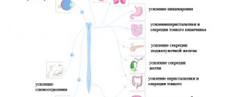
As we have already said, the autonomic (autonomic) nervous
the system regulates the functioning of internal organs.
Functionally, the autonomic (autonomic) nervous system consists of two sections: sympathetic
and
parasympathetic
. Thanks to this, information containing operating instructions reaches the organ in two different ways.
Sympathetic department
ensures the functioning of the body in stressful situations, and the parasympathetic - in a state of rest.
The sympathetic department ensures the functioning of organs during intense work, and the parasympathetic department - during normal physiological stress.
Motor part of the somatic nervous system
consists of one neuron. And in the autonomic (autonomic) nervous system it is represented by two neurons.
The connection of neurons occurs in the nerve ganglion, or g a
England.
,
the neuron that is located between the brain and the ganglion is called preganglionic
, and the neuron that connects the nerve ganglion with the executive organ
is
called
postganglionic .
The central part of the human nervous system, as we have already said, is represented by the spinal cord and brain.
The human spinal cord, like all vertebrates, is located in the spinal canal. It is a white strand 40-45 cm long, 1 to 1.5 cm wide and weighing about 35 grams.
It controls most of the reflexes and provides the ability to move.
At the top, the spinal cord passes into the lower part of the brain - the medulla oblongata
, and below it ends at the level of the first or second lumbar vertebrae.
The spinal cord does not occupy the entire cavity of the spinal canal: between the walls of the canal and the brain there remains a space filled with adipose tissue, blood vessels, meninges and cerebrospinal fluid, which washes the spinal cord and protects it from shocks.
Externally, the brain is covered with three membranes: dura
,
cobwebby
and
soft
.
On the surface of the spinal cord, two longitudinal grooves are clearly visible: the anterior
and
back
. They divide it into symmetrical halves - left and right.
31 pairs of spinal nerves arise from the spinal cord, dividing it into segments. The posterior roots extend from each nerve to the posterior surface, and the anterior roots extend from the anterior surface to each nerve, respectively.
In the internal structure of this section of the central nervous system, the central canal filled with cerebrospinal fluid and two parts that differ in color are clearly visible. In the middle, around the spinal canal, there is the so-called gray matter.
, which in cross section resembles the appearance of a butterfly, and is
white
.
Gray matter is represented by bodies
mi neurons and short branching processes - dendrites
and
tami.
And the white consists of long, non-branching axons that
form nerve fibers.
In the gray matter there are anterior
and
posterior horns
, and in the thoracic region and
lateral
.
Let's look at the direction of passage of the nerve impulse of the somatic reflex.
Along
the sensory neuron
, which enters the spinal cord as part of the dorsal roots, the nerve impulse reaches
the interneuron
, which is located entirely in the gray matter. From it, information is transmitted to the motor neuron, the body of which is located in the anterior horns of the gray matter, and its processes emerge from the spinal cord as part of the anterior roots.
Next, the nerve impulse enters the mixed spinal nerve
. It is called this because it contains both sensory and motor neurons. Along the mixed spinal nerve, the nerve impulse reaches the performing organ.
The nerve impulse enters the spinal cord through the dorsal roots and dorsal horns (through sensory neurons), and exits through the anterior horns and anterior roots (through motor neurons).
The spinal cord performs a reflex and conductive function.
That is, it conducts a nerve impulse to the brain and in the opposite direction.
The spinal cord provides the reflex. If a person unexpectedly pricks his finger, then thanks to a complex system of interactions in the body with the direct participation of the spinal cord, he will immediately withdraw his hand. This means that the reflex function of the spinal cord is that reflex arcs are closed on it.
However, when donating blood in a clinic during an injection, the person will not pull back his hand. Since information from the receptors is transmitted not only to the spinal cord, but also further to the brain, which processes it and in this situation sends instructions to slow down the spinal cord reflex to withdraw the hand.
Thus, the spinal cord conducts nerve impulses to the brain and in the opposite direction, performing a conductor function.
In addition to the spinal cord, the central nervous system also includes the brain.
Brain
located in the skull, the bones of which protect it from mechanical damage.
Externally, the brain resembles a yellowish jelly-like mass. The average weight of the human brain is 1300-1400 grams.
Like the spinal cord, the brain is covered with three membranes: dura
,
cobwebby
and
soft
.
The soft, or vascular, membrane of the brain is directly adjacent to the substance of the brain, extends into all grooves, and covers all convolutions. It consists of loose connective tissue, in which numerous blood vessels branch that feed the brain.
The arachnoid membrane of the brain is thin, translucent, and has no blood vessels.
The dura mater of the brain is the periosteum for the inner cerebral surface of the skull bones. This membrane contains the highest concentration of pain receptors in the human body, while the brain itself has no pain receptors.
Since metabolism is very active in the brain, it is richly supplied with blood vessels
providing it with oxygen and nutrients.
Inside the brain, like the spinal cord, one can distinguish gray
and
white matter
.
But their location is excellent. The cell bodies of neurons that form gray matter are found both on the surface of the brain and within it among the white matter, forming nuclei
.
The human brain is divided into five sections: medulla oblongata, posterior, middle, intermediate, terminal
.
Of these, three large sections are distinguished: posterior
,
middle
, and
front
.
The hindbrain is represented by the medulla oblongata
,
pons
and
cerebellum
.
And the forebrain consists of the intermediate
and
final brain
.
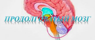
Medulla
is a continuation of the spinal cord, so their structure has a lot in common.
Only the gray matter of the medulla oblongata is located in separate clusters - idrama
.
The functions are also similar: reflex and conductive. Many reflex processes are carried out through the nuclei of the medulla oblongata, such as coughing, sneezing, lacrimation, etc.
The nuclei of the medulla oblongata contain nerve centers responsible for the acts of swallowing and the functioning of the digestive glands.
The medulla oblongata contains vital centers involved in regulating the activity of the heart and blood vessels, as well as breathing (respiratory center).
Over the bridge
All ascending and descending pathways pass through connecting
the forebrain
with the spinal cord, with
the cerebellum
and other structures.
Above the medulla oblongata is the cerebellum
. In principle, the structure of the cerebellum repeats the structure of the entire brain, which is where its name comes from. The surface of the cerebellum (cortex) is represented by gray matter and has folds, convolutions and grooves.
Inside the cerebellum there are also nuclei - accumulations of gray matter.
The cerebellum is a brain center that is extremely important for the coordination and regulation of motor activity. It works reflexively, maintaining the balance of the body and its orientation in space.
Midbrain
is a continuation of the bridge.
On its surface facing the cerebellum there are four tubercles - four about
the cerebellum. The superior colliculus carries out the primary processing of visual information. The lower ones are the centers for primary processing of information from the hearing organs.
Also in the midbrain are the most important motor centers that participate, together with the cerebellum, in coordinating body posture and maintaining muscle tone.
Diencephalon
consists of an upper part -
the thalamus
, or visual hillocks, and a lower part -
the hypothalamus
.
Thalamus
processes all types of information coming from the senses, except olfactory. It receives visual, tactile, gustatory and auditory information. Also in the thalamus are the highest centers of pain sensitivity.
Hypothalamus
, releases special neurohormones that affect the functioning of the
hypnophysis
. Therefore, it can be called the main link in the neurohumoral regulation of body functions.
The hypothalamus contains the hunger and thirst centers.
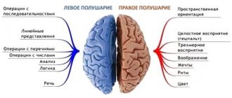
The large hemispheres of the human brain are divided into two halves by a deep longitudinal fissure - left and right.
The left and right hemispheres are connected by a plexus of nerve fibers - the corpus callosum
. Thanks to it, the collected information is transferred from one hemisphere to the cortical and subcortical structures of the other, which ensures an adequate and timely response.
The hemispheres of the brain are covered by a layer of gray matter called the cerebral cortex
. The cortex forms a large number of grooves of varying depth and length, between which convolutions are located.
The cerebral cortex consists of a huge number of neurons and provides higher nervous activity in humans. The number of neurons is between 10 and 11 billion, making up the majority of neurons in the entire human central nervous system.
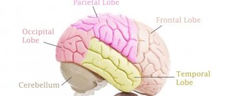
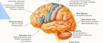
The cortex of each hemisphere is divided into lobes
: frontal, parietal
,
occipital and temporal. The functions of the cortex are associated with different lobes.
The frontal lobes process information about all sensations. Here their summary analysis takes place and a holistic idea of the image is created. This zone of the cortex is called associative and it is with it that the ability to learn is associated. The centers that control muscle movements are also located here.
The parietal lobe is associated with musculocutaneous sensitivity.
The occipital region processes information from the visual organs.
Temporal - auditory information.
Anatomy of the brain
The brain, of course, occupies a dominant place in the central nervous system. Inside the cranium, it is represented by two large hemispheres, dotted with deep and shallow grooves, under which other structural units are located:
- The oblong section is localized on the Blumenbach slope. Downwards it smoothly transforms into the spinal cord. On its front surface there is a longitudinal gap, on the sides of which experts identify 2 peculiar elevations in the form of rollers. They are called pyramids with olives. Whereas a similar groove on the posterior surface of the structure with two posterior cords is usually called pillars.
- Above the medulla oblongata is the hindbrain - in the form of the Varoliev bridge, as well as the cerebellum. Outwardly they are similar to the cerebral hemispheres, but functionally they have their own characteristics. Deep in the tissue there are clusters of nuclei from which the cranial nerves originate.
- The relationship between the medulla oblongata and the higher-lying sections is carried out by the midbrain - represented by the legs, nerve bundles, and also the quadrigeminal. It is impossible to overestimate their importance - it is in this zone that many of the most important nerve pathways lie and the nuclei of several pairs of nerves are located.
- Intermediate area - known as the visual thalamus with the subthalamic region, located further from the center of the brain. They contain the primary cells of the visual system, as well as sensitive conductive fibers. The hypothalamus, also known as the subtubercular region, takes part in metabolic processes.
Each of the listed units of the system - from the hemispheres and cerebellum to the brain stem - has its own significance for human life. If a failure occurs in one area - the lining of the central nervous system, for example, a brain tumor, then the effect will be on all parts of the organ.
Reflex function
An equally significant task facing the spinal cord is the implementation of autonomic and motor reflexes. Impulses coming from the brain along descending pathways are responsible for the movements of the entire torso and limbs. It is thanks to the passage of impulses that motor, food and vasomotor reflexes are performed.
Basic reflex activity of the spinal cord:
- Regulation of muscle tone.
- Formation of normal walking.
- Contraction of the anterior and abdominal muscle wall.
- Reflex movement of the limbs: rhythmic, extension, flexion, posture.
The reflex function of the spinal cord is based on communication with the brain. When a signal is received, the flexion and extension reflexes of the spinal cord are activated. They themselves are quite simple in nature. With repeated stimulation, the strength and duration of the reflex increases significantly. The reflex and conduction function of the spinal cord is controlled by the overlying parts of the central nervous system.
The pathways of the brain and spinal cord are a single system that always works harmoniously. This is what ensures the consistency of all body actions and its normal reaction to a given situation. For example, the receipt of a signal along the ascending pathways from receptors that the street is slippery allows, during the sliding process, impulses to be transmitted along the ascending pathways to ensure balance is maintained.
Spinal cord
It is one of two organs of the central nervous system. The physiology of its work is no different from that in the brain: with the help of complex chemical compounds (neurotransmitters) and the laws of physics (in particular, electricity), information from small branches of nerves is combined into large trunks and either implemented in the form of reflexes in the corresponding part of the spinal cord, or enters the brain for further processing.
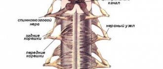
The spinal cord is located in the opening between the arches and vertebral bodies. It is protected, like the head membrane, by three membranes: hard, arachnoid and soft. The space between these tissue sheets is filled with fluid, which nourishes the nervous tissue and also acts as a shock absorber (dampens vibrations during movements). The spinal cord begins from the opening in the occipital bone, at the border with the medulla oblongata, and ends at the level of the first and second lumbar vertebrae. Next are only the membranes, cerebrospinal fluid and long nerve fibers (“cauda equina”). Conventionally, anatomists divide it into departments and segments.
On the sides of each segment (corresponding to the height of the vertebra) sensory and motor nerve fibers called roots extend. These are long processes of neurons, the bodies of which are located directly in the spinal cord. They are a collector of information from other parts of the body.
Anatomy and physiology of the spinal cord
How does the human spinal cord work, where is it located and how does it function? In short, this is the main organ of the central nervous system. With its help, signals from the periphery enter the central part and vice versa. Its anatomy is quite complex; it has many nerve endings, substances and membranes. To better study the features and role played by this body, we suggest staying with us and reading the article.
Anatomical features
A rather thick tourniquet, white in color, located in the spinal canal - this is the human spinal cord. Its diameter is about 1-1.5 cm, and its length almost reaches half a meter (up to 45 cm). This organ weighs about 38 g.
The narrow spinal canal is not only the location of an important organ, but also its protection. The core of the organ consists of a gray substance. It is covered by a white substance, which is also covered with protective and nourishing shells for the core. This is the general plan of the structure of the spinal cord.
Topography
The structure and functions of the spinal cord are quite complex. Neurosurgeon students study it in detail. Experts very carefully consider the development of the spinal cord. Ordinary people are interested in the question of what its topography is and familiarity with the leading role of this organ.
So, it is quite simple to describe the essence and goals that this body serves. The cervical spinal cord at the level of the back of the head in the area of the foramen passes into the cerebellum. The spinal cord ends at the level of the first 2 lumbar vertebrae. The conus spinal cord is located where a pair of vertebrae are located in the lumbar region. Next is the well-known “terminal thread”.
But this fragment is considered atrophied. It is called the “end” region. Nerve endings called “roots” are distributed along the entire circumference of the thread. The filum terminale is equipped with a substance containing a small proportion of nervous system tissue. But the outer part is not even equipped with a similar fabric.
The topography of the organ includes a pair of thickenings where the innervating processes emerge (cervical thickening of the spinal cord and lumbar). The outer and rear surfaces of the bundle are separated by slits called “middle”. The one in front is deeper, the back one is smoothed.
External structure
The general structure of the spinal cord suggests its division into a number of surfaces: posterior, anterior and two lateral. The spinal cord has faint grooves on the lateral surface.
They are located longitudinally, and nerves extend from the grooves. They are also called “roots”. In the lumbar area, together with the terminal filament, they form a tail, which is commonly called a horse's tail.
The grooves divide half of this cord into the following structures:
- front;
- lateral;
- posterior (cords).
The grooves of the spinal cord extend along the canal. The roots are divided into anterior ones - they are formed by efferent neurons, and posterior ones, created by afferent neurons. Their bodies converge into a knot. The roots unite and form a nerve. So, on all sides of the tourniquet there are over 30 nerve endings, forming exactly the same number of pairs. This is the external structure of the spinal cord.
Anatomically, it consists of 2 types of substances: white and gray. The first is the processes of the neural type, and the gray is their bodies.
White matter
All cords are made entirely of white matter of the spinal cord. They consist of longitudinal nerve fibers. These threads converge, forming peculiar conductors. Based on their functional purpose, fibers are divided into 3 types:
- motor;
- associative;
- sensitive.
The first are represented by short bundles and combine all parts into a single system. The second ones are called ascending. They give signals to the centers. Still others are descending. They provide signals from central structures to areas of the horns.
Gray matter
It structurally resembles grouped longitudinal plates consisting of homogeneous neurons. It contains not only neuronal bodies, but also neuropil, glial cells and capillaries. Along the entire spine it forms 2 columnar types, left and right. They are connected by gray adhesions.
The anterior horns contain the largest neurons. They form the motor nuclei of the spinal cord and inhibitory neurons. The structure of the gray matter of the background horns is not the same. It contains a huge number of intercalary type neurons.
The lateral horns of the spinal cord fill the centers of the ANS, the dilation of the pupil, the bases of innervation of the digestive system and other important organs of the human body. In the nucleus of the gray matter of the spinal cord there is a canal that neurosurgeons call “central.” It is filled with liquor. In adults, in some places it is filled with cerebrospinal fluid, and in others it is overgrown.
Shells
Anatomy of the spinal cord describes the membranes of the spinal cord:
- vascular soft;
- hard;
- avascular or arachnoid.
The characteristics of shell 1 are as follows: soft, penetrated by vessels and nerves. It is enveloped by the avascular part. There is some space here called “subarachnoid”. The cerebrospinal fluid generated in one of the systems flows into this niche. The last shell is made up of connective tissue; it is strong and flexible. The membranes of the spinal cord and brain are identical and form a single structure.
Segmental structure
A segment of the spinal cord is a piece of a tourniquet along with associated nerves. There is no morphological separation of one segment of the spinal cord from another. It is extremely functional. Each of the segments innervates a certain region. The designation of spinal cord segments is represented by alphanumeric indices, oriented to a part of the spinal cord and containing segment numbers.
The spinal cord consists of about 33 segments. The spinal cord segments have 4 roots, a pair of anterior and posterior. The spinal column is significantly longer than the cord, so it should be remembered that the segments are not numbered in the same way as the vertebrae. Any nerve consists of motor-sensitive roots. They come out in bunches from this bundle to the openings between the vertebrae.
The nerve ending located behind forms a ganglion and merges with the nerve ending in front. In this case, a mixed nerve is formed, which is divided into branches:
- The meningeal branch innervates in accordance with the nature of the spinal cord membrane and the canal wall.
- Dorsal - the skin in the corresponding areas, as well as deep muscle tissue.
- The connective tissue branch is the connecting link between the tourniquet and the ganglia.
- The abdominal branch is responsible for innervation of the limbs, lateral surfaces of the body and tissue of the abdominal part of the body.
Blood supply
The tourniquet is supplied with blood through the adjacent arteries. Through the fusion of the branches of the vertebral arteries, the anterior artery is formed. It is designed to be located along the front slit of the tourniquet. The blood supply to the spinal cord is also provided by the arteries located there. They are located behind the tourniquet.
They connect to the neck and arteries, which are called the “posterior intercostal, lumbar and lateral sacral arteries.” Between them there is a network of anastomoses, due to which the tourniquet is literally entangled in the branches of the arteries. To supply blood to the spinal cord, in addition to arteries, veins are needed, which also provide blood outflow.
Functions and role in the body
The human spinal cord has two main functions: one normalizes the brain-body connection. It is reflexive, it puts everything into action not without the participation of the will. The second conducts impulses to the main brain in an ascending manner and transmits them back from it. The descending or efferent pathways of the spinal cord are responsible for this activity.
The ascending tracts of the spinal cord are represented by the following tracts:
- spinothalamic;
- spinocerebellar;
- wedge-shaped and thin beams.
The pyramidal tracts, vestibulospinal, tectospinal and red nuclear spinal tracts are classified as special efferent pathways.
The reflex function is aimed at maintaining posture (position reflexes) and the ability to consistently alternate actions (motor programs), for example, walking. This function also provides a reflex defense mechanism (quickly moving the limbs away from hot objects).
Autonomic reflexes of the spinal cord are control signals that ensure the smooth functioning of internal organs. Myomatic reflexes are designed to provide contractile activity of muscles in response to their burning.
Anatomy and physiology of the spinal cord is a whole field of knowledge that describes its structure and features of functioning. It helps to understand how important the organ is and how the spinal cord and brain are connected. Thanks to this description, people receive the necessary ideas about an important organ.
Loading …
Video “Human Anatomy and Physiology”
From this video you will learn about the biological structure of the organ.
Structure of the spinal cord
The spinal cord is actually an extension of the brain, surrounded by the same membranes and cerebrospinal fluid. It makes up two-thirds of the central nervous system and is a kind of conduction system for nerve impulses.
The spinal cord makes up two-thirds of the central nervous system and is a kind of conduction system for nerve impulses. Sensory information (touch sensations, temperature, pressure, pain) goes through it to the brain, and motor commands (motor function) and reflexes pass from the brain through the spinal cord to all parts of the body. The flexible, boney vertebral column protects the spinal cord from external influences. The bones that make up the spine are called vertebrae; their protruding parts can be felt along the back and back of the neck. The different parts of the spine are called departments (levels), there are five in total: cervical ( C
), thoracic (
Th
), lumbar (
L
), sacral (
S
) and coccygeal.
The sections of the spine are designated by Latin symbols based on the initial letters of the corresponding Latin names.
Within each section, the vertebrae are numbered.
A tumor of the spinal cord can form in any part - for example, they say that the tumor is found at the C1-C3
or at level
L5
. Along the entire spinal column, 31 pairs of spinal nerves extend from the spinal cord. They are connected to the spinal cord through nerve roots and pass through openings in the vertebrae to various parts of the body.
Tumors can develop inside the spinal cord (intramedullary tumors) or on the outside of the spinal cord (extramedullary tumors). Since there is very little room inside the spine for the tumor to grow, it begins to compress the spinal cord, and this is what causes the observed symptoms.
With spinal cord tumors, two types of disorders occur. Local (focal) symptoms - pain, weakness or sensory disturbances - are associated with tumor growth in a specific area, when this growth affects the bone and/or spinal nerve roots. More common disorders are associated with disruption of the transmission of nerve impulses through the part of the spinal cord affected by the tumor. Weakness and loss of sensation or muscle control in the area of the body controlled by the spinal cord below the level of the tumor (paralysis or paresis) may occur. Possible problems with urination and defecation (bowel movements).
During surgery to remove a tumor, the surgeon sometimes has to remove a piece of outer bone (the vertebral arch plate, or arch) to get to the tumor.
Important!
This can subsequently provoke curvature of the spine, so such a child should be observed by an orthopedist
.
Localization
Primary tumors
Most neoplasms in adults arise from:
- Forebrain;
- Meninges;
- Nerves leaving the brain or going to it.
In children, the picture is somewhat different - 6 out of 10 (60%) tumors are located in the cerebellum or in the brain stem, only 4 out of 10 (40%) are in the forebrain.
Secondary tumors
Most tumors in adults do not develop from brain cells, but are other types of cancer that have spread to the central nervous system (metastases). These are so-called metastatic brain tumors. Metastases are rare in children.
« previous page | continuation of the article »
Was the material useful?
Cells of the brain and spinal cord
The brain and spinal cord are made up of cells whose names and characteristics are determined by their functions. Cells characteristic only of the nervous system are neurons and neuroglia.
Neurons –
These are the workhorses of the nervous system. They send and receive signals to and from the brain through a network of interconnections so numerous and complex that it is completely impossible to count them or chart them completely. At best, we can roughly say that the brain contains hundreds of billions of neurons and many times more connections between them.
Brain tumors arising from neurons or their precursors include embryonal tumors (previously called primitive neuroectodermal tumors - PNETs)
, such as
medulloblastomas
and
pineoblastomas
.
The second type of brain cells are called neuroglia. In the literal sense, this word means “the glue that holds the nerves together” - thus, the auxiliary role of these cells is already clear from the name itself. Another part of neuroglia contributes to the work of neurons, surrounding them, nourishing them and removing their breakdown products. There are many more neuroglial cells in the brain than neurons, and more than half of brain tumors develop from neuroglia.
Tumors arising from neuroglial (glial) cells are generally called gliomas. However, depending on the specific type of glial cells involved in the tumor, it may have one specific name or another. The most common glial tumors in children are cerebellar and hemispheric astrocytomas, brainstem gliomas, optic pathway gliomas, ependymomas and gangliogliomas. The types of tumors are described in more detail in this article.
Brief Definition
The spinal cord pathways or tracts are collections of nerve fibers located inside the spine that carry impulses from the brain to all parts of the body and back. The nerve endings, the combination of which form the pathways, are distinguished by a similar structure, development and general functions. They are divided among themselves according to the tasks assigned to them. The paths are classified as follows:
- Associative. Their main purpose is to unite gray matter cells from different segments to form their own anterior, lateral or posterior bundles.
- Commissural. These fibers connect the gray matter from the two hemispheres. With their help, the coordinated work of individual areas, nerve centers, and both hemispheres occurs.
- Projection. With the help of such pathways, the work of the overlying and underlying parts of the brain is combined. They are the ones who provide the projection of pictures of the surrounding world, as on a monitor screen.
Projection pathways, in turn, are efferent and afferent. They form the basis of the central nervous system, and are divided into ascending (centripetal or sensitive) and descending (centrifugal, motor).
Important! Nerve fibers provide a constant, unbreakable connection to the brain located in the skull and spine. It is thanks to them that the impulse is quickly transmitted, all body movements are coordinated with each other.
The pathways of the brain and spinal cord differ from each other, but they always act harmoniously, ensuring the passage of an incredibly large number of nerve signals from receptors to the central nervous system. Paths are formed from long axons, special fibers that are capable of creating connections among themselves, thus connecting individual segments of the spinal trunk, providing control of effector organs.
Structural features
In humans, from the moment of fertilization of the egg, the development and formation of the central nervous system begins - the brain and also the spinal cord are formed from the neural tube itself. They are protected by bone frames - the skull and vertebrae. Below there are three membranes - hard with arachnoid and vascular. Within their limits there are liquid media - cerebrospinal fluid with blood.
Traditionally, the structure of the central nervous system implies that cells - neurons - are united into special clusters - nerve centers. The bodies of neurons form the gray matter, while their short and long processes form the white substance, conducting signal impulses.
In addition, the central nervous system contains neuroglia, which consists of glial cells. Their number is several times greater than the number of neurons. Therefore, they make up most of the mass of the central nervous system.
In the head section, it is customary to distinguish several segments - the cerebellum with the cerebral hemispheres, as well as the oblongata, middle, intermediate and posterior sections. Each of them bears its own responsibility for the proper functioning of the organ separately, and of the entire organism and systems as a whole. In the spinal cord, gradation is carried out according to the segments of the spinal trunk - from the cervical, to the thoracic and lumbosacral.
central nervous system
Central nervous system (CNS)
- the main part of the nervous system of animals and humans,
consisting of a collection of nerve cells (neurons) and their processes.
The central nervous system consists of the brain and spinal cord and their protective membranes.
The outermost layer is the dura mater.
, under it is located
the arachnoid (arachnoid
), and then
the pia mater
, fused with the surface of the brain.
Between the pia mater and the arachnoid membrane is the subarachnoid space
, which contains cerebrospinal fluid, in which both the brain and spinal cord literally float. The action of the buoyant force of the fluid leads to the fact that, for example, the adult brain, which has an average mass of 1500 g, actually weighs 50–100 g inside the skull. The meninges and cerebrospinal fluid also play the role of shock absorbers, softening all kinds of shocks and shocks that tests the body and which could lead to damage to the nervous system.
The central nervous system is made up of gray and white matter
.
Gray matter
consist of cell bodies, dendrites and unmyelinated axons, organized into complexes that include countless synapses and serve as information processing centers, providing many functions of the nervous system.
White matter
consists of myelinated and unmyelinated axons that act as conductors transmitting impulses from one center to another. The gray and white matter also contains glial cells.
CNS neurons form many circuits that perform two main functions
: provide reflex activity, as well as complex information processing in higher brain centers. These higher centers, such as the visual cortex (visual cortex), receive incoming information, process it, and transmit a response signal along the axons.
The result of the activity of the nervous system
- this or that activity, which is based on the contraction or relaxation of muscles or the secretion or cessation of secretion of glands.
It is with the work of muscles and glands that any way of our self-expression is connected. Incoming sensory information is processed through a sequence of centers connected by long axons that form specific pathways, for example pain, visual, auditory. Sensitive (ascending
) pathways go in an ascending direction to the centers of the brain.
Motor (descending
) tracts connect the brain with motor neurons of the cranial and spinal nerves. The pathways are usually organized in such a way that information (for example, pain or tactile) from the right side of the body enters the left side of the brain and vice versa. This rule also applies to the descending motor pathways: the right half of the brain controls the movements of the left half of the body, and the left half controls the right. There are, however, a few exceptions to this general rule.
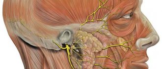
Peripheral nervous system
Nerve fibers stretch from the brain and spinal cord to all organs, muscles and ligaments, making up an extensive nervous network that entangles our entire body. This complex of nerve fibers and ganglia is called the peripheral nervous system. Its main task is to maintain two-way communication between the central nervous system and all parts of our body.
Clusters of sensitive nerve cells - receptors - receive information from the outside world or from internal organs. This information is then sent along receiving nerve fibers (afferent) for processing to the spinal cord and brain. If the most primitive but quick reaction is needed (withdrawing the hand), then the spinal cord is triggered and directs the nerve impulse to the muscles. If more complex information processing and decision-making are required, then the signal enters the brain, where a decision is made and a command is sent to the muscles and ligaments.
For example, there is candy on the table. We see it, that is, the signal reaches the sensitive nerve endings of the visual receptor, then the impulse reaches the visual part of the brain along the afferent nerves, an image appears, the brain comprehends it, makes a decision and gives a command. This signal reaches the muscles of the hand along the transmitting (efferent) nerve fibers, and the hand grabs the candy. Everything is very simple and at the same time incredibly complex, since it is the result of the exchange of nerve impulses between millions of nerve cells, various biochemical reactions, and the activity of different parts of the brain and spinal cord.
The peripheral nervous system is of two types:
- Vegetative, responsible for the functioning of glands, blood vessels and all internal organs.
- Somatic, controlling muscles, reflex and automatic movements.
The peripheral nervous system, unlike the central one, is practically not protected by anything, and therefore suffers more often. For example, for colds and mechanical damage. A variety of neuralgia are fairly common diseases that cause a lot of discomfort.
Midbrain
There is a department in the human central nervous system that is responsible for visual perception. This is the midbrain. It consists of two parts:
- The lower one represents the legs of the brain, in which the pyramidal tracts pass.
- The upper one is the quadrigeminal plate, on which, in fact, the visual and auditory centers are located.
The formations in the upper part are closely connected with the diencephalon, so there is not even an anatomical border between them. Conventionally, we can assume that this is the posterior commissure of the cerebral hemispheres. In the depths of the midbrain there are the nuclei of the third cranial nerve - the oculomotor nerve, and in addition to this there is also the red nucleus (it is responsible for controlling movements), the substantia nigra (initiates movements) and the reticular formation.
The main functions of this area of the central nervous system:
- orientation reflexes (reaction to strong stimuli: light, sound, pain, etc.);
- vision;
- pupil reaction to light and accommodation;
- friendly turn of the head and eyes;
- maintaining skeletal muscle tone.
Diencephalon
This formation is located above the midbrain, just below the corpus callosum. It consists of the thalamic part, the hypothalamus and the third ventricle. The thalamic part includes the thalamus itself (or thalamus), epithalamus and metathalamus.
- The thalamus is the center of all types of sensitivity; it collects all afferent impulses and redistributes them into the appropriate motor pathways.
- The epithalamus (epiphysis, or pineal body) is an endocrine gland. Its main function is the regulation of human biorhythms.
- The metathalamus is formed by the medial and lateral geniculate bodies. The medial bodies represent the subcortical center of hearing, and the lateral bodies represent the center of vision.
The hypothalamus controls the pituitary gland and other endocrine glands. In addition, it partially regulates the autonomic nervous system. We have to thank him for the speed of metabolism and maintaining body temperature. The third ventricle is a narrow cavity that contains the fluid necessary to nourish the central nervous system.