Craniopharyngioma
(
craniopharyngioma
; Greek kranion skull + pharynx pharynx + -oma tumor; synonym:
Rathke's pouch tumor, tumor of the pituitary tract, Erdheim tumor
) - a congenital brain tumor of the epithelial structure, developing from embryonic cells of the pituitary tract - the so-called. Rathke's pocket. Craniopharyngioma is usually a benign tumor; its malignancy is extremely rare. This type of brain tumor was first described in detail by J. Erdheim in 1904.
The incidence of Craniopharyngioma in relation to brain tumors ranges from 2 to 4.5%, and in children - from 7 to 13.4%. Craniopharyngioma occurs at any age, and Wertheimer (P. Wertheimer) indicates a significant frequency of the disease K. (27%) after the age of 40 years.
Schematic representation of the position of the pituitary tract (Rathke's pouch) between the lobes of the pituitary gland: 1—projection of the diaphragm of the sella turcica; 2 - anterior lobe of the pituitary gland; 3 - Rathke pocket; 4—posterior lobe of the pituitary gland.
Based on the direction of growth (Fig.), three groups of craniopharyngiomas are distinguished: 1) those localized under the diaphragm of the sella turcica - endosellar; 2) above the diaphragm - suprasellar; 3) growing subsellarly and penetrating into the sphenoid and ethmoid sinuses towards the cavities of the nose and pharynx.
Causes of craniopharyngioma
Few people can boast that they have never experienced a headache.
This is one of the most common pain syndromes that affects the population. The diagnosis of craniopharyngioma is most often detected in children between 5 and 14 years of age. There are several causes of the disease:
- Hereditary factor.
- Transference of serious illnesses during pregnancy. These include kidney pathologies, tuberculosis, and diabetes.
- Early toxicosis during pregnancy can affect the development of a benign tumor.
- Intrauterine infections that led to the development of fetal intoxication.
- Radioactive radiation to which a woman was exposed during pregnancy.
The complexity of the disease is that the cause that provoked its appearance is almost impossible to establish.
The complexity of the disease is that the cause that provoked its appearance is almost impossible to establish.
Taking certain medications during pregnancy can contribute to the formation of craniopharyngioma in the fetus. What makes the cells of the craniopharyngeal duct remain viable and subsequently multiply? Scientists suggest that this is due to various harmful factors affecting the fetus during the first trimester of pregnancy:
- use of medications;
- exposure to toxins or poisons;
- irradiation;
- intrauterine infections;
- the presence of untreated chronic diseases in the mother;
- severe toxicosis in early pregnancy.
All these factors can lead to improper formation of fetal organs and systems, including the craniopharyngeal duct. However, it should be understood that it is not at all necessary, for example, that after the mother uses any drug, the child she gives birth to will develop craniopharyngioma in the future. It's just that the risk in general will be slightly higher than others.
The main reasons for the development of craniopharyngioma are heredity and various types of mutations. In addition, the development of pathology is influenced by unfavorable factors, especially during the formation of the main organs - the first trimester of pregnancy. The causative factors for the occurrence of craniopharyngioma include the influence of drugs, toxins, poisons, radiation, etc.;
Craniopharyngioma is divided into 2 types: adamantinomatous and papillary. Approximately 60% of craniopharyngiomas are cystic formations, about 15% are solid formations, and the remaining 35% have a mixed structure and structure.
Craniopharyngioma develops from the embryonic remains of Rathke's pouch, a structure from which the adenohypophysis subsequently forms and develops. The neoplasm in most cases has cysts, and in adults it contains calcifications. Due to its localization and progression, craniopharyngioma causes developmental delays in children, visual impairment, and diseases of the endocrine system in adults.
Scientists suggest that germ cells form craniopharyngioma under the influence of harmful factors during the first trimester of pregnancy, such as:
- use of certain medications;
- exposure to toxins, including tobacco and alcohol;
- irradiation;
- intrauterine infections;
- chronic diseases of the mother;
- severe toxicosis in early pregnancy.
1.What are craniopharyngiomas?
Craniopharyngiomas
- benign neoplasms, most often formed in the hypothalamic-pituitary region of the brain, or more precisely, above the sella turcica (suprasellar).
The tumor is formed by embryonic cells of the so-called Rathke's pharyngeal pouch. Craniopharyngiomas are characterized by the presence in their structure of both cystic and solid (solid) components. For this reason, as the tumor grows, cysts may form in it, filled with yellowish-brown liquid contents with a high percentage of cholesterol and protein.
In most cases, craniopharyngioma is found in children 5-14 years old, but can also develop in adult patients.
A must read! Help with treatment and hospitalization!
Symptoms of brain craniopharyngioma
The main sign of the presence of brain craniopharyngioma is compression of the brain structure. This leads to increased intracranial pressure, which causes a number of symptoms, ranging from headaches to the development of mental retardation. Symptoms of a benign tumor differ in adults and children. The main signs are as follows:
- Retarded growth and development is the most striking manifestation of the presence of a tumor in childhood. The child has delayed development both in growth and in physical and mental indicators. Most often, it is these signs that help determine the presence of craniopharyngioma.
Due to tumor growth, the development of the anterior pituitary gland slows down. This leads to a decrease in hormone production. As a result, there is a delay in physical and mental development, and frequent bone fractures in the child. In situations with adolescents, puberty may occur very early or, conversely, too late.
- Headaches – this symptom is observed in all patients, regardless of age. The pain occurs with increasing intensity, while the interval between attacks decreases. Most often, pain is associated with increased intracranial pressure.
- Obesity – 20% of patients with brain tumors have obesity and diabetes insipidus.
- Visual impairment - this symptom refers to the early manifestation of pathology. It occurs in almost all patients in more than 90% of cases. Both children and adults suffer from vision impairment. The danger lies in the fact that symptoms are detected already at the stage of optic nerve atrophy. At this stage, the irreversible process of loss of visual function is already underway.
- Mental disorders are typical for adult patients. In this case, changes occur unnoticed by the patient himself.
The most dangerous phenomenon is the growth of cranipharyngioma into the area of the cerebral ventricles. This can lead to serious sleep disturbance.
Almost all patients whose illness lasts for a long period experience a decrease in intellectual abilities. This is due to the fact that the formation damages the brain and leads to disruption of higher cortical functions.
Symptoms of craniopharyngioma do not appear at the initial stage of tumor formation. After rapid progression, the following signs appear:
- various visual disorders due to compression of the optic nerve;
- severe and prolonged headaches;
- severe nausea;
- endocrine disorders;
- dysfunction of the adrenal glands;
- problems with sex life in men;
- absence of monthly menstruation in women;
- obesity;
- lethargy, slowness;
- memory problems;
- epileptic seizures;
- urinary incontinence.
The patient may experience growth problems (dwarf syndrome), excessive accumulation of fat deposits. The pathological process occurs individually, from several months to ten years.
At the initial stages, signs of lesions may be completely absent, but as the pathological process progresses, symptoms begin to appear:
- Various endocrine disorders such as diabetes insipidus, hypothyroidism or adrenal dysfunction, in men libido may noticeably decrease and erectile dysfunction may develop, and in women menstruation may disappear;
- Intense headaches are usually of an increasing nature and are accompanied by pronounced nausea;
- Visual disturbances occur due to compression of the optic nerve;
- Neuropsychological disorders such as obesity, bulimia, psychomotor retardation, apathy or dementia, lack of emotionality, urinary incontinence and memory problems;
- Hydrocephalus or hydrocephalus, accompanied by headaches, visual disturbances or swelling of the optic nerve.
In children with craniopharyngioma, sexual underdevelopment and dwarfism, obesity are observed, which is explained by hypothalamic disorders. Pathology can develop in a few months, or can slowly develop over 15 years.
The clinical symptoms of craniopharyngiomas are determined by their proximity to the optic nerves and chiasm, the hypothalamic-pituitary system and the cerebrospinal fluid pathways. Complaints include:
- headaches caused by increased intracranial pressure;
- visual disturbances associated with the pressure of the neoplasm on the optic chiasm. Depending on the location of the tumor, various types of narrowing of the visual field occur, less often - complete blindness in one eye.
- external changes. Often in adolescents, craniopharyngioma is accompanied by adiposogenital syndrome (progressive obesity). Less commonly, Frohlich syndrome, in which the development of the skeleton is disrupted and it is formed according to the eunuchoid type;
- hormonal imbalance. The disease may be accompanied by decreased libido and amenorrhea;
- various disorders: insomnia, hearing loss, dysfunction of the oculomotor nerves;
- changes in behavior: confusion, motor agitation, schizophrenia-like states, manic agitation, followed by a catatonic state.
Uncontrolled growth of craniopharyngioma can be fatal.
About 80% of children with pituitary craniopharyngioma or another type of tumor have slower growth and developmental delays. During adolescence, patients experience either late or too early puberty. Often the disease is accompanied by osteoporosis - the destruction of bone tissue with rather slow generation and strengthening.
There are also symptoms that are practically independent of the age and other characteristics of the patient:
- excess weight, which is observed in 20% of cases;
- headaches and high intracranial pressure;
- optic nerve atrophy, decreased quality of vision;
- mental disorders (typical for the older age group).
As soon as you feel at least one of the listed signs, be sure to contact a neurologist or other doctor for diagnostics: x-rays, magnetic resonance imaging, biopsy, hormone tests.
Like many other types of cancer, a malignant brain tumor can develop asymptomatically for a long time. Due to this, brain cancer is most often diagnosed in late stages after examination based on nonspecific symptoms and complaints from the patient. The most typical symptoms of brain cancer include:
- dizziness, visual and speech disorders;
- neurological dysfunction, signs of paralysis;
- coordination disorders, forgetfulness, dementia;
- convulsions and fits, involuntary twitching of parts of the body or limbs;
- frequent headaches (night or morning), worsening when trying to lie down and suddenly stopping;
- nausea and vomiting (gushing) not associated with gastrointestinal diseases, especially in the morning before meals - a sign of increased intracranial pressure;
- personality change;
- immobility of the eye pupil (rare symptom).
The main risk factor is head trauma. Triggered by the development of a brain tumor, the pain becomes stronger as the disease progresses: painkillers are powerless. In some cases, even in the later stages of brain cancer, patients do not feel symptoms such as headaches. Brain tumors are treated only in a modern oncology center.
Craniopharyngiomas can cause a range of symptoms depending on their location. If it compresses the pituitary stalk or the pituitary gland itself, the tumor can lead to partial or complete pituitary hormone deficiency, which can lead to growth problems, delayed puberty, loss of normal menstrual function and sex drive, increased sensitivity to cold, fatigue, constipation, dry skin , nausea, low blood pressure and depression.
Compression of the pituitary stalk can cause diabetes insipidus, as well as an increase in prolactin levels, which causes breast discharge. If the optic chiasm or nerves are compressed, vision loss may result. Involvement of the hypothalamus, an area at the base of the brain, can lead to obesity, increased sleepiness, and problems regulating temperature. Other symptoms, especially if larger, may include personality changes, headache, confusion and vomiting.
Top of page
Symptoms
Craniopharyngioma is a rather insidious tumor, because at first it does not manifest itself in anything. Symptoms appear when the tumor reaches a decent size and begins to compress the surrounding tissue. The signs of this tumor are nonspecific, that is, they do not allow one to suspect this particular type of brain tumor. What are these symptoms? The main signs of craniopharyngioma can be:
- headache. This symptom is a reflection of intracranial hypertension. A tumor is always plus tissue, and the cranial cavity has a constant size, so the appearance of an additional formation inside the cranium leads to an increase in intracranial pressure, which in turn makes itself felt by a headache. The pain can be diffuse, or it can be local in the forehead or temporal areas, and has an aching character. At first, the pain is not constant, but as the tumor grows it becomes more frequent and intense, acquiring a bursting character;
- visual impairment. This symptom is associated with compression of the visual pathways passing near the sella turcica. Depending on which direction the tumor grows, certain visual disturbances appear. This may be partial loss of visual fields (when a person cannot see the right or left parts of the picture - so-called hemianopsia), narrowing of visual fields (when the picture is visible as if through a spyglass), impaired vision in one eye (blindness). With long-term existence of craniopharyngioma, optic nerve atrophy may develop. In this case, even after removal of the tumor, vision restoration is impossible;
- symptoms of intracranial hypertension (increased intracranial pressure). They arise as a result of compression by a growing tumor of the third ventricle of the brain, where cerebrospinal fluid circulates. Due to the disruption of the outflow of fluid from the ventricle, it begins to burst the brain tissue from the inside, thereby causing an increase in headaches, a change in consciousness to the point of stupor or stupor, disturbances in the mental sphere (lethargy or, conversely, agitation, inappropriate behavior). In this case, vision can deteriorate quite quickly (its sharpness decreases), and swelling of the optic disc also occurs (which can be seen when examined by an ophthalmologist). Increased headache is accompanied by persistent vomiting that occurs without warning;
- changes in the mental sphere. Patients may become lethargic, apathetic, and indifferent to what is happening around them. They seem to become emotionally dumb. Memory may deteriorate. Children may lag behind their peers in neuropsychic development. Possible sleep disturbances;
- hormonal disorders. Craniopharyngioma itself has nothing to do with the body’s hormonal levels; it does not produce any hormones. But the location of the tumor near the pituitary gland causes a disruption in its function, and the pituitary gland is the main endocrine organ of humans. Compression of the pituitary gland by a growing tumor can lead to disruption of the activity of the gonads (impaired menstrual cycle, decreased potency and libido, premature puberty in children or delayed sexual development and growth), the thyroid gland (more often a decrease in its function - hypothyroidism, less often - an increase in function - hyperthyroidism ), adrenal glands (tendency to low blood pressure, changes in hair growth, fatigue, intolerance to normal physical activity). In addition, there may be leakage of breast milk from the mammary glands, obesity, or a sharp decrease in body weight. Often the first symptom of craniopharyngioma is diabetes insipidus, when up to 5-10 liters of urine are produced per day. Its occurrence is associated with a violation of the synthesis of antidiuretic hormone by the pituitary gland. As you can see, hormonal disorders are very numerous and varied, and at first glance it may seem that they have nothing to do with the brain. But in fact, a growing brain tumor is to blame;
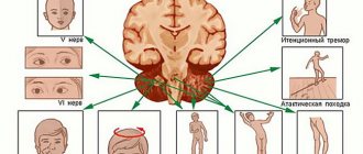
- symptoms of damage to the cranial nerves. Depending on the direction of growth of craniopharyngioma in the cranial cavity, it can compress the I, III, IV, V, VI pairs of cranial nerves. This is manifested by impaired perception of smells, impaired movement of the eyeballs in one or several directions, drooping eyelids, paroxysmal pain in the face;
- epileptic seizures. They rarely become the first symptom of craniopharyngioma, but this situation is also possible.
From all of the above, it becomes clear that craniopharyngioma does not have a single specific clinical manifestation. After all, neither a headache, nor signs of increased intracranial pressure, nor hormonal disorders can serve as reliable evidence of its presence. Therefore, the diagnosis of this disease is based not only on complaints and examination data of the patient, but also on additional research methods, which you will learn about below.
Diagnosis of craniopharyngioma
Craniopharyngiomas are usually diagnosed using magnetic resonance imaging (MRI) of the pituitary gland region of the brain. Many craniopharyngiomas will also be clearly visible on computed tomography (CT), especially since some are partially calcified (contain calcium deposits).
Top of page
Diagnosis of the disease at the first stage includes a neuro-ophthalmological examination, including assessment of visual acuity and fields. If a neuroendocrine tumor is suspected, tests for pituitary hormones are often prescribed, and instrumental visualization is mandatory.
- Computed tomography (CT) is an X-ray method for layer-by-layer examination of the substance of the brain and bones of the cranial vault. In the images obtained using a computed tomograph, both the dense and cystic parts of craniopharyngiomas are clearly visible. Calcifications are detected in 95% of cases in children and in approximately 50% of adults. Sometimes CT with contrast is indicated as more informative. Contraindications to contrast studies are an allergic reaction to iodine, hay fever, severe bronchial asthma, hyperthyroidism, renal failure, pregnancy.
- Magnetic resonance imaging (MRI) is a modern high-precision non-invasive study that provides maximum information about tumors in the brain. This method makes it possible to determine the location, exact size of craniopharyngiomas, and the ratio of their nodular and cystic parts. Images obtained using MRI also provide an idea of the condition of the tissues surrounding the tumor. After the main MRI scan, a clarifying study may be needed, which is carried out after intravenous administration of a contrast agent. Images obtained using contrast not only provide an accurate picture of the size and boundaries of the tumor, but also allow the assessment of functional disorders in organs.
The diagnosis of craniopharyngioma should be the result of a comprehensive examination of the patient. It includes a thorough survey, examination by a neurologist, endocrinologist, ophthalmologist and an instrumental examination.
Neuroendocrine examination is important, since craniopharyngioma leads to the formation of insufficiency of the hypothalamus and pituitary gland, thyroid gland and adrenal glands. To do this, the patient's hormonal profile is checked.
Ophthalmological examination often confirms the presence of intracranial hypertension:
- When examining the fundus, the presence of congestive optic discs is determined;
- during perimetry, narrowing or loss of visual fields, impaired color perception;
- decreased visual acuity, often in one eye.
The main diagnostic method is brain tomography. The most informative is its magnetic resonance subtype. It allows the specialist to see not only the bone structures, but also the soft tissues that make up the patient’s brain. With craniopharyngioma, a tomogram reveals an irregularly shaped tumor conglomerate located in the central structures of the brain.
MRI reflects not only the size, but also the direction of tumor growth. Description of layer-by-layer images will allow us to assess the structural structure of craniopharyngioma, the presence and severity of intracranial hypertension. A reliable symptom that practically confirms the diagnosis is the presence of dense walls and petrification inside the formation.
Sometimes tumors of Rathke's pouch do not contain calcifications, they have homogeneous content, so additional examination must be carried out to clarify the diagnosis. If the craniopharyngioma reaches a large size and puts pressure on the bones, the photographs show atrophic areas of the bones of the skull and the back of the sella turcica.
An X-ray examination of the skull in the area of the sella turcica or above it reveals petrification in 60-75% of patients.
MRI visualizes the location of the tumor, but not calcification, which is present in 50-70% of cases. Solid tumors appear isointense on T1-weighted CT scans. Cystic components appear hyperintense on T1-weighted tomograms.
CT and radiography show calcification, but it is not pathognomonic for craniopharyngioma, as it also occurs in other parachiasmatic lesions such as meningiomas, aneurysms, and chordomas.
Diagnostic methods
The diagnosis of craniopharyngioma is made only after a thorough comprehensive examination of the patient. The patient should receive consultation from a number of specialists, including a cardiologist, neurologist, endocrinologist, and ophthalmologist.
The X-ray method is used to determine the size of the tumor and wall erosion. The patient is required to undergo tests to determine hormone levels and determine the functioning of the thyroid gland. Additional research methods are MRI and CT. With their help, the specialist obtains an image of brain tissue in layers. This allows you to determine the exact location of the tumor.
Symptoms of brain craniopharyngioma
This type of tumor is always benign, but some types can cause very serious complications. As a result of special diagnostics, the neurosurgeon will determine exactly what type of craniopharyngioma you have:
- soft with minor cystic cavities;
- cystic - one or two formations filled with fluid;
- rocky, thoroughly impregnated with calcium salts;
- mixed, combining the characteristics of the above types.
There is also a classification based on the location of the tumor. The infundibular type of tumor is usually located in the third ventricle and most often affects the oculomotor nerve. Suprasellar craniopharyngioma can cause destruction of the skull bones, damage to the internal trunk and optic nerve. Stem craniopharyngioma is located along the stalk of the pituitary gland and causes changes in the structure of the vessels at the base of the brain.
A brain tumor is a malignant neoplasm of various cells. Brain cancer is characterized by the unstoppable spread and growth of tumor cells into adjacent brain tissue. The type and aggressiveness of the tumor directly depends on the cells that form it. Some brain tumors can produce hormones that cause disruptions in the body’s functioning (growth rate, menstruation, etc.).
In general, the statistics on the incidence of primary tumors is small: out of 25 thousand cancer patients, only 500 people have this type of pathology. The main groups of patients are the elderly, as well as children and adolescents; the latter account for the bulk of the disease. And of all types of oncology in children, this type is in first place.
The professionalism of German oncologists in the field of diagnosis and treatment of malignant neoplasms is a medical fact. Fruitful cooperation between specialists from oncology centers in Germany and the conduct of their own research on new options for pharmacological therapy at university clinics increase the effectiveness of drug treatment for brain cancer. Year after year, the number of patients who are completely cured or go into remission is growing.
Brain tumors are difficult to treat: in oncology centers in Germany, in each case, therapeutic tactics are selected individually for each patient. Treatment of brain cancer is expected in stages:
- Surgery to remove the tumor, if it is operable. If the tumor is adjacent to vital areas of the brain, treatment of brain tumors is carried out using alternative methods. The surgery is performed under local anesthesia, the patient remains conscious to control the functioning of important areas of the brain.
- Chemotherapy or radiotherapy. In addition to conventional radiation, brain tumors are also treated using stereotactic radiosurgery. An innovative method that is not used in Russia is the Gamma Knife, which destroys a tumor with the power of 200 radiation sources without affecting the healthy tissue of the most important organ for a person.
In oncology clinics in Germany, a diagnosed brain tumor requires a combined treatment, including all of the above stages.
The cost of treatment for brain cancer depends on the course of therapy prescribed: tumor removal, chemotherapy or radiotherapy, or combination treatment. See the approximate cost of medical services here.
Regarding the organization of treatment for a brain tumor, the cost of cancer treatment in Germany
in Moscow, (24 hours a day, in Russian)
in Germany: 49 152 31930411 (in Russian, German)
Classification
Histologically, there are two types of tumors:
- adamantinomatous craniopharyngioma - found in children, resembles a pulp, contains epithelial cells, filled with protein, oily fluid with cholesterol products, one or more neoplasms are found, calcification is often noted;
- papillary craniopharyngioma - occurs in adults, the epithelial layers extend along the villi of the vascular stroma, forming papillae, calcification is rarely observed.
Externally, a benign tumor is divided into four subgroups:
- compact neoplasms of small shapes with areas of calcification, found in 30% of cases;
- cystic - observed in 47% of cases, consists of several large neoplasms that can reach 12 centimeters, differ in size and shape;
- stony neoplasms in the area of the pituitary gland, consisting entirely of calcareous masses, small in size, rare;
- mixed tumors, which can consist of both soft tissue and calcifications, occur in 17% of patients.
Tumors exhibit exophytic and expansive growth. The exophytic nature of development is manifested in the fact that neoplasms grow towards the preformed cavities, heading into the cavity of the third ventricle of the brain or into the lateral ventricles, into the longitudinal fissure or into the space of the basal cisterns.
Expansive development means that the tumor-like neoplasm does not extend beyond the capsulated form, the parenchyma has no contact with surrounding tissues. The capsular appearance protects the surrounding tissues from what is inside the neoplasm. If the capsule bursts, the contents penetrate into the tissues, and the inflammatory process begins.
The neoplasm is classified depending on its location:
- infundibular group - this type of tumor fills the third ventricle, covering both Monroe foramina, and during development presses the interpeduncular cistern, pinching the oculomotor nerve;
- stable group - develops in the subarachnoid space under the bottom of the third ventricle, pressing on its bottom, the optic nerve is extended, the tumor compresses the pituitary gland and nerve roots, grows on the orbital surface of the frontal lobes;
- the neoplasm resembles an adenoma, is characterized by endosuprasellar growth, spreads in the area of the sella turcica, deforming it, pressing in the contents of the head and optic nerve;
- suprasellar tumors - involve the chiasm and optic nerve, affecting the internal trunk, disorders of the bones of the skull and sella turcica are observed.
Depending on the location of the tumor formation, appropriate surgical measures will be taken.
It is generally accepted at this stage of development of neurosurgery to divide tumors of the third ventricle into tumors of the anterior sections, tumors of the posterior sections (mass intermedia border) and tumors of the bottom of the third ventricle.
Rice. Schematic representation of the localization of tumors of the third ventricle.
Pathological anatomy
There are two main types of tumors: those consisting of dense tissue and cystic.
Rice. 6. Large craniopharyngioma cyst in the area of the sella turcica (the cerebral hemispheres are tilted posteriorly).
Craniopharyngioma is a tumor (usually in the form of a single dense node), growing slowly and expansively. Its size is from 2 to 5 cm in diameter, rarely larger (color fig. 6). Cystically degenerated areas of the tumor contain from 10 to 50 ml (in rare cases up to 200 ml) of thick liquid of yellow, amber or coffee color. K. usually has a dense capsule, quite closely connected with the surrounding brain tissue, membranes and vascular network. The tumor is supplied with blood from the branches of the arterial circle of the cerebrum.
K.'s structure can undergo significant changes over time. In its compact layers, liquefaction necrosis occurs with the formation of cysts containing fluid with a large amount of protein (from 20 to 100 ppm or more), amorphous remains of dead cellular elements and blood, cholesterol crystals, fatty acids with the deposition of salts on the inner surface of the capsule and in the tissue of the tumor itself. Histologically, cells consist of epithelial cells of varying degrees of differentiation. Along with epithelial cells of the embryonic type, epidermal type epithelium is also found. Cellular accumulations in some cases may resemble adamantinomas. In K., dystrophic changes of varying severity are always observed in the form of cyst formation, calcification, or even ossification of the stroma. K.'s capsule consists of connective or glial tissue. The diversity in the structure of the cell is regarded by a number of authors as a result of the phasic nature of its development.
Features of treatment of craniopharyngioma
Treatment for craniopharyngioma involves surgery and radiation therapy. If the location and size of the tumor allow it, then it is more rational to completely excise it. If the tumor is partially removed, a course of radiation exposure is prescribed.
It is possible to drain the cyst by introducing an antibiotic into the tumor cavity, which destroys it. This method is relevant for formations of small sizes. At the initial stage of development of craniopharyngioma, the CyberKnife radiosurgical technology is often used. The procedure is painless, consists of 5-6 sessions and is performed on an outpatient basis.
Life expectancy after timely surgery to remove craniopharyngioma is more than 10 years. Much depends on the location of the tumor, its size and the general health of the patient. There is a risk of relapse, that is, the return of the disease, so observation by a doctor is a must, even if there are no complaints about your health at the moment.
If necessary, make an appointment for a consultation.
Possible complications
The issue of radical surgical intervention is still actively discussed among neurosurgeons. This concerns the possibility of selecting a more optimal method in cases with children and adolescents.
Many neurosurgeons are critical of this method due to the risk of damage to the hypothalamus, as well as a high percentage of relapses. Complications may include infection, bleeding and injury to soft tissue, nerves and blood vessels.
If the craniopharyngioma is located in a very hard-to-reach place or the person has concomitant diseases for which surgical intervention is prohibited, neurosurgeons prescribe only radiation therapy.
Previously, the course of treatment consisted of radiation therapy over several weeks. In this case, the head area was irradiated. The procedures took place five times a week. The disadvantage of this method is the inability to completely remove the tumor. All that happens is that its growth stops.
Today, most clinics use a more innovative method - radiosurgery. This includes Gamma Knife and Cyber Knife. The essence of the method is that the brain is exposed to beams of radiation at different angles using a special apparatus. Unlike surgery, the effect of radiosurgery is not observed immediately, but only after several weeks.
The big advantage of this method is the reduced risk of complications. In addition, the procedures are performed without anesthesia. Radiosurgery is characterized by the precise direction of the beam, which is adjusted taking into account MRI readings. After this treatment method, the patient does not require a recovery period.
Diagnosis and tumor removal
The presence of cerebral craniopharyngioma can be suspected in children with growth retardation, obesity, and bone age lagging behind biological age. With these complaints, the child is often referred to an endocrinologist.
If headaches, vomiting and visual disturbances come to the fore, the patient first turns to a neurologist and ophthalmologist. To clarify the diagnosis, an X-ray examination of the skull is performed, which allows one to detect an increase in volume and changes in the walls of the sella turcica.
Additional research methods for diagnosing a tumor are MRI, which makes it possible to establish the exact location, size and structure of the tumor. In addition, the patient is prescribed tests to study hormonal levels.
The gold standard treatment for craniopharyngioma is surgery, the goal of which is radical surgical removal of the tumor to stop compression of the pituitary gland and restore its functions. It is not always possible to perform the operation in full, which is associated with topographic difficulties of access for a certain localization of the tumor.
When a craniopharyngioma is partially removed, courses of radiation therapy are performed to suppress the growth of remaining cells. In children, the irradiation method is considered the most dangerous and is rarely used due to complications and negative effects on the child’s neuropsychic development. In childhood, preference is given to repeated surgery.
The surgical method has its own risks and complications, including bleeding, infection, nerve and soft tissue damage.
Currently, an innovative non-invasive method is radiosurgery - under CT control, a radiation beam is directed to the tumor with high precision, using a special device (gamma knife, cyber knife). This alternative treatment has virtually no contraindications and is used in all cases where radical surgery is not possible.
Be careful, video of the operation! Click to open
Postoperative period
During the postoperative recovery period, the patient is prescribed:
- Drugs that help relieve swelling and reduce inflammation in the soft tissues of the brain.
- Diuretic drugs.
- Since the presence of a benign tumor cannot exclude the occurrence of relapses, a course of radiation therapy is prescribed after surgery.
If necessary, after the recovery period, hormonal therapy may be prescribed.
Residual effects from a removed tumor may remain. The patient may experience decreased vision, increased daily urine volume, decreased potency, and menstrual irregularities.
Treatment
According to the WHO classification, Craniopharyngioma belongs to tumors of the first degree of malignancy, therefore, if it is detected, it is necessary to take timely measures.
Treatment of craniopharyngioma should be comprehensive, including surgical and conservative measures.
Before surgery, to reduce the risk of complications, the patient is prescribed:
- short course of corticosteroid hormones;
- performing shunting in the presence of severe obstructive hydrocephalus to relieve the brain from high intracranial pressure.
The operation itself is performed using stereotactic methods or by craniotomy. Rathke's pouch is cleared of its contents stereotactically. To remove the remainder of the tumor and exclude recurrence of the disease, the neurosurgeon injects bleomycin into the capsule cavity, which accelerates the process of sclerosis of the tumor. Endoscopic intervention methods cause less damage to the brain and are easier to tolerate by patients. Radiosurgical methods of exposure with the introduction of radioactive isotopes are also used.
If craniopharyngioma is removed using the classical method, then specialists use osteoplastic trepanation with subsequent access to the area of the sella turcica.
In the postoperative period, the patient is prescribed:
- corticosteroids to reduce inflammation and swelling of damaged brain tissue;
- diuretics;
- a course of radiation therapy, since the benign nature of the tumor does not exclude the possibility of malignancy and relapse.
In the long-term postoperative period, hormone replacement therapy is prescribed if necessary. Residual effects of the removed tumor include the following disorders:
- poor vision, up to blindness;
- menstrual irregularities and decreased potency;
- diabetes insipidus (increased daily volume of urine with low specific gravity);
- Signs of infantilism, hypothyroidism and adrenal insufficiency may remain.
Forecast
Depends on the size, location, time of detection of the tumor, and the patient’s health condition. In 40-80% of patients, survival exceeds ten years with timely surgery. But even successful surgery does not save from the dangers of the disease returning; relapses are observed in every fifth operated patient. Radiation therapy gives good results even with incomplete tumor removal.
Considering such serious consequences of craniopharyngioma as visual and mental disorders, hormonal disorders, and the formation of persistent intracranial hypertension, timely consultation with a specialist and examination are necessary. A timely operation will prevent the development of complications, eliminate the possibility of relapses and help maintain health for many years.
Classification of tumors of the third ventricle
Health disorder classified as benign neoplasms
856,968 people diagnosed with Benign tumor of the craniopharyngeal duct
4,048 died with a diagnosis of Benign tumor of the craniopharyngeal duct
0.47% mortality for the disease Benign tumor of the craniopharyngeal duct
The approach to the treatment of craniopharyngioma is purely surgical. If it is impossible to completely remove the formation after surgery, stereotactic or external beam radiation therapy is prescribed.
Through tumor resection, it is possible to get rid of pressure on nearby tissues, and radiation makes it possible to restrain tumor growth in most clinical cases.
Very often, specialists combine radiation and surgical treatment methods, and, if necessary, supplement them with hormone replacement therapy.
When using the radiation method, the entire tumor is exposed to radiation. Megavolt radiation generated by a linear accelerator or a specialized gamma device is used for processing. Irradiation is usually carried out over a course of 5 days in a row.
One of the modern methods of treatment today is considered to be drainage of cystic forms of craniopharyngiomas by draining the formation with the introduction of an antibiotic drug into its cavity. Most often, Bleocin is used for administration, which has a destructive effect on tumor tissue.
For small formations, the Gamma Knife system is often used, which eliminates the development of neuroendocrine postoperative complications.
The degree of postoperative functional impairment is determined by the volume of surgical intervention performed. If the tumor was only partially removed and no radiation therapy was performed, the formation begins to actively grow and soon reaches its original size.
After radical removal of craniopharyngioma, cases of local recurrence are practically excluded, although the risk of complications remains.
Typically, surgical and radiation treatment is supplemented with hormone replacement therapy using gonadotropic and other hormones, depending on the type of reduced pituitary function. If gonadotropic drugs have no effect, injections of androgens are prescribed.
Other types of therapy for craniopharyngioma
Sometimes chemotherapy is prescribed. Drugs are injected into the spinal canal, intravenously, or into the tumor bed after its removal. Some clinics have obtained a positive effect from the injection of interferon drugs into craniopharyngiomas cysts. They caused a cytotoxic effect and triggered the processes of apoptosis (self-destruction) in tumor cells.
For children under three years of age who have large cystic inclusions in the tumor, it is often preferable to implant a catheter into the cyst, which ends in a subcutaneous reservoir. Thus, due to the drainage of the cyst, it is reduced in size, and the pressure on the nearest parts of the brain is significantly reduced. Catheterization of the cyst is performed in a minimally invasive manner, using a stereotactic unit to increase the accuracy of the intervention. Sometimes a drug is injected into the cavity of the cyst, under the influence of which its walls stick together, and further accumulation of fluid stops. As the child grows, the question of radical surgery, partial removal of the tumor, or radiation therapy is decided.
Due to the tendency of craniopharyngiomas to recur, often already in the first year after surgery, even if all visible tumor tissue has been removed, adjuvant (postoperative) radiation therapy is of great importance. It has been proven that the risk of relapse is reduced by almost 2 times. Large neurosurgical centers around the world have the technical capabilities to carry it out with maximum accuracy and efficiency, ensuring that the shape of the ionizing flow matches the complex shape of the tumor within a fraction of a millimeter. Here is held:
- Proton therapy uses heavier charged particles than traditional radiation therapy. They release almost all the energy in the area where the tumor is located. Thanks to the precise planning of each session using computer technology, the zones bordering the craniopharyngioma, including those located behind it along the proton flow, remain not involved in the irradiation process. Therefore, proton therapy is the method of choice for radiation therapy in children;
- intracavitary radiotherapy (a type of interstitial radiotherapy), for which a radiation source is introduced into the tumor tissue, damaging its cells, but without destroying the tissue of the pituitary gland, hypothalamus, cranial nerves and large vessels;
As an alternative to surgical removal of craniopharyngioma in children, clinics around the world use the method of stereotactic radiosurgery with Gamma Knife. This method of high-dose radiation therapy with extreme targeting accuracy makes it possible to eliminate most of the tumor mass without resorting to traditional surgery and without damaging the areas bordering the tumor.
Where does education come from?
Craniopharyngioma is a congenital benign formation that develops from overgrown epithelial cells of the embryonic pharyngeal passage (called Rathke's pouch). Here, at the end of the first month of embryogenesis, the fetal pituitary gland is formed. If for some reason, after the formation of this part of the brain, the division of cells in the epithelium of Rathke's pouch does not stop, then a craniopharyngioma tumor is formed. Why might this happen?
The role of hereditary predisposition to the development of this pathology and various mutations has been reliably established. Adverse factors operating in the first three months of pregnancy (the period of organ formation) have a direct impact on the formation of pathology:
- intrauterine infections caused by viruses (influenza, herpes, hepatitis, rubella), bacteria (syphilis, tuberculosis, sepsis), as well as fungi, chlamydia, toxoplasma;
- radiation exposure;
- the effects of drugs, toxins and poisons;
- chronic maternal diseases (diabetes mellitus, kidney failure or others).
Craniopharyngioma is a benign neoplasm of the pituitary gland, therefore it is characterized by slow but infiltrative growth with separate phases of development, which is reflected in the diversity of the structure of these tumors.
Causes of pathology
Few people can boast that they have never experienced a headache. This is one of the most common pain syndromes that affects the population. The diagnosis of craniopharyngioma is most often detected in children between 5 and 14 years of age.
There are several causes of the disease:
- Hereditary factor.
- Transference of serious illnesses during pregnancy. These include kidney pathologies, tuberculosis, and diabetes.
- Early toxicosis during pregnancy can affect the development of a benign tumor.
- Intrauterine infections that led to the development of fetal intoxication.
- Radioactive radiation to which a woman was exposed during pregnancy.
The complexity of the disease is that the cause that provoked its appearance is almost impossible to establish.
Forecasts
The prognosis for future life after cerebral craniopharyngioma depends on the success of the operation and the size of the tumor. Lethal outcomes during surgical intervention are no more than 10%. Most often this occurs due to damage to the hypothalamus.
Up to 85% of operated patients survive the surgical treatment and recovery period. Statistics are taken over five years. Relapses are possible throughout the entire period. The most dangerous complications are those that can arise within a year after surgery.
Almost all patients should be registered with an endocrinologist and regularly undergo hormonal therapy.
Prevention of harmful effects on pregnant women in the first trimester is used as preventive measures for the occurrence of the disease. Any types of intoxication and exposure to radiation must be excluded. Early registration of a pregnant woman plays an important role. This will allow us to exclude the possibility of a tumor at the initial stage.
If the occurrence of cerebral craniopharyngioma cannot be avoided, it is important to make a diagnosis in the early stages of the disease. This will help to avoid complications and prescribe timely surgical treatment.
Much attention should be paid to the patient’s quality of life after surgery for cerebral craniopharyngioma. Survival can only be ensured if a person undergoes a comprehensive examination by a number of specialized doctors. Monitoring of the patient should be lifelong.


