All types of circulatory disorders in the human body also affect the substance of the brain, which ultimately affects its integrity and the ability to function normally.
And “starvation” of cells, which is provoked by a violation or complete cessation of blood supply (in medicine, this process is called ischemia), causes a change in the brain substance of a dystrophic nature. That is, degeneration, and sometimes, although very rarely, even the disappearance of tissues and a significant deterioration in their function.
We will talk more about this pathological condition in the article.
Types of changes
In medicine, dystrophic manifestations in the brain substance are divided into two types:
- Diffuse.
- Focal.
In the first case, pathological changes spread evenly to the entire brain, and not to its individual areas. They are caused by both general disturbances in the functioning of the blood supply system and concussion or infections (meningitis, encephalitis, etc.).
Diffuse changes are manifested mainly by a decrease in a person’s performance, a dull headache, difficulty switching to another type of activity, a narrowing of the patient’s range of interests, apathy and sleep disorders.
And what a focal change in the brain substance of a dystrophic nature is can be understood by the fact that it can be caused by various minor pathologies:
- cysts (small cavities that form in the brain),
- small foci of necrosis (tissue death in certain areas caused by lack of nutrients);
- gliomesodermal (intracerebral) scars that occur after injuries and concussions;
- minor changes in the structure of the brain matter.
That is, these are pathologies that cause disturbances in the blood supply in a small area. True, they can be either single or multiple.
Symptoms of cerebral atherosclerosis
Among the obvious signs indicating the development of pathological damage to the blood vessels of the brain are:
- Bad memory. A marked decrease in memory, which manifests itself in forgetting events that occurred in the recent past. At first, memory impairments make themselves felt episodically, they are especially noticeable only after mental and physical fatigue. As atherosclerosis progresses, the gaps become more noticeable, but memory of past events remains for a long time. On the topic: 15 substances that speed up brain function and improve memory.
- Decreased mental performance , which manifests itself in the form of fatigue. The patient is unable to focus his attention on a specific object for a long time, or concentrate his own thoughts.
- Emotional lability is another common sign of cerebral atherosclerosis. At the same time, a person’s mood is characterized by instability and variability. That is, even minor events lead to the appearance of a brightly colored emotional reaction. Most often, such people are tearful, prone to depression and increased anxiety, they often have fears for their own health, and lack self-confidence.
- Insomnia. Patients often complain of regular insomnia.
- Headache. Constant companions of this type of cerebrovascular disease are headaches and frequent dizziness. They differ in that they arise during the period when a person changes his position from horizontal to vertical. That is, to put it simply, he gets out of bed, and not even with a jerk, but rather slowly.
- Epilepsy is another striking symptom of progressive cerebral atherosclerosis of cerebral vessels, observed in older people. Expressed in the occurrence of convulsive seizures.
- Senesthopathy , as symptoms of cerebral atherosclerosis, manifests itself in the fact that a person complains of tingling in the face, numbness of the legs, and heat in the occipital region.
- Cheyne-Stokes breathing or periodic breathing, which is characterized by certain cycles: shallow inhalations and exhalations are replaced by deep ones, after the seventh inhalation the breathing becomes rare again, then there is a pause and the cycle repeats. Most often, such breathing is observed when the arteries that supply the medulla oblongata are damaged by atherosclerosis.
- Ataxia is more often observed with damage to the arteries supplying the cerebellum or vestibular apparatus. It is expressed in incoordination of movements of various muscles, loss of balance when walking and standing, inaccuracy and awkwardness of movements, while the strength of the limbs is not lost.
- In addition, patients exhibit speech, auditory and visual impairments, as well as transient paralysis, to varying degrees. It is often difficult for the patient to tolerate loud sounds, and an irritable reaction to light may occur.
It is advisable to divide the symptoms of cerebral atherosclerosis into several periods for a clearer understanding of the progression of the disease:
- First stage
characterized by pseudoneurasthenic manifestations with periodic headaches, tinnitus, sleep disorders, intolerance to bright light, loud sound, decreased performance and other manifestations of a similar nature.
- Second phase
The development of the disease is marked by more pronounced clinical manifestations, with the development of depression, anxiety-delusional symptoms, and confusion.
- Third stage
characterized by the development of dementia with severe memory impairment. Dementia develops against the background of organic brain damage due to atherosclerosis. A person becomes helpless, disorientation in time may occur, however, generally accepted norms of behavior remain intact for a long time.
An important symptom that both patients and their relatives with cerebral atherosclerosis should be aware of is cerebral or hypertensive crisis. It is accompanied by headaches, quite intense ones, and weakness in one upper or lower limb may be observed. Often a crisis is accompanied by visual and speech disturbances. As a rule, it lasts up to 2 days, and then the patient’s condition stabilizes. If the symptoms do not go away after this period of time, then it makes sense to assume that these manifestations are signs of a stroke.
After a crisis, the patient is complacent and may be in a euphoric state, while all interests are reduced to everyday issues, and normal performance is lost.
Causes of dystrophy
The full picture of the appearance of dystrophic changes is not yet clear to researchers. But numerous observations have led to the conclusion that most cases of this pathology have a genetic predisposition. The action of provoking factors only accelerates the development of the process or enhances its manifestation.
Therefore, the reasons that cause focal changes in the brain substance of a dystrophic nature can be safely divided into genetic abnormalities and acquired ones. Although it should be noted that acquired causes are still a very conditional definition in this case, since they begin their destructive effect only if the patient is predisposed to the specified pathology.
Focal changes in the brain substance of a dystrophic nature: symptoms of disease development
Symptoms of changes in the substance of the brain of a dystrophic nature most often appear quite clearly, but, unfortunately, this happens when the disease has already progressed significantly. Therefore, it is important to pay attention to the appearance of even small deviations in health.
- Initially, the described focal changes are manifested by headaches that occur during both physical and emotional stress.
- This disease is also characterized by periodic manifestations of paresthesia - numbness or slight tingling in the extremities.
- The patient complains of dizziness and insomnia, and has impaired coordination of movements (ataxia).
- As the disease progresses, the listed symptoms worsen, hyperkinesis (involuntary movements of the limbs) joins them, and paresis and paralysis develop.
- Further development of the disease leads to memory deterioration, a noticeable decrease in intelligence, and agraphia (loss of the ability to write).
What is brain dystrophy - dyscirculatory changes
20.03.2019
Dystrophy happens:
- Diffuse. The pathology affects all brain tissue evenly; specific local lesions are not formed.
- Focal. Often appear against the background of impaired blood supply. With focal changes, the brain as a whole is not damaged, but individual foci are observed affecting sections and areas of the brain.
Also, discirculatory dystrophy is of the following types:
- atherosclerotic (due to the presence of plaques obstructing blood flow);
- hypertensive;
- venous;
- mixed.
The disease leads to the following consequences:
- Rarefaction of areas of brain tissue: it seems to become loose, like a washcloth.
- Asymptomatic cerebral infarction is an acute disorder of cerebral circulation that has no clinical manifestations and does not give symptoms of neurological disorders.
- Stroke.
Causes
There are the following causes of brain dystrophy:
- Neuroinfections: polio, types of infectious encephalitis, rabies.
- Degenerative diseases: Pick's disease, Alzheimer's disease, Huntington's disease, multiple sclerosis. The diseases are accompanied by gradual destruction of the cortex and pathways.
- Arterial hypertension. High vascular pressure leads to a decrease in minute blood volume. The nerves suffer due to lack of nutrients and oxygen.
- Neoplasms. Tumors can “steal” part of the arteries, taking blood from the neighboring area.
- Vascular atherosclerosis.
- Chronic kidney diseases.
- Spinal cord herniation. They compress the main arteries.
- Diabetes. The disease is accompanied by damage to medium and large arteries.
Symptoms
The disease goes through three stages:
- Stage of asthenia. At this stage, the diagnosis of dystrophy or dyscirculatory encephalopathy is rarely made. Asthenic syndrome is characteristic of many diseases in the clinic of which intoxication syndrome is present. In addition, most mental disorders also begin with asthenic syndrome, and doctors often think in this direction. Asthenia syndrome is manifested by chronic fatigue, lethargy, reluctance to work, drowsiness, irritability, sleep disturbance, dizziness and headaches. There are no specific symptoms.
- Second stage. Characterized by progression of previous symptoms. Mental activity deteriorates: the speed of thinking decreases, the amount of memory and accuracy of information reproduction decreases, and the concentration of attention decreases. Speech becomes disordered: it becomes illegible. The patient mumbles and speaks quietly. Those around him have a hard time hearing him. Coordination is partially impaired: complex manipulations lose precision.
- Outcome of the disease. Discirculatory-dystrophic changes in the brain increase. The clinical picture of the disease includes a stroke - an acute circulatory disorder in the brain, characterized by specific focal symptoms and gross impairment of mental and neurological functions. Often a stroke leads to the death of the patient.
General cerebral symptoms of stroke are manifested by impaired consciousness, lethargy and lethargy. The patient reacts slowly to external stimuli. The patient complains of a severe headache. Nausea and vomiting are observed. Sweating, palpitations, and dry mucous membranes appear.
Focal symptoms are determined by the location of the lesion. For example, if hemorrhage occurs in the motor centers, movement disorders are observed in the clinic: flaccid paresis or paralysis of the limbs. Most often, with any stroke, disturbances in oculomotor functions and sensitivity occur. Speech: words are difficult to pronounce, speech is slurred.
Diagnosis and treatment
Dystrophic changes in the brain are diagnosed using the criterion according to Levin (2007):
- Neurological and psychophysiological disorders are detected using psychological tests. The mental and emotional-volitional sphere of personality is explored. A neurologist checks walking and precision of movements.
- Using a biochemical blood test, metabolic disorders are detected: excessive amounts of fat, sugar.
- Instrumental methods make it possible to visualize foci of dystrophy using computed tomography and magnetic resonance imaging.
Treatment:
- The cause of the disease is eliminated. If it is arterial hypertension, medications that lower blood pressure are prescribed: Obzidan, Metoprolol.
- Pathogenetic treatment aimed at improving blood supply, for example, nootropics: phenotropil, piracetam, glycine.
- Stroke is treated symptomatically: drugs are prescribed to improve the rheological properties of the blood.
Didn't find a suitable answer? Find a doctor and ask him a question!
Source: https://sortmozg.com/zabolevaniya/distrofiya
Is there an age limit for the disease?
It should be noted that single focal changes in the brain substance of a dystrophic nature occur not only in older people, but also in people under fifty years of age.
Stress, injuries, stressful situations, hypertension and other provoking factors can trigger the development of focal changes. The constant overstrain that many able-bodied citizens experience also plays its unseemly role.
Increased brain function against the background of existing vascular spasm in youth, as well as ischemia in old age, can equally lead to the emergence of foci of dystrophic changes with all the ensuing consequences. And it follows from this that timely and properly organized rest is a very important part of the prevention of the described pathology.
What diseases are accompanied by dystrophic changes in the brain?
Focal changes in the substance of the brain of a dystrophic nature, as a rule, are provoked by very common disorders of the functioning of blood vessels. These include:
- vasomotor dystonia,
- atherosclerosis,
- arterial hypertension,
- aneurysm of blood vessels in the brain and spinal cord,
- cardiocerebral syndrome.
Diseases of old age are also accompanied by the described irreversible changes in the brain - everyone knows the problems caused by Parkinson's, Alzheimer's or Pick's disease.
Diagnosis and treatment
Dystrophic changes in the brain are diagnosed using the criterion according to Levin (2007):
- Neurological and psychophysiological disorders are detected using psychological tests. The mental and emotional-volitional sphere of personality is explored. A neurologist checks walking and precision of movements.
- Using a biochemical blood test, metabolic disorders are detected: excessive amounts of fat, sugar.
- Instrumental methods make it possible to visualize foci of dystrophy using computed tomography and magnetic resonance imaging.
- The cause of the disease is eliminated. If it is arterial hypertension, medications that lower blood pressure are prescribed: Obzidan, Metoprolol.
- Pathogenetic treatment aimed at improving blood supply, for example, nootropics: phenotropil, piracetam, glycine.
- Stroke is treated symptomatically: drugs are prescribed to improve the rheological properties of the blood.
How is the diagnosis made?
The diagnosis of “focal changes in the brain substance of a dystrophic nature” is quite difficult to establish. This requires identifying the signs of the pathologies listed above and excluding other somatic diseases and possible neuroses. By the way, people with diabetes and rheumatism are also at risk.
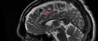
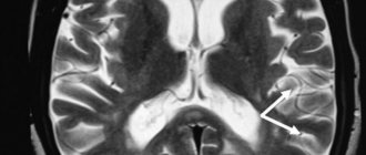
The doctor must assess the patient’s condition, his neurological status, and also conduct the necessary examinations. The most accurate indications are provided by an MRI study, where lesions can be identified, as well as their size and location. Tomography makes it possible to determine changes in the density of brain tissue even in the initial stage of the disease. Correctly reading the MRI results is an important step in starting treatment for the problem described.
Focal changes in the brain substance of a dystrophic nature: treatment
As mentioned earlier, the exact cause of the appearance of this pathology, unfortunately, has not yet been established. And the diseases diagnosed along with it are more likely to be factors that only provoke the onset of its development or intensify processes that have already begun, and not the main cause of the disease.
Therefore, its treatment consists mainly of normalizing the patient’s daily routine and a proper diet, including foods that contain organic acids (baked and fresh apples, cherries, sauerkraut), as well as seafood and walnuts. The consumption of hard cheeses, cottage cheese and milk will have to be limited, since excess calcium causes difficulty in oxygen metabolism in the blood, and this supports ischemia and isolated focal changes in the brain substance of a dystrophic nature.
In addition, the patient cannot do without symptomatic therapy, which involves prescribing drugs that affect cerebral circulation and reduce blood viscosity, taking analgesics, sedatives and B vitamins. However, this is a separate and rather extensive topic.
Treatment of cerebral atherosclerosis
Therapy for this pathology is a long, and most often lifelong, process. This problem is solved by a neurologist whose responsibilities include identifying people with similar problems, assessing the severity of the disease and implementing conservative therapy. First of all, it is designed to improve blood supply to the brain and prevent arterial thrombosis.
Drug treatment
As for therapy with drugs, the modern scheme of action comes down to:
- Antiplatelet therapy, which is aimed at reducing the risk of ischemic stroke. Drugs used for this purpose: acetylsalicylic acid and clopidogrel. However, a hemostasis study is first necessary.
- Reducing cholesterol. In parallel, drugs are used to reduce cholesterol levels in the blood. These can be statins (preventing the synthesis of cholesterol in the liver, reducing LDL and increasing HDL) - lovastatin, atorvastatin, simvastatin, pravastatin, as well as fibrates (lowering cholesterol and triglycerides) - gemfibrozil, fenofibrate, clofibrate. In addition, anion exchange resins or bile acid sequestrants are prescribed to promote the removal of cholesterol; examples of these agents are: hestyramine, colestipol. Ezetimibe, atromide, miscleron, etc. will help reduce the absorption of cholesterol in the small intestine.
- In addition, in complex therapy, patients are prescribed drugs aimed at preventing the development of circulatory disorders. These can be coronary lytics, as well as agents that dilate arteries and anticoagulants in case of a threat of blood clots in the vessels of the brain.
- Iodine preparations and diosponin will help reduce cerebral ischemic disorders. In addition, calcium iodine, potassium iodide or iodine solution can be prescribed for the same purpose. The drugs are taken in courses to eliminate iodism.
More details:
Drugs for the treatment of atherosclerosis
Surgery
When hemodynamically significant stenoses, either complete blockage of the arteries, or unstable plaques are detected, the vascular surgeon decides whether surgical intervention is necessary. Modern medicine has reached significant heights in the field of reconstructive operations on blood vessels, including the brain.
The following types of surgical interventions are currently available:
- Endarterectomy.
It is based on the fact that fatty growth is eliminated using the open method. To do this, a skin incision is made to gain access to the blocked vessel. After this, the surgeon stops the blood flow in this place, the wall of the bloodstream is quickly cut and the fatty deposit is removed. After such manipulations, the surgeon can only sew up the damaged area with a vascular suture. In this way, plaques on extracranial vessels can be removed.
- Stents and balloons.
To eliminate atherosclerotic formation on intracranial vessels, stents and balloons are used. That is, endoscopic removal of the atherosclerotic plaque is necessary. To do this, an endoscope with a stent is inserted into the widest vessel, and then, under constant X-ray monitoring, it is advanced to the place where there is narrowing of the artery due to the presence of plaque. It is there that a stent is installed, which, by increasing the lumen of the vessel, restores the flow of blood through it.
It is worth understanding that cerebral atherosclerosis is classified as a chronic disease, so treatment is most often lifelong. Depending on how timely the diagnosis was made and treatment started, the prognosis will depend. In the practice of neurologists, extensive forms of cerebral atherosclerosis are known, which, nevertheless, allowed people not only to live for a long time, but also to remain functional. However, there are often cases when the first clinical manifestation of this disease ends in a stroke and death for a person. That is why doctors play such an important role in timely diagnosis of the disease and its qualified treatment.