Magnetic resonance imaging is an informative non-invasive way to study cerebral structures. Diagnosis of pathological processes in the brain using other methods is complicated by the presence of the skull, which performs a protective function. Scanning using an induction field allows you to identify the slightest changes. Foci of gliosis are most accurately detected on MRI of the brain; a specialist can determine the nature and localization of the process.
Various forms of gliosis on MRI images of the brain
The study is carried out using a tomograph consisting of a mobile table and a wide tunnel. The device generates a magnetic field, under the influence of which the hydrogen atoms in water molecules are aligned in a special way. Sensitive sensors read the response of scanned tissues, obtaining information about the degree of fluid saturation of brain cells.
A computer program converts the signal into a series of layer-by-layer images taken in axial, sagittal and frontal projections. Based on the images obtained, the doctor can, if necessary, reconstruct a 3D model of the brain.
Causes
Gliosis of the white matter of the brain interferes with the full functioning of the nervous system, but it is necessary to fight this disease not directly, but by investigating its cause.
Basically, the catalysts for the appearance of glial accumulations are infectious or other causes of diseases of the nervous system, such as:
- hereditary diseases associated with the death of neurons;
- multiple sclerosis - destruction of nerve tissue fibers in the brain and spinal cord;
- tuberous sclerosis is a genetic disease in which benign tumors develop;
- epilepsy;
- trauma at birth (in infants);
- head and back injuries;
- high blood pressure and encephalopathy;
- cerebral edema;
- chronic or acute cerebrovascular accident (CVA/CVA);
- hypoxia – acute lack of oxygen in tissues;
- neuroinfections such as leukoencephalitis, encephalomyelitis, etc., caused by viruses or bacteria;
- low blood sugar;
- high consumption of animal fats;
- previous operations;
Glial accumulations are often observed in athletes who have suffered head concussions, as well as in those who are exposed to bad habits such as alcohol and drugs that contribute to the destruction of neurons. These changes may also occur in patients taking narcotic-based medications.
Complications and consequences
The presence of extensive areas of replacement of neurons with gliocytes threatens with dangerous consequences:
- persistent headaches that cannot be controlled by medications;
- mental disorders;
- permanent loss of speech, vision or hearing;
- convulsions and epileptic seizures;
- paralysis (partial or complete);
- mental disorders;
- impaired coordination of movements;
In extremely rare cases, gliosis of non-genetic origin causes death.
Symptoms
Gliosis is a disease that can masquerade as a number of problems associated with the cardiovascular and nervous systems. Its most common symptoms are:
- constant headaches, migraines, dizziness;
- sudden changes in blood pressure;
- the appearance of problems with vision or hearing;
- memory and attention disorder;
- the appearance of convulsions, paralysis.
These problems can also appear in a number of other diseases that are completely different from gliosis, so for an accurate diagnosis you need to contact a specialist. Sometimes cerebral gliosis is detected already with an MRI of the brain, although the patient does not feel any negative changes.
Kinds
By the extent of the process:
- focal, single or multiple;
- diffuse, more common.
By structure and structure , visualized using MR tomography:
- isomorphic (foci of vascular origin have a homogeneous structure formed by astrocytes and glial fibers);
- anisomorphic (“variegated” gliosis, which is not characterized by morphological homogeneity);
- fibrous (mainly represented by glial elements with single astrocytes).
Types
The distribution of glial cells occurs in different ways. Depending on their location in the body, their foci are divided into:
- anisomorphic – irregular order of glia distribution;
- isomorphic – correct formation of glial cells;
- marginal – growth of glial cells in the intrathecal spaces of the brain;
- diffuse – accelerated spread of gliosis both in the brain and spinal cord;
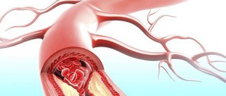
- perivascular or vascular - gliosis located along the vessels. Most often it appears after atherosclerosis.
- fibrous – processes of glial cells exceed the size of their bodies;
- marginal - glial elements are located on the surface of the brain;
MRI results: cerebral gliosis
The study protocol includes a description of the state of the sub- and supratentorial space. Based on the results of an MRI of the brain, the doctor evaluates:
- location of midline structures;
- perivascular spaces of the frontal, parietal lobes; areas of the basal ganglia;
- homogeneity of the white matter structure;
- localization, size of focal changes, presence of perifocal (marginal) infiltration;
- condition of the pituitary gland, ventricles of the brain;
- the presence of pathological formations;
- functionality of blood vessels in the area of interest.
If there are characteristic foci of gliosis, the doctor clarifies the genesis of the changes, indicating the main diagnosis in the conclusion.
Foci of gliosis (shown by arrows) on an MRI image of the brain in axial projection
If you suspect any cerebral pathologies, you can undergo an MRI at the Magnit diagnostic center.
Diagnostics
Detection of this disease is impossible without the use of special electronic equipment. Diagnosis can be carried out using one or more methods:
- Magnetic resonance imaging (MRI) – obtaining images of internal organs and tissues by exposing the object of study to electromagnetic waves. This method is used to find abnormalities in the functioning of organs, tumors and improper tissue regeneration;
- computed tomography (CT) – obtaining images of internal organs using x-rays and subsequent data processing on a computer. It helps to identify changes associated with blood vessels, for example, obstructed blood circulation, thrombosis, etc.;
- electroencephalography (EEG) - measurement of brain activity using electrodes and computer data processing. It is applicable when it is necessary to record problems of the nervous system, such as seizures or epilepsy.
All these methods are applicable in specialized clinics equipped with modern medical devices.
Causes
Among the most likely causes of gliosis are:
- birth injuries;
- ischemic stroke of the brain (leads to the occurrence of a post-ischemic form of gliosis);
- multiple sclerosis;
- hypertensive encephalopathy;
- microangiopathy;
- consequences of epilepsy;
- VSD;
- cerebral tuberculosis;
- encephalitis;
- hereditary lipid metabolism disorders;
- traumatic brain injury (TBI);
- infectious diseases that provoke the formation of a focus of inflammation;
- parasitic infestations.
Recently, doctors have especially noted two factors that have the most active effect on the state of the brain:
- Drinking alcoholic beverages. In moderation, alcohol helps improve blood circulation and stimulates metabolic processes in tissues. However, the slightest excess of the minimum dosage is fraught with irreversible changes in the integrity of neural connections.
- Drug use. This leads to atrophy of brain tissue, its necrosis (demyelination) and vascular inflammation. Even if the drug was used for medical purposes, the person who used it is still at risk.
In fact, gliosis transformation of the brain is completely natural and occurs during the aging process. This is why in old age people have problems with coordination, memory and fine motor skills.
Treatment
Cerebral gliosis itself is not a disease, but a complication that was caused by chronic or acquired diseases of the nervous system. Therefore, there is no specific medicine or procedure to eliminate such tumors.
Treatment is aimed at the specific disease that caused the development of gliosis. It should be noted that medications are prescribed directly by a doctor.
During drug treatment, it is necessary to take special agents that can maintain and improve the condition of blood vessels. Also, with this disease, the brain may experience a lack of oxygen, so patients are often prescribed antioxidants that neutralize oxidative processes, and nootropics that help improve brain activity.
Surgery
Surgical intervention is used when large single foci of gliosis appear and in case of their negative impact on an organ or system that cannot be ignored, for example, during seizures. But most often, surgery is resorted to if it is impossible to control the patient’s well-being with the help of medications.
Complementary and alternative treatments at home
In addition to traditional methods of treatment, a patient suffering from this disease must eat in accordance with a special diet and take preventive measures to maintain normal functioning of the body and prevent the development of pathologies against the background of gliosis.
Nutrition and supplements
With gliosis of the brain, it is necessary to normalize your daily nutrition. The most important condition here is the exclusion of fatty foods and dishes from the diet, because fatty compounds disrupt the functioning of neurons and lead to their death.
Alcohol and herbal infusions
As mentioned earlier, a patient with this disease may have problems with cerebral circulation.
Traditional treatment
- To inhibit the processes of sclerosis of blood vessels and neurons:
400 gr. mix crushed leaves of 5-year-old aloe with 650 mg of honey and 650 g. red Cahors. Let it brew in a dark place for at least 5 days. Take 1 teaspoon 3 times before meals. The course of treatment is 2-4 weeks.
- To dilate blood vessels in the brain:
2 tbsp. Mix dill seeds and a glass of crushed valerian root with 1.5-2 glasses of honey, pour 1 liter of boiling water. Let it brew for a day. Before meals, take a tablespoon of the mixture for 4-6 weeks .
150-250 gr. Pour boiling water over hawthorn fruits. Let it brew for 30 minutes. Take the infusion 1 tbsp. half an hour before meals. The course of treatment is up to 1.5 months .
Prognosis (how long people with the disease live)
Brain gliosis can be caused by diseases associated with the cardiovascular and nervous systems, but it can also provoke the appearance of diseases of this nature. Regardless of age, both adults and children have the same conditions for dealing with this problem.
But as a rule, such people do not live more than 2-3 years.
Of course, only a doctor can determine how life-threatening glial tumors are, so first of all you should contact a specialized clinic as early as possible and identify the cause of their appearance, which will help you understand further actions and develop the necessary therapy.
Types of gliosis
Gliotic changes have several forms, depending on the location of cell concentration and the type of transformation.
| Form | Description |
| Anisomorphic | Education is growing haphazardly in different departments hemispheres , and its structure is predominantly cellular. |
| Fibrous | It is distinguished pronounced signs of the predominance of fibrous structures. |
| Diffuse (fine-focal) | There are no clearly demarcated affected areas; scarring occurs not only in the brain, but also in the spinal cord . The number of small lesions is difficult to count. |
| Focal | The place of tissue changes is clearly demarcated by the affected area. Most often, such gliosis is a consequence of an acute inflammatory process. |
| Regional | The superficial part of the brain, located directly under the membrane, is destroyed. |
| Perivascular | Clusters of glia ring blood vessels that have undergone sclerosis. This may be a consequence of systemic vasculitis . |
| Periventricular | Gliotic formations are located in the cerebral ventricles. , cystic-gliotic changes in the brain are observed . |
| Subependymal | The affected area is located near the ventricle of the brain. |
| Supratentorial | The formations are located above the tentorium of the cerebellum (a process of the hard membranes). Associated with the upper parts of the brain. This area is most often affected by bruises. heads or birth injuries. Also, glial formations of vascular origin most often occur there. |
If the process is limited, then its localization is determined very clearly (left temporal gyrus or right superior parietal lobe). In some cases, patients have single supratentorial foci of gliosis of vascular origin, which arise as a result of birth trauma or natural aging.
Symptoms of vascular origin.
General signs accompanying vascular genesis:
- arrhythmia _ These are noticeable (up to ninety beats per minute) pulse disturbances even during a period of complete rest;
- causeless episodic or regular high blood pressure (more than one hundred and forty mm Hg);
- unreasonable weakness in the limbs;
- headaches or dizziness. It is worth noting that their nature directly depends on the type of circulatory disorder;
- attention disorder. Patients cannot concentrate and highlight the main thing from a large amount of information;
- increased fatigue.
The diagnosis can be made accurately by the pain experienced by the patient. Therefore, it is necessary to pay attention to its character.
Increasing ringing in the head, throbbing pain and a sensation of pulse appear with changes in the craniocerebral arteries. Most often, symptoms appear against the background of high blood pressure . At the last stage of the disease, the pain begins to acquire a dull character, and nausea often appears.
When the veins of the brain are very full, a person feels heaviness in the back of the head , which indicates the source of the disorder in this area. Headaches in the morning are explained by experts that in an upright position, the outflow of blood occurs more efficiently. It often happens the other way around - in this situation, circulation slows down, which leads to pain and insomnia .
One of the main signs of vascular origin are some mental disorders . The most important manifestation of the presence of the disease is a superficial and short period of sleep. The patient always feels tired and weak after waking up. In this case, only physical activity can help.
Various manifestations of vascular genesis of this nature include:
- sensitivity to bright light or sound;
- increased irritability;
- attention and memory disorders;
- tearfulness.
It is worth noting that the patient in this case understands his condition perfectly. In case of mental disorder, it is necessary to pay attention to the fact that it is difficult for the patient to remember the event, its date and chronology.
In the case when the disease progresses, the asthenic state also intensifies, which means the following appear:
- anxiety,
- uncertainty,
- constant dissatisfaction and irritability without any good reason.
Treatment is carried out with medication.
Prevention
At the initial stages of the formation of pathology, the body of a sick person is able to independently neutralize negative manifestations. To maintain health, a person must lead a healthy lifestyle, completely remove provoking factors (physical inactivity, bad habits, poor diet, stress, etc.).
It is important to adhere to a diet - exclude fatty, fried and other foods rich in cholesterol. It is recommended to normalize the work rhythm, devote time to rest and proper sleep, reduce physical activity and regularly undergo preventive examinations from specialized specialists.

