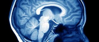
In the first days of a newborn’s life, various tests are taken, vaccinations are given, and examinations are also carried out in order to obtain complete information about the general condition of the child. One of the main procedures is ultrasound of the brain. It allows you to find out not only about any abnormalities and the degree of development of the brain, but also to check the overall dimensions of the ventricles of the brain in a newborn, the norm of which is a certain value. Examination of a baby is an important stage in his life, since disorders and pathologies that are not immediately identified can negatively affect the future life and development of the baby.
What to do if suddenly an ultrasound showed an enlargement of the ventricles of the brain in a newborn? If newborns with enlarged ventricles of the brain are in normal condition and do not have any serious neuropathological abnormalities, then a specialist can schedule regular visits to a neurologist to monitor and monitor the condition. But if the deviations from the norm are quite serious, and the neuropathological symptoms are pronounced, then the child needs special treatment, which is prescribed by a neurologist.
Causes of deviations in the development of the ventricles of the brain
At the moment, many factors are known that influence the appearance of pathologies of the ventricles of the brain in children. All of them can be divided into two categories: acquired and congenital. Acquired causes include those reasons that could arise during the pregnancy of the child’s mother:
- Infectious diseases that a woman suffers from during pregnancy.
- Infections and sepsis inside the womb.
- Penetration of foreign bodies into the brain.
- Chronic diseases of the mother that affect the normal course of pregnancy.
- Delivery ahead of schedule.
- Hypoxia of the fetus inside the womb (insufficient or, conversely, increased blood supply to the placenta).
- Abnormal duration of the dry period.
- Injury to the baby during childbirth (suffocation by the umbilical cord or deformation of the skull).
- Stormy birth.
Congenital causes include a genetic predisposition to enlarged ventricles; abnormalities occurring in chromosomes, as well as various neoplasms (cysts, malignant or benign tumors, hematomas). Along with the listed reasons, characteristic changes in the size of the ventricles of the brain can be provoked by traumatic brain injury, cerebral hemorrhage, or stroke.
Functional impairment
Age-related changes such as cerebral atherosclerosis; vascular lesions caused by toxic causes or diseases such as diabetes mellitus, dysfunction of the thyroid gland, can lead to the death of a large number of capillaries of the choroid and their replacement by growing connective tissue. Such growths are scars, which are always larger than the original area before the lesion. As a result, large areas of the brain will suffer from deterioration in blood supply and nutrition.
The surface area of affected vessels is always less than that of normally functioning vessels. In this regard, the speed and quality of metabolic processes between blood and cerebrospinal fluid decrease. Because of this, the properties of the cerebrospinal fluid change, its chemical composition and viscosity change. It becomes thicker, disrupting the activity of nerve pathways, and even puts pressure on the areas of the brain bordering the 4th ventricle. One type of such condition is hydrocephalus, or dropsy. It spreads to all areas of the liquor supply, thereby affecting the cortex, expanding the gap between the grooves, exerting a pressing effect on them. At the same time, the volume of gray matter is significantly reduced, and a person’s thinking abilities are impaired. Dropsy, affecting the structures of the midbrain, cerebellum and medulla oblongata, can affect vital centers of the nervous system, such as the respiratory, vascular and other zones of regulation of biological processes in the body, which causes an immediate threat to life.
First of all, disorders manifest themselves at the local level, as indicated by the symptoms of damage to those same pairs of cranial nerves from the fifth to the twelfth. Which, accordingly, is manifested by local neurological symptoms: changes in facial expressions, impaired peripheral vision, hearing impairment, impaired coordination of movements, speech defects, taste abnormalities, problems with spoken language, secretion and swallowing of saliva. There may be disturbances in the activity of the muscles of the upper shoulder girdle.
The causes of dropsy may lie not only at the cellular level. There are tumor diseases (primary from nervous or vascular tissue, secondary - metastasis). If the tumor occurs near the boundaries of the 4th ventricle, then the result of an increase in size will be a change in its shape, which again will lead to hydrocephalus.
Anatomy of the ventricles of the brain
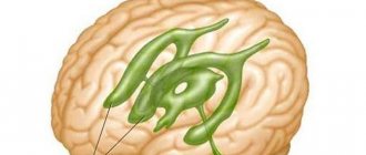
The human brain is a very complex structure, in which each substructure and each component part is responsible for fulfilling certain goals. In humans, there is a special structure in the brain that contains cerebrospinal fluid (CSF). The purpose of this structure is the circulation and production of cerebrospinal fluid. Each child and adult has 3 types of brain ventricles, and their total number is 4. They are connected to each other through channels and openings, valves. So, the ventricles are distinguished:
- Lateral.
- Third.
- Fourth.
The lateral ventricles are located symmetrically relative to each other. The left is designated first, the right is designated second, they are connected to the third. The third ventricle is the anterior one and houses the centers of the autonomic nervous system. The fourth is the posterior one, it is shaped like a pyramid and is connected to the spinal cord. Changes in the size of the ventricles entail a disorder in the production and circulation of cerebrospinal fluid, which can lead to an increase in the volume of fluid in the spinal cord and disruption of the working condition of a vital organ.
How it manifests itself
Ventriculomegaly is determined by how much intracranial pressure is increased and how the patient’s adaptive mechanisms fight this change. Mild dilatation manifests itself:
- Frequent headaches and dizziness. May be worse before bed or upon awakening.
- Nausea.
- Apathy, lethargy.
Severe dilatation manifests itself:
- Headache and dizziness.
- Nausea and vomiting.
- Drowsiness, loss of strength, lack of desire to explore the world around us.
- Sleep disturbance.
- Protrusion of a vein on the forehead due to obstructed venous outflow.
- Changes in muscle tone: tone can weaken or increase.
- Trembling of hands and feet.
- Large head, disproportionate to the body.
Ventriculomegaly in a child can manifest itself with general symptoms, for example:
- head circumference is growing too quickly;
- the fontanel bulges and pulsates;
- “setting sun” symptom: when the eyelids are raised, the child’s pupils are lowered;
- the head is often thrown back;
- in places of the skull where the bones do not meet at the seams, pulsating round protrusions are observed;
- nystagmus is a synchronous and rhythmic movement of the eyes in one direction (up to 200 movements per minute).
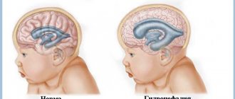
In children, severe expansion of the ventricles of the brain causes hypertensive-hydrocephalic syndrome. The essence of the pathology is a gross increase in intracranial pressure and an increase in the volume of cerebrospinal fluid. Signs of the syndrome:
- Lack of appetite, apathy, no interest in toys.
- Decreased muscle tone.
- Weakness of the basic innate reflexes of swallowing, sucking and grasping.
- The child spits up like a fountain.
- Strabismus and nystagmus.
- The sutures of the skull are not connected; pulsating protrusions protrude between them.
- Child retardation at psychosomatic age.
- Reduced intelligence and all mental abilities.
Hypertensive-hydrocephalic syndrome that does not respond to treatment leads to the following consequences:
- paresis or paralysis;
- mental retardation;
- speech disorders;
- deterioration or loss of vision.
What is “asymmetry of the lateral ventricles of the brain”? First you need to understand what the ventricles of the human brain are.
The “ventricles of the brain” are a system of special anastomizing cavities that communicate with the subarachnoid space, as well as the canal of the human spinal cord. The ventricles contain what is called cerebrospinal fluid. The reverse surface of the walls of these ventricles is covered with ependyma.
Enlarged ventricles: manifestation
As is known, one of the functions of the ventricles is the secretion of cerebrospinal fluid into the cavity between the meninges and spinal membranes (subarachnoid space). Therefore, disturbances in the secretion and outflow of fluid lead to an increase in the volume of the ventricles.
But not every increase and change in size is considered a pathology. If both lateral ventricles become symmetrically larger, then there is no need to worry. If the increase occurs asymmetrically, that is, the horn of one of the lateral ventricles increases, but the horn of the other does not, then pathological development is detected.
Enlargement of the head ventricles is called ventriculomegaly. It exists in 3 types:
- Lateral (dilatation of the right or left ventricles, enlargement of the posterior ventricle).
- Cerebellar (the size of the cerebellum and medulla oblongata changes).
- Pathological release of cerebrospinal fluid in the frontal region.
There are 3 degrees of the disease:
- Easy.
- Average.
- Heavy.
Sometimes the disease is accompanied by disruption of the central nervous system. Enlargement of the ventricles in large children with a non-standard skull shape is considered normal.
Four Hill Cistern
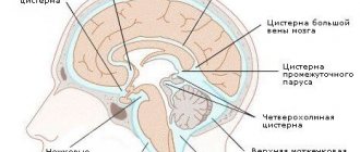
The quadrigeminal cistern is the space between the arachnoid membrane and the medulla covered by the pial membrane, filled with cerebrospinal fluid (Fig. 5).
Fig.5.
Quadrigeminal cistern (a) and subarachnoid fissures of the quadrigeminal cistern (b). 12 - arteries, 22 - vein of Galen, 149 - cerebellum, 150 - corpus callosum, 188 - quadrigeminal cistern, 215 - occipital lobe, 232 - choroid, 234 - choroid of the cerebellum, 236 - tela choroidea ventriculi tertii, 254 - connective tissue strings, 261 - subarachnoid cells, 295 - quadrigeminal plate, 297 - pineal body, (Baron M.A., Mayorova N.Functional stereo-morphology of the meninges. M. Medicine, 1982.)
Large vessels of the pineal region pass through it, surrounded by arachnoid trabeculae or strings. At the points of attachment of the strings to the great vein of the brain there are conical extensions. The strings transmit the rhythmic pulsations of the artery to the vein and protect the vein from collapsing during changes in cerebrospinal fluid pressure.
The quadrigeminal cistern is located posterior to the quadrigeminal plate and communicates superiorly with the posterior pericallosal cistern, inferiorly with the cerebellomesencephalic cistern (“precentral cerebellar cistern”), inferiorly and laterally with the posterior parts of the enveloping cistern, which is located between the midbrain and the parahypocampal gyrus, and laterally - with the retrothalamic cistern, which encircles the posterior surface of the optic thalamus cushion to the crus of the fornix.
Interpretation of the appearance of dilated ventricles
Deviation from the normal size of the ventricles does not always indicate the occurrence of pathological processes. Most often, these changes are a consequence of the anthropological characteristics of the baby. Almost all newborns up to one year of age have ventriculomegaly. It appears as a result of impaired fluid outflow or excessive accumulation of cerebrospinal fluid.
According to statistics, enlargement of the lateral ventricles is more common in children born prematurely. In them, unlike babies born at the right time, the sizes of the first and second cavities are more enlarged. If there is a suspicion of asymmetry, measurements, diagnostics and qualitative characteristics should be determined.
Symptoms of venticulomegaly
With venticulomegaly, due to a large amount of cerebrospinal fluid, the pressure inside the baby’s skull rises; swelling of the cortex, gray matter, and tissues appears. The pressure disrupts the blood supply to the brain, and deterioration and disruption of the central nervous system are also observed.
The following symptoms are observed with enlarged ventricles:
- Increased muscle activity.
- Deterioration of vision (defocus, squint, downcast gaze).
- Trembling of limbs.
- Strange gait (movement on tiptoes).
- Inactive reflexive manifestations.
- Lethargic, apathetic behavior.
- Increased moodiness and irritability.
- Insomnia, sleepwalking.
- Lack of appetite.
An obvious symptom of venticulomegaly is regurgitation and vomiting, the amount of which exceeds the norm. This occurs due to irritation of the vomiting center in the fourth ventricle, which is located at the bottom of the diamond-shaped fossa.
Bark
It is difficult to imagine that the cortex, which provides the main characteristics of consciousness and intelligence, is only 3 mm thick. This thinnest layer reliably covers both hemispheres. It is composed of the same nerve cells and their processes, which are located vertically.
The layering of the crust is horizontal. It consists of 6 layers. The cortex contains many vertical nerve bundles with long processes. There are more than 10 billion nerve cells here.
The cortex is assigned various functions that are differentiated between its different sections:
- temporal – smell, hearing;
- occipital – vision;
- parietal – taste, touch;
- frontal – complex thinking, movement, speech.
In the hemispheres themselves there are basal ganglia - these are large clusters that consist of gray matter. It is the basal ganglia that transmit information. Between the cortex and the basal ganglia are the processes of neurons - the white matter.
It is the nerve fibers that form the white matter; they connect the cortex and those formations that are under it. The subcortex contains the subcortical nuclei.
Diagnosis of the disease
Diagnostics are carried out to clarify the diagnosis. A doctor can notice a chronic form of venticulomegaly as early as three months of age using an ultrasound. The examination includes the following procedures:
- Examination by an ophthalmologist (this will reveal swelling of the eyes and hydrocephalus).
- Magnetic resonance imaging (the MRI procedure helps monitor the growth of the ventricles after the fusion of the cranial bone. To carry out the examination, which takes from 20 to 40 minutes, the baby is put to sleep with the help of drugs).
- CT scan. In this case, medicated sleep is not required because the procedure does not take much time. So CT is the best option for children who cannot tolerate anesthesia.
Ultrasound is prescribed for children born after a pregnancy during which there were complications. It is done in the first year of life, and if there are no neurological abnormalities, then it is repeated after three months.
Indicators of normal sizes
Each ventricle has certain sizes that are considered normal. Deviation from them is a pathology. So, the normal depth of the third ventricle is no more than 5 mm, the fourth ventricle is no more than 4 mm. When taking side measurements, the following values are taken into account:
- Side cavities - depth should not exceed 4 mm.
- Horns in the occipital part – 10 – 15 mm.
- The horns in the front part are 2–4 mm.
The depth of a large tank is no more than 3–6 mm. All cavities and structures of the brain must have a gradual development, consistent and linearly dependent on the size of the skull.
Treatment of the disease
Treatment can only be prescribed by a neurosurgeon or neurologist. Drug therapy is usually used. Not all episodes require treatment, but it is used in cases of pronounced neuropathological abnormalities. The main medications are:
- Diuretics are used to reduce cerebral edema, normalize and accelerate fluid excretion.
- Potassium-containing preparations compensate for the deficiency of the required amount of potassium while accelerating the process of urination.
- Vitamin complexes are used to replenish lost vitamins, as well as to restore the patient’s body.
- Nootropics improve blood supply to the brain, circulation in microtissues and vascular elasticity.
- Sedatives have a calming effect and reduce neurological signs such as tearfulness, moodiness, and irritability.
If the cause of deviations in the size of the brain cavities is mechanical damage to the head, then surgical intervention is required.

