The brain is a closed system of the body that needs protection from the external environment. The main barrier is the bones of the skull, under which several layers of shells are hidden. Their function is to create a buffer zone between the inside of the skull and the brain itself.
In addition, between the 2nd and 3rd membranes there is a functional cavity - the subarachnoid or subarachnoid space, in which cerebrospinal fluid - cerebrospinal fluid - constantly circulates. With its help, the brain receives the necessary amount of nutrients and hormones, and also removes metabolic products and toxins.
The synthesis and control of the release of cerebrospinal fluid is carried out by the ventricles of the brain, which are an open system of cavities lined from the inside with a layer of functional cells.
What is a cerebral ventricle
Anatomically, the ventricular system of the brain is a collection of cisterns of brain sections through which cerebrospinal fluid circulates through the subarachnoid space and the central spinal canal. This process is carried out due to a thin layer of ependymocytes, which, with the help of cilia, provoke fluid movement and control the filling of the ventricular system. They also produce myelin, which serves as a sheath for the myelinated fibers of white matter.
The ventricles are also responsible for performing secretory and cleansing functions: the ependyma cavity lining them not only produces cerebrospinal fluid, but also filters it from metabolic products, toxic and medicinal substances.
How much cerebrospinal fluid the ventricles secrete and their size are influenced by many factors: the shape of the skull, the volume of the brain, the physical condition of the person and the presence of concomitant diseases of the central nervous system, for example, hydrocephalus or ventriculomegaly.
Experts have calculated that in a healthy person, the volume of cerebrospinal fluid released per hour is approximately 150-160 ml, and it is completely renewed after 7-8 hours. In total, the ventricular system secretes about 400-600 ml of cerebrospinal fluid per day, but this figure may vary depending on the blood pressure and psycho-emotional state of the person.
Modern methods of studying the structure of the brain make it possible to study its internal structures without resorting to direct opening of the skull. If a specialist needs to obtain information about the size of the child’s lateral ventricles, he will give a referral for neurosonography, a method of examining the brain using ultrasound equipment. If an examination is required for an adult, then he is given an MRI or CT scan of the relevant departments.
Table of norms for the size of structures of the ventricular system of an adult when examining the brain using X-ray computed tomography
| Structure | Normal, mm. |
| front horns of the side tanks | 2-5 |
| lateral groove | 3-5 |
| III ventricle | 2,5-4,5 |
| IV ventricle | 12-14 |
Also, to assess the condition of the ventricular system of an adult, the condition index of each of its parts is calculated separately.
Table of indices of the IV ventricle, bodies and anterior horns of the lateral ventricles
| Age | Body of the lateral ventricles | Anterior horns of the lateral ventricles | IV ventricle |
| Up to 50 years | 18,4-22 | — | — |
| After 50 years | 22,6-26 | — | — |
| Up to 60 years old | — | 24-26,3 | 11,3-13 |
| After 60 years | — | 28,2-29,4 | Doesn't change |
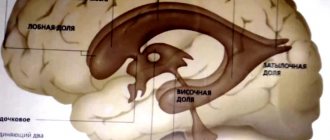
How many ventricles does a person have, their structure and functions
The ventricular system of the brain consists of 4 cavities through which cerebrospinal fluid is produced and circulates between the structures of the central nervous system. Sometimes specialists, when examining the structures of the central nervous system, discover the 5th ventricle, which is not one - it is a slit-like hypoechoic expansion located in the midline of the brain. Such an abnormal structure of the ventricular system requires attention from doctors: often patients with a 5th ventricle are at increased risk of developing mental disorders.
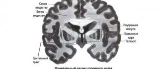
Anatomically, the first and second ventricles are located in the lower part of the left and right hemispheres, respectively. Each of them is a C-shaped cavity located below the corpus callosum and encircling the posterior part of the cluster of nerve ganglia of the subcortical structures of the brain. Normally, the volume and, accordingly, the size of the lateral ventricle of an adult should not exceed 25 ml. These cavities do not communicate with each other, but each has a channel through which the cerebrospinal fluid enters the third ventricle.
The third ventricle has the shape of a ring, the walls of which are the thalamus and hypothalamus. In the brain, it is located between the optic thalamus, and in its center is the intermediate mass of the visual thalamus. Through the aqueduct of Sylvius it communicates with the cavity of the 4th ventricle, and through the interventricular foramina with the 1st and 2nd ventricles.
Topographically, the 4th ventricle is located between the structures of the posterior section and the so-called rhomboid fossa, the posteroinferior angle of which opens into the central canal of the spinal cord.
The structure of the internal layer of the structures of the ventricular system is also heterogeneous: in the first and second ventricles it is a single-layer ependymal membrane, and in the third and fourth there may be several layers of it.
The cytological composition of the ependyma is uniform throughout: it consists of specific neuroglial cells - ependymocytes. They are cylindrical cells, the free end of which is covered with cilia. With the help of vibration of the cilia, the flow of cerebrospinal fluid through the structures of the central nervous system is carried out.
Not so long ago, at the bottom of the third ventricle, experts discovered another type of ependymocyte - tanycytes, which differ from the previous ones in the absence of cilia and the ability to transmit data on the chemical composition of the cerebrospinal fluid to the capillaries of the pituitary portal system.
Lateral ventricles 1 and 2
Anatomically, the lateral or lateral ventricles of the brain consist of a body, anterior, posterior and inferior horn.
The central part of the lateral ventricle looks like a horizontal slit. Its upper wall is formed by the corpus callosum, and in the lower part there is the caudate nucleus, the dorsum of the thalamus and the posterior peduncle of the fornix. Inside the cavity of the lateral ventricles there is a choroid plexus, through which cerebrospinal fluid is synthesized.
Outwardly, it resembles a strip of dark red color 4 mm wide. From the central part, the choroid plexus is directed to the posterior horn, the upper wall of which is formed by the fibers of the large forceps of the corpus callosum, and the rest is the white matter of the occipital part of the telencephalon.
The inferior horn of the lateral ventricle is located in the temporal lobe and is directed downward, anterior and medial to the central line. On the side and above it is limited by the white matter of the temporal lobe; the medial wall and part of the lower one forms the hippocampus.
Anatomically, the anterior horn is a continuation of the body of the lateral cavity. It is directed laterally forward relative to the central cavity of the ventricle, and on the medial side it is limited by the wall of the transparent septum, and on the side by the head of the caudate nucleus. The remaining sides of the anterior horn form the fibers of the corpus callosum.
In addition to the main functions - synthesis and circulation of cerebrospinal fluid, the lateral ventricles are involved in the restoration of brain structures. Until recently, it was believed that nerve cells are not capable of renewal, but this is not entirely true: between the lateral ventricle and the olfactory bulb of one hemisphere there is a channel, inside which scientists have discovered an accumulation of stem cells. They are able to migrate inside the olfactory bulb and take part in restoring the number of neurons.
Physiometric indicators of the lateral ventricles (namely their size) can be taken in several ways. Thus, in children of the first year of life, examination is carried out using neurosonography (NSG), and in adults - using MRI or CT. Then the obtained data is processed and compared with standard indicators.
The lateral ventricles of the brain are normal in a child:
| Ventricular structure | Newborn, mm | 3 month old baby, mm |
| Body | Up to 4 | 2-4 |
| Front horns | 2-4 | Up to 4 |
| Occipital horns | 10-15 | Up to 15 |
These indicators are taken into account when diagnosing brain pathologies, for example, hydrocephalus or hydrocephalus of the medulla - a disease that is characterized by increased secretion of cerebrospinal fluid and disruption of its outflow, which leads to increased pressure on the walls of the ventricles and expansion of their cavities.
To reduce the risk of developing pathology, the first examination of the child’s brain is carried out during his intrauterine development during screening examinations. This makes it possible to identify central nervous system diseases at an early stage. For example, during such a study, asymmetry of the lateral ventricles of the embryo may be detected. This approach allows specialists to prepare and immediately begin carrying out therapeutic measures immediately after the birth of the child.
3rd ventricle of the brain
Topographically, the third ventricle of the brain is located at the level of the intermediate section, between the visual thalamus, surrounding the intermediate mass of the visual thalamus with a ring. Has 6 walls:
- Roof. It is formed by a strip of epithelium and a vascular tegmentum, which is a continuation of the pia mater, which serves as the basis of the choroid plexus of the 3rd ventricle. This structure penetrates the lateral cisterns through the interventricular foramina in the upper part, forming its own choroid plexus in them.
- The surface of the visual tuberosities serves as the lateral walls, while the inner part of the ventricle is formed due to the germination of the intermediate mass.
- The anterior upper wall is formed by the columns of the fornix of the brain and its white anterior commissure, and the lower wall is formed by the terminal gray plate, which is located between the columns of the fornix.
- From the back, the third ventricle is limited by a commissure located above the opening of the entrance to the Sylvian aqueduct. At the same time, the rear part on top is formed by a pineal-shaped depression and a soldering of wires.
- The bottom of the third ventricle is the base of the brain in the area of the posterior perforated substance, mastoid bodies, gray tubercle and optic chiasm.
The physiological significance of the third ventricle is that it is a cavity whose walls contain autonomic centers. For this reason, an increase in its volume and abnormal structure can cause deviations in the processes of excitation and inhibition of the autonomic nervous system, which is responsible for the physical condition of a person. For example, if the third ventricle of the brain is dilated, this affects the functioning of the structures of the circulatory, respiratory and endocrine systems.
Standards for the size of the third ventricle in a child:
| Structure | Newborn | 3 month old baby |
| III ventricle | Up to 3 mm | Up to 3.3 mm |
4th ventricle of the brain
Anatomically, the fourth ventricle is located between the cerebellum, the posterior surface of the pons and the medulla oblongata, in the so-called rhomboid fossa. At the embryonic stage of child development, it is formed from the remains of the hindbrain vesicle, and therefore serves as a common cavity for all parts of the hindbrain.
Visually, the IV ventricle resembles a triangle, the bottom of which is the structures of the medulla oblongata and the pons, and the roof is the upper and lower velum. The superior velum is a thin membrane stretched between the superior cerebellar peduncles, and the inferior velum is adjacent to the peduncles of the cerebellum and is complemented by a plate of soft membrane, which forms the choroid plexus.
The functional purpose of the IV ventricle, in addition to producing and storing cerebrospinal fluid, is to redistribute its flow between the subarachnoid space and the central canal of the spinal cord. In addition, in the thickness of its bottom are located the nuclei of the V-XII cranial nerves, which are responsible for the work of the muscles of the corresponding muscles of the head, for example, oculomotor, facial, swallowing, etc.
5th ventricle of the brain
Sometimes in medical practice there are patients who have a V ventricle. Its presence is considered a structural feature of the ventricular system of an individual and is more of a pathology than a variant of the norm.
The walls of the fifth ventricle are formed by fusion of the internal parts of the membranes of the cerebral hemispheres, while its cavity does not communicate with other structures of the ventricular system. For this reason, it would be more correct to call the resulting niche the cavity of a “transparent partition”. Although the fifth ventricle does not have a choroid plexus, it is filled with cerebrospinal fluid, which enters through the pores of the septa.
The size of the V ventricle is strictly individual for each patient. In some, it is a closed and autonomous cavity, and sometimes in its upper part there is a gap up to 4.5 cm long.
Despite the fact that the existence of a cavity of the septum pellucidum is an anomaly in the structure of the adult brain, its presence is obligatory at the embryonic stage of fetal development. Moreover, in 85% of clinical cases it heals by the age of six months.
What diseases can affect the ventricles
Diseases of the ventricular system of the brain can be either congenital or acquired. Experts include hydrocephalus (water on the brain) and ventriculomegaly to the first type. These diseases are often the result of improper development of the child’s brain structures during the embryonic period due to a previous chromosomal malfunction or infection of the fetus with infections.
Hydrocephalus
Dropsy of the brain is characterized by improper functioning of the ventricular system of the head - excessive secretion of cerebrospinal fluid and its insufficient absorption into the bloodstream by the structures of the occipital-parietal zone. As a result, all cavities and the subarachnoid space are filled and, accordingly, put pressure on other structures, causing encephalopathic destruction of the brain.
In addition, due to increased intracranial pressure, the bones of the skull are displaced, which is visually expressed in an increase in head circumference. The strength of the manifestations of the symptomatic signs of hydrocephalus depends on how strong the deviation is in the system of production and absorption of cerebrospinal fluid: the more pronounced this discrepancy, the stronger the manifestations of the disease and the destruction of brain matter.
Sometimes, if untreated, the head grows so quickly that the patient cannot cope with its severity and remains bedridden for the rest of his life.
A person can get hydrocele at any age, but most often it occurs in children, being a congenital disease. In the adult population, pathology usually occurs due to a violation of the outflow of cerebrospinal fluid due to head trauma, infection of the meninges, the occurrence of a tumor and toxic poisoning of the body.
Clinical manifestations of hydrocephalus include the development of neurological disorders of varying severity in the patient and a change in the volume of the cranium, which is noticeable to the naked eye:
Since the bones of the head of a child in the first year of life are plastic, an increase in the amount of cerebrospinal fluid deforms it, which is visually expressed not only in an increase in the volume of the head due to the divergence of the seams of the bones of the cranial vault, but also in the enlargement of the frontal bone.
A child with hydrocephalus usually experiences swelling and bulging of the fontanelles due to increased intracranial pressure.
There are also other external signs of hydrocephalus:
- lack of appetite;
- pronounced vascular network on the bridge of the nose;
- hand tremors;
- premature extinction of the sucking and swallowing reflex;
- profuse and frequent regurgitation;
- swelling and protrusion of the fontanelles.
Neurological disorders manifest themselves in the development of strabismus, nystagmus of the eyeballs, deterioration in clarity of vision, hearing, headaches, weakness of the muscles of the limbs in combination with hypertonicity.
In adults and children over 2 years of age, the development of dropsy is signaled by the appearance of morning headaches, vomiting, severe swelling of the optic discs, paresis and other disturbances in coordination of movements.
Hydrocephalus is diagnosed using modern neuroimaging methods. Typically, enlargement of the cerebral ventricles in the fetus is noticed during a screening ultrasound and then confirmed after birth by neurosonography.
In adults, the diagnosis is made during an examination of brain structures using MRI or CT, and in this case, the X-ray examination method will be more informative, since it allows, if necessary, to identify the place of bleeding in the cavity of the ventricles, due to damage or rupture of the blood vessels of the ventricular wall.
Treatment tactics for dropsy of the brain depend on the severity. For small to moderate accumulations of cerebrospinal fluid, specialists carry out drug therapy aimed at reducing the amount of fluid in the brain by taking diuretics.
The work of the nerve centers is also stimulated using physiotherapeutic procedures. Severe pathology requires immediate surgical intervention, which is aimed at reducing intracranial pressure and draining excess fluid from brain structures
Ventriculomegaly
Ventriculomegaly or pathological expansion of the lateral ventricles of the brain is a congenital disease, the true causes of which are still unknown. However, it is believed that the risk of having a child with this disorder increases in women over 35 years of age.
The impetus for the development of pathology can be intrauterine infection of the fetus, trauma to the abdomen of a pregnant woman and uterine bleeding, due to which the child ceases to receive the required amount of nutrients. Often, pathological enlargement of the ventricles of the brain in the fetus is a concomitant disease of other defects of the child’s central nervous system.
Clinically, the expansion (dilatation) of the lateral ventricles manifests itself in the development of neurological abnormalities, since the increased volume of cerebrospinal fluid constrains and puts pressure on the internal structures of the brain. The patient may also experience psycho-emotional disorders, schizophrenia and bipolar disorder.
Ventriculomegaly can be unilateral or bilateral, while a symmetrical and slight increase in the lateral cisterns can be a normal variant and be a feature of the structure of the child’s brain. For newborns, this diagnosis is made only when the dimensions of the diagonal sections of the ventricles at the level of the foramen of Monroe exceed 0.5 cm from the accepted norms.
Pronounced asymmetry of the ventricles requires close attention from specialists - after all, an enlarged cistern on one side upsets the balance of cerebrospinal fluid production. Typically, a child with ventriculomegaly lags behind in development: he begins to speak and walk later, has poor fine motor skills, and also experiences constant headaches. The volume of the skull also grows, and the difference between it and the chest can be more than 3 cm.
Treatment tactics for a child with ventriculomegaly depend on the severity of the disease. Thus, with a mild deviation, the child remains under the supervision of the attending physician; a moderate degree of pathology requires drug treatment and physiotherapeutic procedures aimed at compensating and correcting the neurological manifestations of the disease.
To normalize brain function, the child is prescribed nootropic drugs that improve brain activity, diuretics that reduce intracranial pressure, antihypoxants, potassium-sparing drugs and vitamin complexes.
In case of severe ventriculomegaly, the child requires surgical treatment, which consists of inserting a drainage tube into the ventricles of the brain.
Other causes of pathology of the ventricles of the brain
Dilatation of the cavities of the ventricular system can be caused by damage to brain structures by tumor-like neoplasms or inflammation of its individual parts.
For example, adequate outflow of cerebrospinal fluid may be impaired due to inflammation of part of the soft membrane due to brain damage from meningococcal infection. The basis for damage to the central nervous system by this disease is first the poisoning of the brain vessels with toxins that are released by the infectious agent.
Against this background, tissue edema develops, while bacteria penetrate into all structures of the brain, causing its purulent inflammation. As a result, the membranes of the medulla swell, the convolutions become flattened, and blood clots form inside the vessels, which block the flow of blood, causing multiple cerebral bleeding.
And although this disease is fatal, timely treatment can stop the process of destruction of white matter by infectious agents. Unfortunately, even after a person has completely recovered, there is a risk of developing cerebral hydrocele and, accordingly, enlargement of the cavities of the ventricles of the brain.
One of the complications of meningococcal infection is the development of ependymatitis, or inflammation of the inner lining of the ventricles. It can occur at any stage of the infectious-inflammatory process, regardless of the stage of treatment.
In this case, the clinical course of the disease is no different from the manifestations of meningoencephalitis: the patient experiences drowsiness, prostration, stoppage, or falls into a coma. He also has muscle hypertonicity, tremors of the limbs, convulsions, and vomiting.
In young children, the accumulation of cerebrospinal fluid causes increased intracranial pressure and secondary hydrocephalus of the brain. To make an accurate diagnosis and identify the pathogen, specialists take a puncture of the contents of the ventricles, and in children this procedure is carried out through the fontanelle, and in adults they perform craniotomy
The cerebrospinal fluid puncture specimen for ependymitis is yellow, it contains a large number of pathogenic bacteria, proteins and polynuclear cells. If in the future the disease cannot be treated, then due to the accumulation of a large amount of fluid, all structures and autonomic centers of the brain are subject to compression, which can lead to respiratory paralysis and death of the patient.
The appearance of tumors in the structures of the brain can also cause disruption of the secretion of cerebrospinal fluid and abnormalities in the functioning of the ventricles of the brain. Thus, on the inside of the cisterns and along the outflow pathways of cerebrospinal fluid, ependymoma may appear - a malignant tumor of the central nervous system, which is formed from atypical cells of the ependymal layer. The situation is complicated by the fact that this type of neoplasm can metastasize to other parts of the brain through the cerebrospinal fluid circulation channels.
The clinical picture of the disease depends on where the tumor is located. So, if it is in the lateral tanks, then this manifests itself in increased intracranial pressure, apathy, excessive drowsiness, etc.
As the situation worsens, the patient becomes disoriented, memory impairment, mental disorders, and hallucinations occur. If the tumor is located close to the interventricular foramen or covers it, then the patient may develop unilateral hydrocele of the brain, since the affected ventricle ceases to participate in the circulation of cerebrospinal fluid.
When the fourth ventricle is affected by ependymoma, the patient experiences pronounced neurological abnormalities, since the resulting tumor puts pressure on the cranial nuclei located in its bottom. Visually, this manifests itself in eye nystagmus, paralysis of the facial muscles and impaired swallowing. The patient also experiences headache, vomiting, tonic convulsions or decerebrate rigidity.
In older people, disruption of the ventricular system can be caused by atherosclerotic changes, since as a result of the formation of cholesterol plaques and thinning of the vessel walls, there is a risk of developing cerebral bleeding, including in the ventricular cavity.
In this case, a burst vessel provokes the penetration of blood into the cerebrospinal fluid, which will cause a violation of its chemical composition. Excessive intraventricular hemorrhage can provoke the development of cerebral edema in the sick person with all the ensuing consequences: increasing headache, nausea, vomiting, decreased visual acuity and the appearance of a veil before the eyes.
In the absence of medical care, the patient's condition quickly deteriorates, convulsions appear, and he falls into a coma.
Colloid cyst of the 3rd ventricle
Colloid cyst of the 3rd ventricle is a disease common among adults 20-40 years old. It is characterized by the appearance of a benign round formation in the cavity of the 3rd ventricle, which is not prone to rapid growth and metastasis.
The colloid cyst itself does not pose any danger to human health. Problems begin if it reaches a large size and interferes with the outflow of cerebrospinal fluid. In this case, the patient experiences neurological symptoms associated with increased intracranial pressure:
- severe headache;
- vomit;
- visual impairment;
- convulsions.
Diagnosis and treatment of colloid cyst of the third ventricle is carried out jointly by a neurologist and a neurosurgeon. If the size of the formation is pronounced, determined on CT or MRI, surgical treatment of the cyst is prescribed. After the operation, the normal flow of cerebrospinal fluid is quickly restored, and all symptoms of the disease disappear.
Features of the third ventricle
The 3rd ventricle of the brain is the connecting link between the lateral cisterns and the lower part of the human ventricular system. The cytological composition of its walls is no different from the structure of similar brain structures.
However, its functioning is of particular concern to doctors, since the walls of this cavity contain a large number of autonomic nerve nodes, the functioning of which determines the functioning of all internal systems of the human body, be it breathing or blood circulation. They also maintain the state of the body’s internal environment and participate in shaping the body’s response to external stimuli.
If a neurologist suspects the development of a pathology of the third ventricle, he will refer the patient for a detailed examination of the brain. In children, this process will take place as part of a neurosonological study, and in adults, with the help of more accurate neuroimaging methods - MRI or CT scan of the brain.
Normally, the width of the third ventricle at the level of the aqueduct of Sylvius in an adult should not exceed 4-6 mm, and in a newborn – 3-5 mm. If the subject exceeds this value, then experts note an increase or expansion of the ventricular cavity.
Depending on the severity of the pathology, the patient is prescribed treatment, which may consist of medicinal attenuation of the neurological manifestations of the pathology or the use of surgical treatment methods - shunting the cavity in order to restore the outflow of cerebrospinal fluid.
Indications for NSG
Neurosonography in newborns is performed in maternity hospitals. The study is prescribed if the following indications exist:
- Prematurity. This term is used to refer to the condition of a fetus born before the end of the normal period of intrauterine development. A baby born is considered premature if the gestational age (the period lasting from the first day of the last menstrual period until birth) is less than 36 weeks.
- Low results of assessing the condition of the newborn. It is performed using the Apgar score at 1-5 minutes of life. Within normal limits, this indicator should be equal to 7 points. NSG is carried out in cases where the child receives less than 7 points 5 minutes after birth.
- Low body weight. In newborns, this figure can be within the normal range from 3 to 3.5 kg. Minor deviations are allowed. Body weight not exceeding 2800 g indicates the possible presence of serious pathologies. With this indicator value, NSG is carried out.
Indications for the study are also a history of chronic intrauterine hypoxia, infectious diseases in the newborn child and his mother, and asphyxia that occurred during childbirth. Neurosonography is also necessary in the presence of clinical signs of dysfunction of the central nervous system (constant trembling, tremors of the limbs and chin, decreased motor activity), multiple stigmas of disembryogenesis (small deviations in the anatomical structure of any organs).
Neurosonography of the brain
After discharge from the maternity hospital, NSG is prescribed to children in the 1st month of life. The study is carried out in a children's clinic. After the 1st month of life, neurosonography in children is performed for the same indications as in newborns (prematurity, low birth weight, signs of central nervous system damage and multiple stigmata of disembryogenesis). Repeated examinations are prescribed if indicated and to assess the effectiveness of treatment.