The course of neuropathy of the peroneal nerve is characterized by impaired sensitivity in the lower leg area. With such a lesion, the patient is unable to bend the foot and its toes. Tunnel syndromes of the lower extremities develop due to compression of local nerve fibers. Compression occurs against the background of injuries or other damage to the legs, as well as under the influence of pathological processes. Neuropathy is treated with medications, physical therapy exercises, or joint surgery.
Description of the disease
Peroneal neuropathy, or peronial neuropathy, is one of the most common neurological diseases. Disease code according to ICD-10 G57 – mononeuropathies of the lower extremities.
Neuropathy occurs equally frequently on both the right and left peroneal nerves.
The myelin sheath of a thickened dense nerve is much thicker than that of others. It arises from the lower 1/3 of the ischial, descending through the popliteal fossa and passing on the anterior side of the leg, dividing into internal and external branches, innervating the dorsum of the foot. The anterior branch is divided into motor and cutaneous branches, innervating the skin of the leg, foot, interdigital spaces and fingers.
Thanks to them, the foot and toes are extended and its outer edge is raised. Often, trauma to the nerve occurs at the point where it enters the foot - in the area of the head of the fibula.
With acute or chronic hypoxia or compression, damage to the nervous tissue and demyelination occurs, as a result of which the passage of impulses through the fibers is disrupted until they are completely absent. Due to their absence, the functioning of the foot is disrupted: it is impossible to bend and straighten it - foot drop syndrome. The disease is also accompanied by impaired sensitivity of the back of the foot and the skin of the lower leg in front.
According to statistics, women are more susceptible to the disease. Pathology is more often detected in girls and girls aged 10-19 years.
Causes of neuralgia
The development of neuritis of the peroneal nerve is caused by exposure to the external environment or the course of diseases.
Based on these features, the disease is classified into primary or secondary neuropathy, respectively.
The most common causes of peroneal nerve syndrome include:
- bruises;
- fractures;
- blows;
- fiber compression.
More often, neuropathy develops against the background of damage to the upper outer part of the leg, since the peroneal nerve lies directly under the skin. Compression of local fibers (tunnel syndrome) is also considered a common cause of neuritis. Such violations arise under the influence of various reasons. Lower limb tunnel syndrome is diagnosed in people who often sit with their legs crossed or have worn a cast for a long time.
In addition to post-traumatic neuropathy of the peroneal nerve, neuritis is caused by:
- nerve ischemia (impaired blood supply);
- prolonged immobilization (for example, long periods of lying down);
- infectious diseases;
- general articular pathologies that provoke compression of nerve canals;
- course of tumor processes;
- toxic damage to the body caused by renal failure and other factors.
The appearance of neuropathy can be caused by errors during intramuscular injections, when the needle touches the peroneal or sciatic nerves.
Cause of occurrence
In most cases, acute oxygen starvation up to anoxia leads to destructive processes in the myelin sheath, disrupting tissue metabolism. Often, this occurs for the following reasons:
- injuries;
- compression;
- vascular pathology;
- infections;
- toxins.
Peroneal nerve neuropathy occurs after injury to the knee, ankle, fibula, or lower leg. These can range from minor bruises to severe dislocations or fractures.
Compression occurs due to compression of the fiber by musculoskeletal structures. Often the compression form of the pathology occurs in people whose work activity requires prolonged squatting. For example, paving slab or parquet layers, berry and vegetable pickers, and others. In this position of the body, compression and disruption of trophism occurs. Another name for compression neuropathy is “tunnel syndrome.”
With vascular pathologies, a lack of oxygen and nutrients depletes the tissues of the lower limb.
Infections and toxins destroy the myelin sheath and tissue.
Reference . In some cases, damage occurs during surgery unrelated to neuritis. This complication is one of the most common when performing operations on the knee joint, lower leg and ankle.
Symptoms
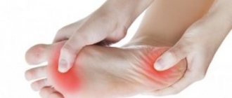
This is what a foot with damaged peroneal nerve looks like.
Clinical manifestations of peroneal nerve neuropathy depend on the location of the lesion and the form of the disease. Thanks to specific signs, doctors are able to accurately determine the location of the pathological process.
Signs of high compression
A characteristic sign of compression of a nerve before its branching (in the area of the popliteal fossa) is inhibition of all its functions at once, since the impulse does not pass through any of its branches. The most frequently identified complaints are:
- pain on the lateral part of the lower leg, intensifying during squats;
- inability to straighten the foot and toes;
- violation of abduction of the outer edge of the foot;
- the foot droops down and bends inwards - “horse foot” syndrome;
- the patient cannot stand or walk on his heels, stepping only on his toes;
- loss of sensation on the front surface of the leg;
- Chronic compression leads to atrophy of the leg and foot muscles, causing the affected leg to lose weight.
Their severity and intensity depend on the intensity of compression. So, with strong compression, the nerve impulse does not pass through the tissues, completely stopping the performance of those functions for which the nerve is responsible. The gait changes completely, and a characteristic lameness appears. To move your leg, you have to bend your knee strongly to avoid damaging your heel.
Compression of the external cutaneous nerve
Symptoms are mild due to the additional innervation of this area by the tibial nerve. Patients complain of decreased sensitivity of the skin of the lower leg and may not perceive minor touches. There is slight numbness of the skin.
Damage to the superficial peroneal nerve
The main symptom is the appearance of pain and burning on the lower leg and the back of the foot and the first four toes. Due to decreased sensitivity, it is difficult to lift and abduct the heel, their gait takes on a characteristic appearance - in order not to catch the heel, the patient strongly bends the leg at the knee joint, moves it forward and stands first on the toes, and then on the heel.
Deep branch lesion
It is difficult for the patient to straighten the foot and its toes due to severe muscle weakness, which is accompanied by their drooping. There is a significant decrease in sensitivity on the back of the skin and the surface of the fingers. Light touches and tingling sensations are not felt, depression of sensitivity is accompanied by a feeling of numbness. Muscle atrophy and a decrease in leg size indicate a long period of disease.
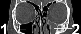
What diseases are visible on MRI of nerves?
If we consider MRI as a method for studying nerve trunks, it can be used to recognize both the problem of the state of a specific nerve or nerve plexus, and more global problems of surrounding structures (brain, spinal cord) that affect the functioning of the periphery. So these could be:
- Abnormalities of nerve or vascular development
- Trauma, nerve damage
- Hemorrhages, spinal cord compression
- Pinching and inflammation (neuritis) of nerve endings
- Atrophies, vascular structure disorders (malformations, cavernous angiomas, aneurysms)
- Disturbances of normal blood circulation
- Hydrocephalus of the brain
- Infectious diseases, their consequences
- Oncological neoplasms (including in the early stages, before the development of clinical manifestations)
Diagnostics
A neurologist can make a diagnosis based on the collected history, complaints and symptoms, and the results of instrumental and laboratory examinations. The following examination methods are often used:
- electromyography;
- electroneurography;
- Ultrasound.
Also, the doctor must conduct a series of tests using a special needle to determine the preservation of reflexes, the level of sensitivity reduction, the speed of impulse transmission, and others.
A patient with trauma is additionally given x-rays of bones and joints.
Treatment
Therapy is aimed at eliminating neuropathy, normalizing the functioning of muscle tissue and relieving symptoms. Often, this requires eliminating the cause of the pathology. Depending on the nature and course of the disease, doctors determine the tactics for managing the patient. In some cases, symptomatic drug therapy is sufficient, but an integrated approach is required to achieve the desired result.
Important! To improve the condition and prevent exacerbation, he recommends wearing only comfortable orthopedic shoes that ensure the anatomically correct position of the foot.
Drug therapy
With the help of drugs, it is possible to relieve inflammation and swelling after injury, improve blood circulation in the lower extremities and ensure normal trophism and oxygen supply to the nerve. Most often, patients are prescribed the following groups of drugs:
- non-steroidal anti-inflammatory drugs - by relieving inflammation, they eliminate swelling and pain;
- B vitamins – improve trophism of the nervous system;
- drugs that improve the passage of nerve impulses - help restore limb function;
- vascular agents – improve the condition of the vascular wall, improving blood circulation;
- antioxidants – necessary during the recovery period during rehabilitation.
Only the attending physician can prescribe medications and a regimen for their use after a thorough examination.
Physiotherapy
With the help of various physiotherapeutic procedures, it is possible to achieve a significant improvement in the condition of the nervous tissue and its functioning. The following physiotherapy procedures are most effective:
- therapeutic massage – improves blood circulation and oxygen saturation of tissues. Helps restore skin sensitivity, strengthens and restores atrophied muscles;
- magnetic therapy - activates the microvasculature and metabolic processes, helping to restore nerve conduction. Reduces pain, improves muscle condition;
- electrophoresis - used to achieve greater effect from drug therapy. Medicines are injected directly into the affected area using an electric current;
- electrical stimulation - electric current excites the cells of the neuromuscular system, helping to improve their functioning.
Mud, healing baths and other methods are also used.
Exercise therapy
Therapeutic exercises are necessary for patients with peronial neuropathy during the rehabilitation period. Active muscle contraction promotes increased blood circulation and saturation of the affected tissues with oxygen and nutrients. Thanks to this, inflammatory processes are eliminated, pain is reduced and skin sensitivity is improved. Enriching the MBN with oxygen improves its condition and ensures normal conduction of impulses.
Physical exercise is indispensable for muscle atrophy. By activating their work, they will help restore muscle mass.
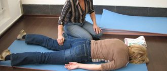
Depending on the severity of the disease, the exercises are performed lying down or standing. One of the simple exercises while lying down is to imitate walking.
A rehabilitation specialist will help you choose the most optimal set of exercises, taking into account your physical fitness and general health.
In case of severe damage to the leg and severe muscle atrophy, patients are prescribed to wear special orthopedic braces: orthoses.
Watch a video about therapeutic exercises for peroneal nerve neuropathy.
Surgical treatment
Surgical correction of the pathology is carried out only in severe cases, when the innervation of the leg below the knee has completely ceased. Surgery is also indicated when other treatment methods are ineffective and for outdated neuropathies.
Surgery is also performed for post-traumatic neuropathy.
The purpose of the operation is to restore the integrity of the nerve structure if it is ruptured. If there is a tendency to compression, the location of the tendons or nerve may change.
Structure of the human nervous system
Before delving into the pathological physiology and diagnosis of nerve damage, let us explain the features of the structure of the human nervous system as a whole.
Moving from the general to the specific, we distinguish between its central and peripheral parts. The central includes the brain and spinal cord, the peripheral consists of somatic and autonomic components. From the point of view of physics, the brain and spinal cord are the main computer of actions (idea of action), and the periphery provides communication with the body’s receptors, muscles, joints (implementation of the idea of action).
The somatic nervous system is consciously controlled and implements the transmission of a nerve command signal from an external stimulus to the central computer via ascending sensory fibers, thereby triggering a whole cascade of chemical and biological reactions. And vice versa, from the command center along descending motor fibers to a limb or its specific element (example: having touched a hot object, we feel pain, the signal quickly arrives and is processed by the central nervous system, transmitting an impulse about the necessary action - to pull the hand away from the source of danger). For us in everyday life this happens reflexively. We don’t even think about how well everything works in our body, and what valuable work is carried out in a matter of milliseconds.
In addition to consciously controlled functions, the nervous system is capable of operating on “autopilot”, controlling those processes that we are not able to decide consciously. First of all, this is the work of internal organs. Even when we sleep, the body continues to function: blood vessels, heart, respiratory and digestive organs, urination and internal secretion. All this is controlled by the autonomic nervous system, its sympathetic and parasympathetic parts. Sympathetic is a self-defense weapon, and parasympathetic is a mode of rest and relaxation.
Complications and consequences
Peroneal nerve neuropathy is not life-threatening for the patient and does not affect its duration. But its manifestations significantly worsen the quality of life.
Complications arise in the absence of proper comprehensive treatment of the problem. If innervation is not restored, muscle weakness will remain, a symptom of dropped leg. Progressive muscle atrophy can cause severe lameness and pain.
Important! The sooner you start restoring atrophied muscles, the greater the chance of returning them to their normal state. Completely atrophied muscles are extremely difficult to restore.
Disruption of innervation for a long time can be complicated by damage to the musculoskeletal system. The development of arthrosis significantly worsens the prognosis of recovery, accompanied by persistent severe joint deformation and severe pain.
Anatomy of the peroneal nerve
The peroneal nerve originates from the sacral plexus. Nerve fibers are part of the sciatic nerve; at the level of the knee joint, the nerve bundle is divided into two: the tibial and peroneal nerves, which connect in the lower third of the leg into the sural nerve.
Peroneal nerve neuritis is an inflammatory disease that occurs most often after hypothermia or injury to the lower leg.
The most common symptoms are impaired movement with atrophy of the lower leg muscles, sagging of the foot and the inability to stand on the heel, and changes in sensitivity. An experienced neurologist needs only an examination to make a diagnosis.
Combined drug and physiotherapeutic treatment is used.
The peroneal nerve consists of several trunks and innervates the extensor muscles, the muscles that allow external rotation of the foot, and the muscles of the toes.
What do you need to remember?
- Peroneal nerve neuropathy is a lesion of the nerve tissue in any of its areas.
- Most often, pathology occurs due to injury and compression of the nerve.
- The main manifestations are impaired expansion of the foot and fingers, impaired sensitivity of the anterior surface of the lower leg.
- The diagnosis is established after a thorough examination of the patient and a series of neurological tests.
- Complex treatment with drug therapy, physical therapy, exercise therapy and surgical correction allows you to completely restore the functioning of the affected structure.
- Often, the disease can be completely eliminated.
Literature
- Averochkin A.I., Shtulman D.R. Tunnel neuropathies // Journal. neuropathol. and psychiatrist, named after. S.S. Korsakov. -1991. No. 4. — P.3-6.
- Akimov G.A., Odinak M.M. Differential diagnosis of nervous diseases: A guide for doctors. St. Petersburg: Hippocrates, 2000. - 664 p.
- Voznesenskaya T.G. Pain syndromes in neurological practice //ed. AM V;yn. M.: Medpress, 2001. - 368 p.
- Flores LP, Koerbel A., Tatagiba M. Peroneal nerve compression resulting from fibular head osteophyte-like lesions. Surg Neurol 2005; 64(3):249-52.
- Perkins AT, Morgenlander JC Endocrinologic causes of peripheral neuropathy. Pins and needles in a stocking-and-glove pattern and other symptoms. Postgrad Med 1997;102(3):81-2, 90-2, 102-6.
