Atrophy is a decrease in the volume of functioning tissues or organs that were originally formed normally. The reduction occurs due to a decrease in the size of one cell, and then the remaining number of cells that make up the tissue.
The development of atrophy leads to the complete cessation of the vital functions of the organ. Before this, its function decreases.
Local atrophy is divided into several types, depending on the reasons for its development:
- Dysfunctional atrophy (from inactivity).
- Ischemic atrophy.
- Neurotic atrophy.
- Pressure atrophy.
Dysfunctional atrophy develops from inactivity. There is a decrease in the function of a certain organ. For example, prolonged bed rest after a fracture leads to muscle atrophy.
When the lumen of the arteries that supply the organ narrows, ischemia-induced atrophy develops. This narrowing of the arteries is accompanied by hypoxia. Hypoxia provokes a decrease in cell volume, and then a decrease in organ function.
Neurotic atrophy tends to develop with denervation.
In the presence of an encapsulated benign tumor, pressure occurs on the bone tissue, which can cause atrophy.
Causes of general atrophy
1. Cancerous exhaustion.
2. Lack of nutrients.
3. Endocrine cachexia.
4. Cachexia in chronic infectious diseases.
5. Cerebral cachexia.
First, during general atrophy, fat disappears from the fat depots, and then the development of skeletal muscle atrophy begins.
By identifying the cause of atrophy and eliminating it, it is possible to partially and sometimes completely restore the structure and function of the damaged organ. If the sclerosing processes have gone too far, recovery is impossible.
Causes of the disease
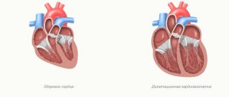
To understand the features of left ventricular atrophy and determine what it is, you need to look at the anatomy of the heart.
The heart is one of the most important human organs, whose functions include blood supply to the entire body. Its basis is the myocardium, consisting of striated muscle tissue. It allows the heart to contract, causing blood to be pumped between the atria and ventricles. In its structure, the myocardium resembles skeletal muscles, but, unlike it, the heart muscle does not obey the cerebral cortex. Signals are sent to it by the vasomotor center, located in the medulla oblongata.
The heart consists of 4 chambers, namely 2 atria and 2 two ventricles. Thanks to their alternating contractions, blood circulates in the small and large circles, providing nutrition to all tissues of the body.
In general, the cardiac cycle looks like this:
- Atrial systole (contraction). They fill with blood and contract. From the right atrium, blood flow moves into the same ventricle, and the same happens with the left.
- Contraction of the ventricles. Blood entering the left cardiac stomach is sent to the systemic circulation through the aorta. Contraction of the right ventricle leads it to the pulmonary artery (pulmonary circulation).
- Diastole (relaxation). This phase represents the relaxation of all chambers of the heart to fill the atria with blood. There is little oxygen in the bloodstream from the systemic circulation. This blood is called venous and enters the right atrium. The blood flow from the pulmonary circle is rich in oxygen, thanks to the pulmonary vessels. It is called arterial and goes to the left atrium.
The time it takes for the contraction phase to take place is slightly longer than for the relaxation phase. If everything goes well, then the person does not have problems with blood circulation. In the case of atrophy of the left ventricle of the heart, the body does not receive all the substances it needs due to the inability of the affected tissues to fully perform their functions. This disrupts blood flow, causing shortness of breath and swelling. In the congenital form of the pathology, developmental delays are often observed.
Left ventricular atrophy may be congenital or an abnormality acquired over time.
In the first case, the resulting dystrophy is a secondary phenomenon of disorder of myocardial cells. Its development is provoked by congenital heart defects and cardiomyopathies. Anomalies in the structure of the heart muscle mainly affect the connective tissue of the myocardium and the valvular component. Doctors still don’t know much about cardiomyopathy, but they often eliminate the consequences of this disease, among which are left ventricular atrophy.
The acquired form of the disease occurs in approximately 70% of patients. The reasons for its development are mainly the following:
- Viral infections. Among them, influenza and Coxsackie virus prevail. Atrophy of the left cardiac ventricle is a consequence of the effect of infection on the myosymplast. It is a fusion of cells, so it has several nuclei. After a virus attack, the myosymplast remains intact, but failures occur at the genetic level, as a result of which dystrophic changes in the myocardium can begin. The degree of atrophy depends on the number of affected cells.
- Bacterial infections. Doctors distinguish scarlet fever from this group. This is due to the fact that the bacterium that causes the disease is very similar in its gene code to cardiomyocytes, which are the muscle cells of the heart. The immune system, after the heart muscle is damaged by a bacterial infection, perceives its tissues as foreign and attacks them. This process leads to dystrophic changes in the myocardium.
- Lack of nutrition (ischemia) of the myocardium. In adults suffering from left ventricular atrophy, this is the main cause. This is usually associated with chronic ischemia, which gradually leads to dystrophic changes in the heart muscle.
- Intoxication. Long-term exposure to toxins in the body negatively affects the heart muscle. The main culprit of intoxication is alcoholic drinks. In just 2-3 years of their intensive use, the myocardium undergoes significant atrophy.
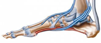
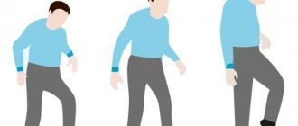
Amyotrophy. Signs
During the process of muscle atrophy, a decrease in volume and degeneration of muscle fibers occurs. They become thinner than usual, and if the case is particularly severe, the number of muscles is reduced to zero.
With the development of muscle atrophy, tissue deformation occurs, and muscle is replaced by connective tissue, in which motor function is impossible.
This disease is divided into two types:
- primary atrophy;
- secondary atrophy.
In case of damage to the muscle itself, the primary form is diagnosed. Most often it develops as a result of injury, intoxication, bruise or physical fatigue.
In case of post-traumatic complications or any infections, a secondary form of the disease may develop. This form is much more common than the first.
All types of muscle atrophy have a common feature - the damaged muscle decreases in volume, and this is especially noticeable when compared with a healthy one.
Causes of muscle atrophy
Besides the lack of physical activity that leads to muscle wasting and the diseases mentioned above, there are other reasons that can deteriorate muscle health.
Some other diseases and injuries that interfere with or limit movement can cause wasting. There are even cases where muscle wasting is a symptom of severe malnutrition or muscle disease associated with alcohol abuse. Even astronauts who spend a lot of time traveling in space can also begin to develop muscle atrophy due to the lack of gravity.
Causes of atrophy due to lack of movement
Possible causes of muscle atrophy due to lack of movement:
- hospitalization or long-term leave due to illness or surgery;
- temporary injuries such as a broken arm or leg;
- malnutrition, in which the muscles do not have enough nutrients, resulting in gradual weakening and inability to use the muscles properly;
- dermatomyositis, which causes muscle inflammation, manifested by a rash;
- muscular dystrophy, an inherited disease that causes progressive loss of muscle tissue and produces muscle weakness;
- osteoarthritis, a type of arthritis that causes pain and difficulty moving;
- rheumatoid arthritis, a chronic autoimmune disease that causes inflammation in the joints;
- polymyositis, a generalized inflammation that causes muscle weakness;
- lack of physical activity;
- severe burns.
Causes of neurogenic atrophy
Possible causes of neurogenic muscle atrophy:
- myopathy associated with alcohol abuse;
- multiple sclerosis, which affects the nervous system, brain and spinal cord, causing imbalance, lack of coordination, weakness and other symptoms;
- Amyotrophic lateral sclerosis or Lou Gehrig's disease, a severe neuromuscular condition that causes muscle weakness and difficulty controlling voluntary muscle movement;
- diabetic neuropathy, a complication of diabetes associated with high blood sugar;
- damage to the neck, peripheral nerves, or spinal cord;
- spinal cord atrophy, a genetic disorder that causes decreased muscle function;
- exposure to toxins or compounds harmful to the body in the form of poisons;
- Guillain-Barré syndrome, an autoimmune nerve disorder that causes nerve inflammation and muscle weakness;
- spinal muscular atrophy, or Werdnig-Hoffman disease, a genetic disorder that weakens and atrophies muscles and impairs movement.
Other reasons
- natural aging process;
- long-term use of corticosteroids;
- vascular damage to the brain;
- problems with swallowing;
- neuropathy, a disease that causes damage to one or more nerves;
- polio, a viral disease that affects muscle tissue and causes paralysis.
Muscle atrophy: treatment with the Bubnovsky method
The causes of muscle atrophy are as follows:
- aging of the body;
- impaired metabolism;
— parasitic and infectious diseases;
- enzyme deficiency.
The Bubnovsky method is a unique technique that is protected by a patent. According to this method, a full myofascial examination is first carried out, after which the condition of the muscles and joints in the complex is assessed, and then the location of the trigger zones is indicated.
The recovery program is established after all studies have been completed. The recovery program is formed strictly individually. The treatment is complemented by balneotherapy and cryotherapy.
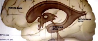
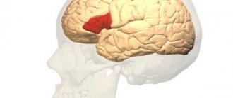
Symptoms of brain atrophy
The disease begins with changes that occur in the personality itself. The patient becomes lethargic, inactive, and indifferent. The ability to think productively gradually decreases, and vocabulary depletion is observed. Motor skills also deteriorate.
After a while, the patient does not recognize the objects, cannot understand why they are needed, and, accordingly, cannot use them. An orientation problem develops due to memory impairment. After a few years, brain atrophy provokes complete moral and physical disintegration of the personality - insanity.
Symptoms of pathology
Atrophy of the left ventricle of the heart can be identified by the clinical picture characteristic of this pathological process:
- Developmental delay. The problem is characteristic of the congenital form of the disease. The patient's tissues do not receive the required amount of necessary substances for full growth, as a result of which developmental retardation is observed.
- Dyspnea. Atrophic changes in the left ventricle disrupt blood flow, which is why the body does not receive the required amount of oxygen. This phenomenon leads to ischemia of internal organs and tissues. To correct the situation, the respiratory center receives a signal to increase the amount of air inhaled. Shortness of breath is a consequence of this entire process and is designed to increase the concentration of oxyhemoglobin in the blood. It is a combination of hemoglobin and air in red blood cells.
- Edema. They represent the second clear sign of acquired ventricular atrophy and indicate heart failure. Edema occurs due to stagnation of venous blood, since the heart cannot fully perform its functions. Initially, the problem begins in the lower extremities. As swelling progresses upward, it can be concluded that heart failure is worsening.
- Irregularities in heart rhythm. Chronic lack of nutrition leads to the death of cardiomyocytes. They perform not only the functions assigned to them, but also regulate the heart rhythm. With atrophic changes in the myocardium, the number of cardiomyocytes is reduced, which leads to disruptions in the process of heart contraction.
Treatment of brain atrophy
The patient should be kept in familiar living conditions as soon as atrophy begins to appear. This will help him maintain life stereotypes for a longer time. Conditions should be created for the patient so that he is busy with his usual activities, and also lies less in the daylight and moves more. When moving to a hospital, the patient's condition may deteriorate sharply, but if there is no possibility of constant supervision at home, moving to a hospital or a special boarding school is necessary.