Find out more interesting information about other types of diseases starting with the letter “E”: Extramedullary tumors of the spinal cord; Encephalitis; Rasmussen's encephalitis; Encephalitis St. Louis; Encephalopathy; Hashimoto's encephalopathy; Ependymoma of the brain; Ependymoma of the spinal cord; Epidural hematoma; Epilepsy; Epilepsy in pregnant women; Status epilepticus; Erythromelalgia; Essential tremor; Esthesioneuroblastoma; Echinococcosis of the brain.
What are extramedullary spinal cord tumors?
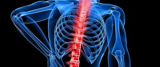
Extramedullary tumors are a disease that manifests itself in the formation of neoplasms in the spinal region, which do not have the ability to grow to the spinal cord, but only affect the area under the dura mater. Often, the disease is diagnosed when an anomaly occurs in the spinal root, as a result, compression of the brain begins with 50% damage to one part, and subsequently the disorder spreads to its transverse region. The rate of progression of the anomaly depends on the type of formation.
As diagnostic techniques, doctors resort to the use of magnetic resonance imaging or computer examination. Treatment consists of surgical intervention - radical elimination. When the tumor transforms into a malignant form, a course of chemotherapy and radiotherapy is prescribed.
Causes and risk factors
The etiology of the disease has not been established, but doctors have found that the following causes contribute to its occurrence:
- bad heredity: close relatives had spinal cord cancer;
- the presence of spinal cord lymphoma, leukemia, or HIV, Hippel-Lindau disease;
- there is already a history of other cancers;
- presence of neurofibromatosis;
- spinal cord injuries;
- unhealthy lifestyle, decreased immunity.
Provoking factors are:
- radioactive radiation, including solar radiation;
- influence of microwave and electromagnetic devices;
- prolonged exposure to harmful chemicals;
- unfavorable environmental conditions;
- repeated stressful situations.
brief information
The disease begins its development in individual structures that are located near the spinal cord (vessels, surface layer, paraspinal fiber, nerve endings). In general, this form of tumors accounts for up to 80% of the entire class, and neoplasms of intramedullary origin account for 20%. The anomaly affects any age category of the population. Sometimes doctors observe a multiple type of disease (formation of metastases, Recklinghausen disease, etc.). Often, the disorder is of benign origin, but in some situations a harmful element can provoke the progression of irreversible degenerative deformations. They begin due to high load. The course of treatment is controlled by doctors from the neurological, neurosurgical and oncological fields.
Classification of extramedullary tumors of the spinal cord
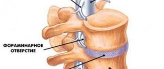
The division is based on localization, which creates the cervical, thoracic, lumbosacral and cauda equina groups. Depending on the presence of a hard surface layer, medical staff have created two forms of extramedullary neoplasms: subdural (intradural) and epidural. In adults, the first option occurs in 65% of lesions, and the second - up to 15%.
Like any tumor, the neoplasm presented is divided into benign and malignant types. According to the source of origin, there are primary and secondary forms (oncological elements of a different location, often breast cancer, prostate cancer, uterine cancer, etc.). To presumptively determine the class, you will need to undergo neuroimaging techniques. Accurate data is provided by histological diagnosis. Let's consider what types of neoplasms exist. These include:
- Meningioma is a neoplasm with a soft membrane. According to average statistical indicators, up to 70% of cases of its formation occur in patients in the thoracic area, and only 20% are localized in the cervical region. The type of occurrence is subdural (in less than 15% it may be of epidural origin). Scientists have found that the disease occurs most often in females. It is formed as a result of the influence of female hormones.
- Neuroma - develops in the cells of the Schwann membrane of the crust, as a result it has the second name schwannoma. It is shaped like a capsule and is usually detached from the nerve tissue, which helps perform surgeries for patients without completely cutting the process.
- Neurofibroma - often occurs at sensory endings. Has a diffuse method of germination and provokes thickening of the root. The tumor can be eliminated only by completely cutting it off. The disease can become malignant - neurofibrosarcoma.
- Lipomas are quite rare anomalies that progress in the tissue near the spinal cord. Sometimes doctors can diagnose epidural lipomatosis when a buildup of fat begins to affect the affected area. It can occur in patients with obesity, hypothyroidism and long-term use of corticosteroid pharmacological drugs.
- Hemangioma - begins to grow from vessels that are localized near the spinal area. It is represented by capillary growths or spongy particles filled with blood cells.
- Chondroma and osteoma - affect cartilaginous sections and bone areas. They can transform into malignant ones.
Symptoms of pathology
Symptoms are signs of a pathological process. Many factors influence the manifestation of symptoms:
- tumor type;
- tumor size;
- location;
- patient's age;
- stage of pathology;
- health status;
- chronic pathologies;
- other factors.
Depending on the factors described above, the patient may begin to experience a number of negative manifestations from the affected organ. For example, similar symptoms:
- Intense pain (the main location of pain is the spine at the site of the neoplasm). The reason is the effect on the nerve centers from the tumor. But pain will also begin to spread to the arms, hips, and legs. The spread of pain from the back to the limbs is the main symptom indicating the presence of a tumor in the human spinal cord.
- Exhaustion of the body. As a tumor develops, the hormonal structure changes, which leads to a change in body mass index. With primary manifestations, rapid weight loss occurs (up to 10 kilograms per month). Such loss leads to exhaustion and can cause associated pathologies.
- Problems with spatial orientation and coordination. When the organs of the central nervous system are damaged, the functioning of all spatial landmarks of a person is disrupted, which leads to impaired coordination in space.
- Loss of tactile sensitivity in the patient's limbs. This symptom initially manifests itself as tingling in the tips of the fingers and toes. With the passage of time and the progression of the tumor, the negative factor develops to the extent of limb numbness and paralysis.
- Muscle weakness. The appearance of weakness is directly related to disruption of the passage of nerve impulses from the brain to the limb.
- Deformation changes in the structure of the spine. It is typical for the later stages of the tumor process, when the tumor is so developed that it is not hidden inside the body, but is visible to the naked eye. The area of the spinal column has been visually changed.
- Disturbances in the activity of the urinary system. In this case, a person may experience an increased urge to empty the bladder or face the problem of poor urine formation.
- Disturbances in the gastrointestinal tract. Often causes the development of chronic diarrhea (increased frequency of the urge to empty the bowel), which is replaced by constipation and obstruction. This is due either to hormonal imbalances or the spread of the metastasis system to the human intestine.
- Pain when maintaining a stationary body position. When staying in one position for a long time in space, abnormally severe pain occurs in the body. It is often detected when standing in one place for a long time.
- Increased body temperature within subfebrile range (37.1-37.8 degrees Celsius). It manifests itself when there is a slight inflammatory process inside the human body, which leads to the likelihood of such a fever.
To study The dural sac is deformed, what is it?
The further development of symptoms will depend on where the secondary foci of tumor growth (metastases) begin to spread. When metastases enter the lungs, depression of respiratory functions is observed. If it enters the myocardium, symptoms will manifest themselves in the form of necrosis of cardiac tissue (infarction). If it enters the brain, it results in impaired consciousness up to loss and deep coma.
Also, the symptoms will depend on the emerging chronic concomitant pathologies that accompany the development of the oncological process. The fact is that with oncology, immune processes are suppressed and it is easier for pathologies to affect the body. Depending on what pathology arose and the development of symptoms occurs.
What are the manifestations of extramedullary spinal cord tumors?
The onset of the disease is indicated by the appearance of painful sensations in the form of “lumbago”, which is limited by the area of innervation of a particular root. During this interval, disruptions in the functioning of sensitivity and a decrease in the level of muscle strength can be observed. If the neck or chest area is affected, the patient develops symptoms of radiculitis. The first stage of the anomaly lasts from several months to 5 years. It all depends on the class of the tumor.
As the lesion increases in size, it causes compression, which causes the sequential appearance of additional stages: half and full transverse lesion. The first method causes the patient to experience a disorder of dissociated motor and sensory function. On the infected area of the body below the location of the harmful element, paresis of central origin and disruption of deep sensitivity are formed, and on the other, the sensitive area to pain and temperature decreases. The full stage provokes a symmetrical neurological deficit.
Forecast
If the diagnosis is made at an early stage, then with proper treatment there is a high probability of recovery. The best prognosis can be given with early diagnosis of the primary tumor, the development of which can occur very slowly and take years. Secondary tumors, on the contrary, can sometimes grow within a few weeks, and if the cancer has gone too far, it becomes impossible to defeat it. Moreover, in such cases the overall picture is complicated by the presence of metastases to other organs. Although even with secondary tumors, there are cases where the patient and doctors had months of time left to carry out treatment. It is very important how the patient’s environment and, most importantly, the patient himself perceive the diagnosis.
Diagnostic techniques
Detection of tumors in the first stages of their development is quite difficult, since they can be masked by symptoms of radiculitis or the presence of somatic anomalies. Doctors begin to sound the alarm if there is no effect after medical therapy. To examine the patient, a referral is issued to undergo:
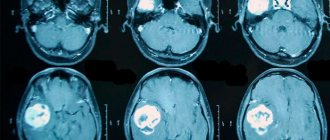
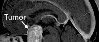
- Magnetic resonance scanning of the spinal region is the most effective diagnostic method, helping to detect the exact location, extent of distribution, shape, expected type and level of spinal compression.
- If it is not possible to carry out MRI testing, it is allowed to attend radiography, which will reveal signs of osteochondrosis, but will not be able to accurately indicate the presence of a neoplasm.
- Additionally, electroneuromyography is performed, which allows you to determine the location of the lesion without its appearance.
- Checking the cerebrospinal fluid provides information to rule out an infectious disease.
If the patient has limitations for undergoing a magnetic resonance examination, computed tomography (myelography) is performed. In the presence of neoplasms of vascular origin, angiography will be required. The exact shape of the tumor can only be determined after a histological examination.
Diagnostics
Once the presence of a tumor is suspected, a comprehensive examination should be carried out. To refute or prove the presence of the disease, it is necessary to find out the location and structure of the tumor, its degree, and its severity. It is necessary to undergo a number of tests and diagnostics:
- Thorough examination of the neurological system.
- Mandatory magnetic resonance therapy (MRI).
- Mandatory tomography.
- Taking an x-ray.
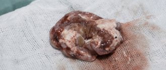
- Biopsy of the diagnosed site.
- Ultrasound.
- Analyzes.
- Cerebrospinal fluid puncture.
Collection of cerebrospinal fluid
After diagnosis, doctors make a prognosis.
Treatment options
Often, medical staff resort to radical treatment - surgery. Laminectomy becomes a surgical intervention. During the procedure, the doctor touches a minimal area of the spinal cord to prevent the formation of injuries. To remove intradural tumors from the patient’s body, a more complex operation will be required, as a result of the need to make an incision on the hard surface and act close to the brain. If there is a formation with two components, its extradural area is first resected.
If the malignant nature of the disease is detected, the patient is prescribed courses of radiation therapy in addition to surgery. Limitations for laminectomy may include a large number of metastases and common malignant neoplasms. In this case, doctors resort to a palliative format, which is aimed at pain relief and decompression. Chemotherapy and radiotherapy are prescribed.
Radiation therapy
This technique may be the primary treatment for inoperable tumors and aggressive lesions such as anaplastic astrocytomas and glioblastomas.
Radiation therapy may be beneficial for residual tumor after surgery or recurrent tumor, but controversy exists. A dose of 50 Gy is delivered to the tumor in daily fractions of 1.5-2 Gy. This dose was not curative in most studies. Some series have reported a reduction in local failure rates when delivering more than 50 Gy.
Side effects of radiotherapy include:
- acute and delayed myelopathy;
- decreased skeletal growth in young children;
- increased difficulties with subsequent surgical removal of the tumor;
This is especially important if radiation therapy does not control the growth of the lesion.
Further forecast
With timely detection of extramedullary tumors of the spinal cord, the prognostic plan of the disease is considered positive than with the intramedullary form. Completing a full course of treatment using microsurgery techniques shows a stable result with rapid regression of pain and deficits of neurological origin. Postoperative consequences include liquorrhea, spinal arachnoiditis, meningitis and mobility. The most negative prognosis is attributed to neoplasms transformed into a malignant type.