Hydrocephalus in adults can be an independent disease or a manifestation of pathology of other organs and systems. In patients with hydrocephalus, the process of excessive accumulation of cerebrospinal fluid in the cerebrospinal fluid spaces, caused by disturbances in its production, circulation or absorption, is actively progressing. It is manifested by an enlargement of the ventricles of the brain, a decrease in the density of the brain matter due to its saturation with cerebrospinal fluid and a narrowing of the subarachnoid spaces. At the Yusupov Hospital, neurologists treat hydrocephalus with the most effective drugs that have a minimal range of side effects. Patients are examined using modern research methods performed by experienced specialists from the neurology clinic.
Classification
Hydrocephalus can be either acquired or congenital. The latter begins to manifest itself in infancy, while the acquired form is typical for adults and even elderly patients. Depending on the prerequisites for the appearance of hydrocephalus, the following types are distinguished:
- Closed, occlusive hydrocephalus occurs due to disruption of the flow of cerebrospinal fluid due to blockage of the fluid-conducting pathways. Most often, blockage occurs due to a tumor, the presence of a blood clot or adhesions.
- Open, or dysresorptive, hydrocephalus. It develops against the background of a violation of the structures that are involved in the process of absorption of cerebrospinal fluid into the system of cerebral veins (arachnoid villi, venous sinuses, cells and pachyonic granulations).
- Hypersecretory hydrocephalus. Production of large amounts of cerebrospinal fluid into the plexus of ventricular vessels.
- Replacement mixed hydrocephalus of the brain. It develops as a result of an increase in the amount of cerebrospinal fluid both in the subarachnoid space and in the cerebral ventricles. In this case, atrophy of brain tissue occurs.
The last option is considered the most dangerous to the life and health of the patient.
Classification of hydrocephalus
This disease is divided into 3 main types:
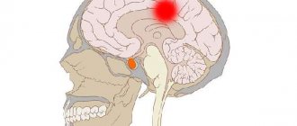
- Internal hydrocephalus of the brain . It is characterized by the accumulation of cerebrospinal fluid directly in the ventricles of the brain, as a result of which intracranial pressure increases. The cerebrospinal fluid begins to put pressure on various parts of the brain, causing disruption of the organs and entire systems for which these areas are responsible. This form is provoked by inflammatory processes in the body, disruption of the central nervous system in the patient, tumors and neoplasms.
- External hydrocephalus. Previously it was a full-fledged type of disease, but now it is not considered as such. This type of dropsy has a different origin and completely different consequences. With this disorder, the volume of the brain decreases and it may even partially atrophy. In this case, all the space freed up in the cranium is occupied by cerebrospinal fluid.
- Mixed replacement hydrocephalus . It is characterized by a decrease in brain volume and filling with cerebrospinal fluid not only of the cerebral ventricles, but also of the resulting empty space in the skull. This form of the disease is called replacement. Elderly people are most susceptible to this type of disease.
Based on the nature of its course, hydrocephalus is divided into:
- Spicy . The acute form of the disease is characterized by a rapid course and is a serious threat to the life and health of the patient. As a rule, it requires urgent surgical intervention. It involves craniotomy and installation of drains to drain cerebrospinal fluid from the brain cavities.
- Chronic . The chronic form does not pose such a serious danger, but undoubtedly requires treatment. It involves shunting - the installation of a complex system of catheters that ensure the movement of cerebrospinal fluid from the cranium to other cavities of the body - the abdominal or pelvic cavity, where rapid absorption of cerebrospinal fluid will occur.
There is also subacute hydrocephalus - a combination of acute and chronic forms of the disease. It is the most dangerous form of the disease and poses a great threat to health and normal life. However, subacute dropsy is diagnosed extremely rarely.
There is also vicarious hydrocephalus of the brain, which occurs as a result of atrophy of the cerebral cortex.
Causes
Severe replacement mixed hydrocephalus is most typical for newborns. In adults, this pathology is less common, but also occurs. Research in the field of medicine has shown that any disturbance in the functioning of the human nervous system can provoke the development of hydrocephalus. Acquired causes of hydrocephalus can be:
- Ruptures of hematomas or hemorrhages in the brain.
- Severe traumatic brain injuries.
- Birth injuries.
- Severe cerebrovascular accident.
- Previously suffered infectious-inflammatory diseases, including encephalitis, meningitis, arachnoiditis, etc.
- Infection of brain structures by various parasites.
- Germinomas, astrocytomas, tumors in blood vessels and other neoplasms.
- Metastasis of tumors of other organs to the brain.
- Formation of cetacean cavities in the third ventricle.
- The occurrence of vascular malformations.
- Atrophy of brain tissue as a result of organ damage by encephalopathy.
Dropsy of the brain can have a negative effect on all organ systems. Therefore, to eliminate hydrocephalus, it is necessary to accurately determine the cause of the pathology.
What are the signs of mixed replacement hydrocephalus?
Causes of the disease
Hydrocephalus, or dropsy of the brain, is most often diagnosed in newborns. The fact is that the brain, like other organs, continues to grow and develop rapidly after birth, which provokes disruptions in the body. In this case, a diagnosis of hydrocephalus of the brain is made. It is considered congenital and, often, a moderate form is not a threat to health. The disease will go away as soon as the skull begins to grow and the ratio of brain mass to skull size is restored.
However, in some cases, increased intracranial pressure and hydrocephalus diagnosed in a newborn can be a serious cause for concern. Quite often, the disease is detected before the fetus is born.
The development of the disease in young children can be provoked by the following factors:
- Birth and intrauterine injuries.
- Infections suffered during pregnancy.
Most often, hydrocephalus occurs for the following reasons:
- Serious traumatic brain and spinal injuries.
- Previous infections and meningitis.
- Intoxication.
Elderly people are also susceptible to hydrocephalus, including mixed hydrocephalus. We can say that they are the ones at risk. The fact is that in old age the spine becomes weak, and any injuries, especially to the cervical spine, and displacements can provoke a violation of the outflow of cerebrospinal fluid. Causes of dropsy in older people:
- Weakness and fragility of the spine, its injuries.
- Atherosclerosis.
- Hypertension.
The most dangerous thing about this disease is that it may not make itself felt for a long time. At first, a person may not experience discomfort, since signs of the disease do not appear immediately. Regardless of the cause, advanced forms of hydrocephalus, both in children and adults, can have serious consequences. For example, partial or complete disability in adults and severe developmental delay in children.
Clinical picture
The characteristic clinical picture of mixed replacement hydrocephalus is the following:
- Constant pain in the head and a feeling of heaviness, intensifying during sleep and immediately after waking up. Inability to determine the source of pain. When a person is in a lying position, the symptom becomes more pronounced as the pressure of the cerebrospinal fluid increases.
- Nausea and vomiting in the morning, regardless of food and time of consumption.
- Feeling of pressure on the eyes.
- Persistent hiccups.
- Constant weakness, increased fatigue, drowsiness.
- Difficulties with concentration and attentiveness; it is difficult for a person to perform basic actions.
- Apathy, nervousness, decreased intellectual abilities.
- A sharp drop in blood pressure, slowing or increasing heart rate.
- Permanent dark circles under the eyes. When the skin of the lower eyelids is stretched, blood-filled capillaries are clearly visible.
- Significant increase in sweat, tendency to faint.
These symptoms indicate intracranial increased pressure.
Symptoms
The severity of neurological symptoms depends on the age of the patient and the degree of development of the pathology. In children, the most well-known visual symptom is an abnormally large circle of the ready and the appearance of a convex fontanel on the parietal part. Other childhood manifestations of the disease include:
- Bad dream.
- Vomit.
- Frequent bouts of crying.
- Refusal to eat.
- Seizures, convulsions.
- Rolling eyes.
- Hearing loss.
Impaired circulation of cerebrospinal fluid negatively affects the mental development and growth of the child. There is low motor activity, memory problems, lack of response to sounds and speech, sluggish expression of emotions, etc.
There can be many reasons for developmental delays in children. To exclude the presence of hydrocephalus, you need to contact your pediatrician and examine your baby.
In adults, the disease manifests itself differently, but no less pronounced. The patient feels constant muscle weakness, nausea, vomiting in the morning, and drowsiness. Consistently high intracranial pressure contributes to an enlargement of the skull and thinning of its bone tissue. Under constant pressure, the brain contracts more intensely. This causes severe cutting and stabbing headaches. The patient's hearing and vision are impaired. As the disease progresses, atypical emotional reactions to reality, loss of coordination when walking, forgetfulness, slow reactions, memory impairment, and frequent mood swings are observed.
In the later stages of development of the pathology, muscle weakness becomes the cause of uncontrolled bowel movements and involuntary urination. In this state, the patient loses the ability to self-care and needs constant help and attention.
Manifestation of neurological disorders
Neurological disorders with mixed replacement hydrocephalus of the brain in adults will manifest themselves as follows:
- Decreased quality of vision – bifurcation and difficulty focusing the gaze on one object.
- Loss of visual fields.
- Against the background of constant compression, atrophy of the optic nerve occurs, which in the future can cause complete loss of vision.
- Strabismus.
- Lack of pupillary response to bright light.
- Dysfunction of the vestibular system. Dizziness, unsteadiness in gait, tinnitus, and involuntary vibrations of the eyeballs appear.
- Paralysis of limbs.
- Increased reflexes and muscle tone.
- Decreased or complete loss of sensitivity.
- Involuntary fixation of the arms and legs, when due to increased tone it is impossible to straighten the limbs.
- Symptoms of cerebellar ataxia, accompanied by impaired motor function and splayed handwriting.
- Unstable emotional state, sudden mood swings.
- With a sharp increase in cerebrospinal fluid pressure, aggression occurs.
The combination of symptoms and diagnostic measures taken indicate mixed hydrocephalus.
Concept of hydrocephalus
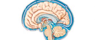
What is hydrocephalus? Translated from Greek - “dropsy of the head.” This disease is manifested by the accumulation of liquor (cerebrospinal, cerebrospinal) fluid in the ventricles of the brain. It can occur due to pathologies of resorption or movement of cerebrospinal fluid from the ventricles to the site of its absorption into the circulatory system, when the entrance to the vessel is blocked or when the ventricle produces cerebrospinal fluid in excess.
Cerebrospinal fluid is formed in the ventricles of the brain, from which it enters the subarachnoid space. It is located between the brain and the cranial surface, and the cerebrospinal fluid plays the role of a conductor and shock absorber there. The ventricular system of the brain is represented by two ventricles, which are located symmetrically on the sides, a third ventricle located in the middle of the brain, and a fourth located in the brain stem. Liquor is produced in the lateral ventricles, partially in the fourth. It should be noted that cerebrospinal fluid is constantly produced and its amount is approximately 120 or 140 ml in an adult and about 50 ml in a child. During normal function, it circulates continuously about three times a day. With this pathology, the amount of cerebrospinal fluid is much higher than normal, which occurs either as a result of its excessive formation in the ventricles, or due to reduced absorption into the venous system.
Diagnostics
Confirmation of the diagnosis of mixed replacement hydrocephalus of the brain in adults occurs on the basis of a study using both laboratory and instrumental techniques. The latter are the most informative in the case of brain disorders. Laboratory tests show the patient's general condition and how the disease has affected his or her health.
To identify severe and moderate mixed replacement hydrocephalus of the brain, the following studies are prescribed:
- Measuring head circumference with a tape. If hydrocephalus is in question in a child, then an upward change in head circumference of more than 1.5 cm per month indicates the presence of the disease. In adulthood, any enlargement of the head is considered pathological, regardless of the period during which it occurred.
- Fundus examination. If swelling of the optic disc is detected, we can talk about increased intracranial pressure, which means hydrocephalus cannot be ruled out.
- Ultrasound examination of the skull, or neurosonography. This study is not prescribed for adult patients, as it is not very informative. In childhood, an ultrasound is performed through the child's fontanel.
- Magnetic resonance imaging. This is the most accurate method for detecting hydrocephalus. Tomography will not only confirm the suspicion of the presence of pathology, but also determine the causes of its development, and assess the damage to brain structures and blood vessels. If MRI shows the presence of periventricular edema, the diagnosis of hydrocephalus is confirmed.
- CT scan. This is similar to an x-ray; the information content of the method is somewhat lower than in the previous version.
- Rheoencephalography and echoencephalography.
- Lumbar puncture. It is a collection of material for a histological examination of the composition and condition of the cerebrospinal fluid.
- Radiography. Makes it possible to identify thinning of bone structures.
The main criteria that a specialist relies on to clarify the diagnosis are the results of fundus examination and magnetic resonance imaging. After confirmation of the diagnosis, treatment for mixed replacement hydrocephalus of the brain is prescribed.
General information
The medical term “hydrocephalus” refers to a condition in which an accumulation of cerebrospinal fluid (CSF) occurs in the brain.
Mixed hydrocephalus in adults is manifested by headaches, nausea, drowsiness, and speech disorders. This condition may indicate the development of a tumor in the head, or be a consequence of a head injury and other health and life-threatening pathologies.
To prescribe treatment, qualified assistance from a neurologist and examination are required.
Normally, it freely penetrates into and out of the brain, ensuring normal brain activity. But when exposed to certain factors, the outflow of cerebrospinal fluid is disrupted and it begins to accumulate in the cavities of the brain.
In addition to mixed, occlusive, communicating and hypersecretory hydrocephalus are distinguished.
With occlusion, the flow of cerebrospinal fluid is disrupted due to the closure of the cerebrospinal fluid pathways; with communication, the processes of resorption of cerebrospinal fluid worsen; hypersecretory develops due to excess production of cerebrospinal fluid.
Neurologist, reflexologist, hirudotherapist
Kislitsyna Ekaterina Nikolaevna
10 years of experience
With a mixed type of hydrocephalus, the accumulation of cerebrospinal fluid occurs inside and outside the brain, as well as in its ventricles and subarachnoid space. Since the development of this disease affects almost all parts of the brain, the pathology progresses very quickly and is accompanied by a pronounced symptomatic picture.
The main point in the treatment of mixed hydrocephalus is timely diagnosis of the disease. Since only in the initial stages of its development it is possible not only to cure the pathology, but also to prevent the occurrence of serious complications.
Drug treatment
Mixed hydrocephalus is a dangerous and severe pathology. Treatment of hydrocephalus should include not only a set of measures related to taking medications, but also surgical intervention. Depending on the identified form of pathology, the selection of a separate therapeutic regimen is required.
Some patients try to use traditional medicine methods, but most specialists reject such methods of treating hydrocephalus of any type or allow their use only as auxiliaries.
How is mixed hydrocephalus treated?
The volume and nature of treatment depend on the stage of disease progression and the age of the patient. Doctors use conservative and surgical therapeutic methods.
Drug therapy
With a moderate form of mixed hydrocephalus, specialists resort to conservative methods. Sometimes physiotherapeutic procedures and the participation of a chiropractor are sufficient to stabilize the patient’s condition. Drug treatment is sought in cases where the accumulation of cerebrospinal fluid exceeds normal levels. Medicines reduce the volume of cerebrospinal fluid production, normalize intracranial pressure, and alleviate neurological symptoms. The patient is prescribed:
- Diuretics.
- Drugs to improve cerebral circulation (Cavinton, neurometabolic stimulator Phenotropil, Cerebrolysin).
- Agents that replenish magnesium and potassium deficiency (Asparkam and Panangin).
- Blood thinning anticoagulants and antiplatelet agents (Ecotrin, Clexane Warfarin).
- Venotonics that help improve the condition of blood vessels (Detralex, Phlebodia).
- Sedatives (Diazepam, Relanium, Seduxen)
- Multivitamin complexes.
- Antibiotics (when identifying infectious and inflammatory processes).
Drug therapy is a temporary measure to eliminate unpleasant symptoms and alleviate the patient’s condition. Mixed hydrocephalus can be stabilized, but even with adequate drug treatment, deviations in psychomotor development (mental disability, neurological disorders, etc.) cannot be ruled out. For this reason, surgery may be the only and most effective treatment method.
Surgery
The main and most effective method of treating mixed hydrocephalus is surgery. To carry out the operation, specialists use several methods:
- Shunting (installation of shunt systems in the brain). This technique has been used for a long time. The operation is complex, effective, but dangerous due to complications. The tube or catheter inserted into the brain becomes bent and clogged over time. There is a possibility of infection, the development of inflammatory processes and other consequences that threaten the patient’s life.
- Drainage systems. They are used in cases where there is an urgent need to stabilize pressure and free the brain cavities from excess cerebrospinal fluid. The drainage absorbs cerebrospinal fluid, which reduces the risk of infection and the development of serious complications.
- Endoscopic surgery. This is the most minimally traumatic and gentle therapeutic method. Using a neuroendoscope with a video camera, the surgeon creates an opening at the bottom of the ventricle for the fluid to exit. This allows you to relieve the ventricular systems and normalize intracranial pressure. The risk of infection during endoscopic procedures is unlikely.
Don't risk your health! Traditional medicine is powerless in the treatment of mixed hydrocephalus. Any procedures and prescriptions cannot cope with physical and anatomical problems, so self-medication must be immediately excluded. Only a doctor can help.
Drugs
Drug therapy is carried out using the following drugs:
- Diuretics. The diuretic effect of these drugs allows you to stop the production of cerebrospinal fluid. Most often the choice falls on Piracetam, Diacarb and Glyserol. All these drugs are administered by injection. Along with diuretics, the patient is prescribed vitamin complexes, since diuretics wash out magnesium, potassium and sodium from the body.
- Antibacterial drugs. Prescribed to eliminate infectious pathogens.
- Medicines that promote blood circulation in the brain.
- Vitamin and mineral complexes. The most commonly prescribed vitamins are C, B and E. They improve cellular metabolism throughout the body.
- Enzymes of animal origin. Pyrogenal and lidase help break down and remove excess cerebrospinal fluid.
- Glucocorticoids.
If drug therapy for mixed replacement hydrocephalus in adults does not show positive dynamics in the patient’s condition, surgical intervention is prescribed.
Signs
Moderate hydrocephalus of the brain can develop asymptomatically for a long time.
For several years, the only sign of the disease may be periodic headaches, which often occur in the morning.
The disease can be detected during a diagnostic examination of the brain completely by accident.
Sometimes headaches do not occur, while intracranial pressure remains normal.
However, at a certain period, the consequences of excess accumulation of cerebrospinal fluid still manifest themselves - for example, in the form of impaired blood supply to the brain and its hypoxia (oxygen deficiency).
This condition can lead to any number of consequences, including stroke and dementia.
Severe symptoms that occur with the developed form of the disease include:
- problems with vision and hearing;
- dizziness;
- decreased intellectual abilities and partial memory loss;
- loss of attention and ability to concentrate;
- loss of spatial orientation (patients may go for a walk and get lost);
- impaired coordination of movements (patients may experience a change in gait);
- chronic fatigue;
- sleep disorders;
- irritability.
In the acute stage, external, internal and mixed forms of hydrocephalus give such manifestations as vomiting, urinary incontinence, loss of consciousness, and cerebral edema.
This phenomenon is called an occlusive crisis and requires immediate hospitalization: the cause of the crisis is a complete block of the outflow of cerebrospinal fluid.
Surgery
If hydrocephalus occurs in acute or chronic form, the patient is prescribed surgery. A contraindication for such treatment of mixed replacement hydrocephalus may be an infectious-inflammatory process that has spread throughout the body. Therefore, the infectious focus is first eliminated, and then surgery is prescribed.
Surgical treatment involves the formation of pathways for the outflow of cerebrospinal fluid. This process is called bypass surgery and requires preliminary removal of adhesions and tumors.
Treatment of hydrocephalus
For moderate mixed hydrocephalus of the brain, which does not pose a great danger to human health, there are the following treatment methods:
- Medication.
- Manual therapy.
- Physiotherapy.
As a rule, the patient is prescribed diuretics that stimulate accelerated circulation of fluid in the body, as well as antibiotics if the infectious cause of the disease is confirmed. In addition, treatment is carried out with drugs that accelerate blood circulation in the brain.
Surgery
However, even moderate replacement mixed hydrocephalus of the brain can develop into an acute form at any time, so it is important to constantly monitor the doctor who will collect data. If an acute form of the disease occurs, immediate neurosurgical intervention is performed.
There are several types of operations for hydrocephalus:
- Shunting. Shunting, used for chronic hydrocephalus, is being used less and less. The fact is that this operation is quite traumatic and threatens serious complications if performed incorrectly. Even after the installation of shunts, the complex catheter system requires constant careful maintenance, replacement of elements, and so on. In such conditions, it is quite easy to introduce an infection into the body and get a serious infection.
- Installation of drains. Drainage is used when it is urgently necessary to reduce intracranial pressure and remove part of the cerebrospinal fluid from the cavities of the brain. This can happen when the tumor completely blocks the channel for the outflow of cerebrospinal fluid from the skull and its rapid accumulation occurs in the cavities of the brain.
- Endoscopy. Endoscopy is performed with minimal trauma to the body. Using a special device - an endoscope - the doctor penetrates the ventricles of the brain and makes holes to ensure the outflow of cerebrospinal fluid directly into the brain cisterns. Intracranial pressure decreases and cerebrospinal fluid is absorbed.
Bypass surgery and the installation of drains are a somewhat outdated form of surgery, although they are still used in practice. The most modern way to combat hydrocephalus of the brain is endoscopic surgery.
Mixed hydrocephalus of the brain is a very serious and dangerous disease that affects all age groups of people. Treatment of this disease should not be delayed, since with an advanced form of the disease, irreversible changes in the body are possible, including a decrease in intellectual level and complete incapacity. Nowadays, successful medical and neurosurgical treatment of this disease is carried out.
Methods
In addition, hydrocephalus is eliminated by the following methods:
- Palliative intervention. It is carried out through puncture if hydrocephalus is characterized as open.
- Radical surgery. To eliminate excess fluid, special shunts are installed in the spinal canals. Internal drainage allows you to remove cerebrospinal fluid into an adjacent organ or system.
Most often, operations to eliminate hydrocephalus go well and allow you to get rid of the problem. If dropsy is caused by a tumor in the brain, surgical removal can prolong the patient's life by several years.
What is mixed type hydrocephalus?
A complex neurological disease has a simple non-medical definition. Hydrocephalus is called dropsy. The disease is provoked by a violation of the circulation of cerebrospinal fluid (CSF). It is produced daily and distributed in the ventricles, subarachnoid fissures and cisterns of the brain. Circulating in tissues, cerebrospinal fluid nourishes brain cells and removes metabolic products. In addition, cerebrospinal fluid ensures a stable location of the brain in the skull and performs a shock-absorbing function. This is a kind of anatomical protective barrier that protects soft, vulnerable brain tissue from internal and external influences.
Normally, the volume of fluid is 120-150 ml, but for constant renewal the body produces more, approximately 500-600 ml per day. If the cerebrospinal fluid levels are normal, there are no abnormalities in brain function. But for various reasons its volume may increase. The intensively produced cerebrospinal fluid does not have time to be absorbed, accumulates in large quantities and blocks the work of vital centers in the brain area. Violation of the dynamic balance leads to the development of foci of the disease and the inevitable deterioration in such cases of the physical and psycho-emotional state of the patient.
Causes and forms of hydrocephalus in children
Since the onset of the disease could begin at the stage of intrauterine maturation or debut immediately after birth due to certain reasons, hydrocephalus in newborns is usually divided into antenatal and intranatal.
Antenatal HC, formed in the womb , is congenital.
It is due to:
- The formation of defects in the vascular bed and malformations of the central nervous system (spina bifida and cranial hernia, aneurysm of the vein of Galen, intracranial or arachnoid cyst);
- Chromosomal damage and aberrations;
- Neoplasms in the fetal brain (extremely rare);
- Infections the mother has previously suffered or currently has (cytomegalovirus, rubella, herpes, syphilis, mycoplasma, influenza, and even a seemingly harmless ARVI).
Video: antenatal GC on ultrasound during pregnancy
Intrapartum HC is the result of trauma, inflammatory processes in the fetal meninges, intraventricular and subarachnoid hemorrhages in the newborn, which are a consequence and complication of a difficult birth. Obviously, intrapartum hydrocephalus can safely be called acquired
, since it occurs at the end of the period of intrauterine development.
In addition, HC in infants is classified depending on:
- Mechanism of development (occlusive or closed, communicating or open, mixed);
- Areas of cerebrospinal fluid accumulation (internal, external, combined);
- Activity of the pathological process (active, passive);
- Clinical course (acute, chronic);
- Indicators of the level of intracranial pressure (hypertensive, normotensive, hypotensive);
- Degree of severity, the basis of which is the ventricular-hemispheric index;
- Stages (compensated, subcompensated, decompensated).
By the way, a similar division into forms is often used in the classification of HC in adult patients.
Treatment
The tactics of the rehabilitation course depend on the form of the disease, the patient’s age group and other characteristics of the condition (the presence or absence of complications, genetic diseases). Comprehensive treatment is effective, involving conservative and surgical procedures. You need to learn about each of the tactics separately.
Conservative
If hydrocephalus does not have obvious and aggressive symptoms, and is not associated with increased intracranial pressure, therapy aimed at improving the condition of the body and brain activity will be more than sufficient. To this end:
- Massage of the temples and collar area is provided against the background of moderate activity and the use of means that normalize cerebral circulation. This could be Glycine, Phenibut, Cerebrolysin.
- The use of diuretic names in situations where the patient’s blood pressure is elevated. Furosemide and Diacarb will allow you to remove excess fluid and reduce the volume of cerebrospinal fluid.
- Herbal remedies are used if the use of diuretics is not possible. Corn silk, nettle, and poplar buds can be used.
- It is necessary to introduce potassium and magnesium supplements into treatment. This is necessary provided that diuretic tablets are regularly used, since trace elements are excreted in significant quantities in urine.
Special attention is paid to nutrition. It should be healthy and complete, preferably fractional (5-6 times a day, in small portions). Nuts and legumes, bananas, spinach, lettuce, dark chocolate will be beneficial for a patient with hydrocephalus, because they improve immunity and physiological reactions.
Surgical
Today, bypass surgery and endoscopic operations are practiced. The first intervention involves creating a bypass for the fluid. For this purpose, flexible tubes are placed into the cavity inside the skull, which are carried out into the peritoneum or other internal environments of the human body.
Bypass surgery has a serious drawback - the likelihood of complications. This is due to clogging of the shunts, their kinks and simply failure. As a result, repeated surgery may be required to replace the shunt, which not every person can endure.
The priority direction in modern treatment is neurosurgical endoscopic interventions. Wherein:
- a hole is formed in the cerebral ventricles through which the outflow of fluid is ensured;
- monitoring of the intervention is possible on the monitor screen;
- the main device, the endoscope, is equipped with a mini-video camera that provides detailed observation.
Such treatment makes it possible to do without extensive intervention and is practically free of complications. The quality of life of the patient, who will not require regular revision of shunts, also improves. There will also be no chance of unexpected clogging of the outlet tube.
Clinical signs of HC in infants
If a child was born with a seemingly normal head circumference (or slightly increased), then as the baby grows, in this case the following signs of hydrocephalus appear:
- The head begins to grow disproportionately due to the expansion of parts of the brain (ventricles), however, if it grows evenly on both sides, which happens more often, then the GC is called bilateral, and in the case of expansion of one ventricle and, accordingly, one part of the head - asymmetrical;
- The enlarging brain no longer fits in the cranium, so in search of space it begins to move apart the sutures (fontanelles), which prevents their natural fusion;
- The fontanel, instead of narrowing and overgrown by the year of life, on the contrary, moves apart, giving way to the brain, becomes tense and visible to the eye;
- An isolated fontanel in a one-year-old child is a sign of hydrocephalus and in such circumstances can only close up by the age of two or even three;
- A disproportionately enlarged frontal part of the skull with blood vessels protruding under the skin also indicates hydrocephalus in a newborn;
- The presence of neurological symptoms - strabismus, sunken eyeballs partially or completely (only the whites are visible), nystagmus, high tone of the limbs, convulsive twitching;
- Delayed psychomotor development of the child also occurs: the head often throws back, it is difficult for the baby to hold it, he does not acquire skills appropriate to his age, cannot sit down and stand on his legs, does not show interest in the world around him, at the slightest reason (or without it) he is capricious and cries .
All these symptoms are characteristic of severe hydrocephalus, which can be noticed not only by doctors, but also by parents who constantly monitor their newborn child. Therefore, it is very important to notice dropsy of the brain at the earliest stages of development , because its consequences can be quite sad:
- Visual and hearing disorders;
- Delayed mental and physical development;
- Seizures;
- Early death.
Classification: origin, course, pathogenesis
Cerebrospinal fluid or cerebrospinal fluid (CSF) is constantly produced, circulates in parts of the brain (ventricles), is absorbed and renewed. All these processes are interconnected and are in a certain balance. If disturbances occur at any stage, cerebrospinal fluid may accumulate in the ventricles or subarachnoid space and increase intracranial pressure. This condition is called hydrocephalus (HC), which can be of congenital or acquired origin.
According to the nature of the course, the disease is divided into stages:
- Acute, rapidly developing over 3 days and carrying information about the causes of the disease;
- Chronic, having only symptoms of the underlying pathology, but not indicating factors remote in time (up to six months) that provoked it;
- Compensated, characterized by the restoration of normal intracranial pressure, although the cavities containing cerebrospinal fluid are still in an expanded state;
- The decompensated form, in which dropsy with increased intracranial pressure under certain circumstances in the form of injuries, infectious processes and other factors, returns again.
The pathogenesis of hydrocephalus also has its own variants:
- Communicating or open hydrocephalus with its inherent free movement of excess cerebrospinal fluid through the departments, which could be formed as a result of excess production or malabsorption;
- Closed form , which occurs in cases where cerebrospinal fluid cannot circulate freely throughout the departments. It is also called occlusal. In its acute course, it results in severe symptoms and manifests itself as: nausea and vomiting;
- severe morning headache;
- drowsiness (an important prognostic criterion for neurologists, since it is a sign of impending deterioration);
- depression of consciousness with transition to a comatose state lasting a short time;
- congestion on the optic nerve head;
- rapid increase in respiratory and cardiovascular failure, leading to the death of the patient;
Possible consequences
The lack of proper treatment leads to progression of the disease. In this light, the development of complications, including critical and fatal ones, is likely. The main complications of hydrocephalus are cerebral edema and its displacement inside the skull.
The development of epilepsy with frequent seizures is likely. In especially severe cases, coma, stroke, and acute respiratory failure occur. It is much more difficult to stop the presented anomalies than to prevent them in the first place.
Everything is fine, but for some reason my eyes are drooping...
Pediatricians call the phenomenon when a baby’s eyes squint and often seem to droop downward a symptom of the “setting sun,” but special attention should be paid to this sign , since sometimes it is the only one with mild hydrocephalus.
In the maternity hospital, the mother can be assured that everything is fine, the local doctor will not find any special deviations either, and a neurologist who accidentally came to the call (the reason is ARVI) on Saturday will suspect something is wrong, that is, increased intracranial pressure, the cause of which is sometimes the baby’s overwork during passage through the birth canal or application of exit forceps (recently practically not used).
It should be noted that such children can grow and develop normally, since they do not have obvious symptoms of hydrocephalus, however, the convulsive syndrome that appears at the age of 17-18 will force one to remember the day and hour of the person’s birth.
Treatment in such cases is very simple - a solution of magnesium , prepared in a pharmacy according to a prescription, will put the child’s eyes in place within 1 month, and, therefore, normalize intracranial pressure and help avoid consequences in the future.
Mild hydrocephalus can only be treated with medications (prescription of diacarb, which reduces the production of cerebrospinal fluid) subject to constant monitoring of the child: regular measurement of head circumference, computed tomography (CT) and neurosonography. However, treating severe hydrocephalus in this way is unreasonable and useless. Drug treatment in severe cases
only complements surgery, but does not replace it.
Discirculatory encephalopathy - what is it and what does it threaten?
Being a syndrome and not a separate nosological form, DEP, however, has 3 degrees of severity. The first degree has mild symptoms and manifests itself:
- General weakness, fatigue, insomnia at night and drowsiness during the day;
- Headache, tinnitus, dizziness;
- Disorder of the motor function of the lower extremities, the appearance of a shuffling “skier’s gait” (walking for the patient presents certain difficulties, he is afraid to take the first step, moves slowly, in small steps, and sometimes it is also difficult to abruptly interrupt the movement;
- Instability of the psycho-emotional state - causeless tearfulness, irritability, aggression, depressive syndrome. These manifestations are practically not corrected by psychotropic drugs and antidepressants. In addition, patients complain (often unfounded) of pain in various parts of the body;
- Impaired cognitive functions: absent-mindedness, memory impairment, poor tolerance of mental work, decreased mental activity. Patients may forget important dates in their lives, and sometimes cannot correctly indicate their age.
The second degree of dyscirculatory encephalopathy is characterized by the formation of a clear clinical picture of DEP:
- Worsening of cognitive impairment - further decline in memory, thinking abilities, criticism and self-criticism (the patient cannot give a correct assessment of his actions);
- Irritability, depression and mental disorders;
- Limitation of a person’s functional abilities, especially when trying to take several steps. In some cases, there is a decrease in pelvic organ function (frequent urination at night). At this stage, the patient, although with difficulty, serves himself; however, due to the disappearance of professional skills and loss of general ability to work, depending on the severity of symptoms, he is assigned disability group 2 or 3.
With DEP of the 3rd degree, the symptoms of the second worsen further: dementia (dementia) sets in, signs of decreased criticism and self-criticism are significantly pronounced, the patient almost completely loses the ability to think. Motor functions suffer, the patient stops walking because he cannot maintain balance and stand on his feet, so he constantly falls. Also, in the third stage, as a rule, there are symptoms such as speech disorders, trembling of the limbs, reminiscent of parkinsonism, paresis, seizures, and urinary incontinence. A person cannot take care of himself, so he needs outside care. He is assigned 2 or 1 disability group.