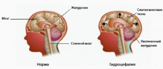
Hydrocephalus is a disease characterized by excessive accumulation of spinal life in the human brain. The disease is otherwise called dropsy. What is moderate external hydrocephalus of the brain? This is one of the most common brain pathologies that can develop in both newborns and adults.
What does the diagnosis of “moderate hydrocephalus of the brain” mean?
Hydrocephalus cerebri is a disease in which an excessive amount of cerebrospinal fluid forms in the brain. Moderate hydrocephalus of the brain is one of the varieties of this pathology.
In a healthy state, fluid (cerebrospinal fluid) washes the brain and helps protect it from concussions, and also serves as nutrition. When the outflow of cerebrospinal fluid is disrupted, as well as when it forms excessively, hydrocephalus occurs (also known as dropsy).
Classification
Moderate hydrocephalus comes in several types:
- moderate external hydrocephalus of the brain is a disease in which impaired circulation of fluid leads to its accumulation mainly in the subarachnoid space;
- moderate internal hydrocephalus of the brain is a type of pathology in which cerebrospinal fluid accumulates in the ventricles of the brain;
- moderate mixed hydrocephalus of the brain - cerebrospinal fluid accumulates both in the ventricles and in the space under the membranes of the brain.
Moderate replacement hydrocephalus of the brain is also diagnosed: in this case, the brain matter gradually decreases in volume, and its place is taken by cerebrospinal fluid. Replacement hydrocephalus usually develops in older people.
Sometimes replacement hydrocephalus can accompany Alzheimer's disease or other degenerative diseases of the central nervous system.
The disease can be acquired or congenital.
In the first case, internal pathology can be the result of external influences - injuries, as well as systemic diseases.
In the second, it can be caused by a difficult birth or infectious diseases acquired during intrauterine development.
Medical therapy
First of all, treatment of pathology is aimed at restoring the functionality of the blood vessels of the brain and central nervous system. Moderate external hydrocephalus of the brain responds well to drug therapy. The attending physician prescribes a course of diuretic medications that help remove excess fluid from the body. Solutions of plasma expanders, saluretics, drugs that help strengthen venous tone, glucocorticosteroids, and barbiturates are used. To eliminate pain, painkillers are prescribed. During treatment, patients are prescribed to follow a special low-fat diet. Complex therapy involves regular performance of therapeutic gymnastic exercises.
If drug treatment does not produce positive results over a certain period of time, then a decision is made about the need for urgent surgical intervention.
Today, hydrocephalus of any form is treated using minimally invasive surgical techniques. Many clinics have at their disposal all the necessary equipment to perform neuroendoscopic operations. During such an intervention, the neurosurgeon performs various types of shunting in the brain tissue, which allows drainage of cerebrospinal fluid into other cavities. All manipulations are performed through small incisions using miniature surgical instruments and a neuroendoscope. Such manipulations last only a few hours and are performed in a hospital under general anesthesia. The procedure is quite safe for the patient's life.
Treatment of hydrocephalus is always an individual process, which has its own prognosis depending on the characteristics of the patient’s body. However, in most cases, timely diagnosis and treatment give positive results, which allows the patient to return to normal life in a short time.
Causes
The immediate cause of the disease is a violation of the outflow of cerebrospinal fluid from the ventricles of the brain through the appropriate channels into the cisterns and further into the spinal canal, where fluid is absorbed into the circulatory system.
But the factors that can lead to this situation may be different.
Moderate hydrocephalus is usually a consequence of other diseases:
- strokes;
- atherosclerosis;
- brain tumors and cysts that compress the ventricles or obstruct the outflow of fluid;
- hypertension;
- infectious diseases (both current and those suffered in the past - this could be meningitis, encephalitis);
- osteochondrosis and hernias of the spine in the cervical region.
In addition, internal moderate dropsy of the brain can be caused by a concussion, falls and head contusions, and injuries received in car accidents.
Internal, external and mixed types of dropsy of the brain can also be caused by alcoholism.
Causes of hydrocephalus
Liquor performs several functions at once: it cushions the brain, protects it from the penetration of pathogenic organisms, nourishes the cells of the organ, and participates in blood circulation and metabolic processes. Cerebrospinal fluid ensures the preservation of the internal environment through coordinated reactions aimed at maintaining dynamic balance due to intracranial pressure.
The amount of cerebrospinal fluid in an adult is 120-150 ml. It is constantly updated. Synthesis occurs in the ventricles of the brain due to the cells of the glandular tissue that lines their inner surface. Then, from the 1st and 2nd ventricles, cerebrospinal fluid penetrates into the 3rd cavity through the foramen of Monroe. From it it enters the 4th ventricle through the aqueduct of Sylvius, and from there, through the foramina of Magendie and Luschka, into the subarachnoid space.
On the surface of the brain, the subarachnoid membrane forms cavities filled with cerebrospinal fluid. They are called tanks. From them, the fluid is directed to the periphery of the brain and surrounds it on all sides.
The cerebrospinal fluid is removed through the venous vessels of the brain with the help of arachnoid cells and villi. Their accumulation around the sinuses is called pachion granulations. Some of the cerebrospinal fluid is eliminated through the lymphatic system by nerve sheath cells.
It follows that cerebrospinal fluid is synthesized inside the ventricles, moves around the brain, and then is excreted through the bloodstream in equal quantities. This process does not stop for a second.
If at one of the stages there is a failure (with production or with excretion), then excess cerebrospinal fluid will accumulate in the brain cavity of the skull, and hydrocephalus will occur.
Dropsy of the brain in adults is provoked by:
- Acute disorders in the blood circulation of the organ. This condition provokes vascular thrombosis, hemorrhagic or ischemic stroke, bleeding caused by rupture of an aneurysm, subarachnoid and intraventricular bleeding.
- Intracerebral inflammation of the central nervous system structures, meninges: meningitis, ventreculitis, encephalitis, tuberculosis.
- Chronic hypoxia, which provokes atrophy of nervous tissue. This condition is typical for poisoning, injuries, and alcohol addiction.
- Neoplasms of various etiologies. Formed and growing neoplasias can cause hydrocephalus because they interfere with the movement of cerebrospinal fluid along the cerebrospinal fluid pathways.
- Head injuries and post-traumatic complications. Intracranial damage to the structures of the central nervous system leads to swelling of the organ tissue and subsequent rupture of blood vessels.
- Complications after surgery: cerebral edema, strangulation and deformation of the cerebrospinal fluid ducts or blood vessels.
- Genetic diseases, malformations of the central nervous system - Bickers-Adams, Dandy-Walker syndromes.
The presence of even one of the above factors increases the risk of hydrocephalus as a complication of the disease or pathology. At the first symptoms, it is recommended to visit a specialized specialist to confirm or refute the diagnosis.
Signs
Moderate hydrocephalus of the brain can develop asymptomatically for a long time.
For several years, the only sign of the disease may be periodic headaches, which often occur in the morning.
The disease can be detected during a diagnostic examination of the brain completely by accident.
Sometimes headaches do not occur, while intracranial pressure remains normal.
However, at a certain period, the consequences of excess accumulation of cerebrospinal fluid still manifest themselves - for example, in the form of impaired blood supply to the brain and its hypoxia (oxygen deficiency).
This condition can lead to any number of consequences, including stroke and dementia.
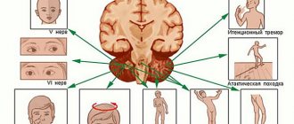
Severe symptoms that occur with the developed form of the disease include:
- problems with vision and hearing;
- dizziness;
- decreased intellectual abilities and partial memory loss;
- loss of attention and ability to concentrate;
- loss of spatial orientation (patients may go for a walk and get lost);
- impaired coordination of movements (patients may experience a change in gait);
- chronic fatigue;
- sleep disorders;
- irritability.
In the acute stage, external, internal and mixed forms of hydrocephalus give such manifestations as vomiting, urinary incontinence, loss of consciousness, and cerebral edema.
This phenomenon is called an occlusive crisis and requires immediate hospitalization: the cause of the crisis is a complete block of the outflow of cerebrospinal fluid.
Diagnostics
Like all other types of hydrocephalus, a moderate form of the disease is detected using radiography of the skull in two projections and magnetic resonance imaging.
The second method is preferable in modern medicine, as it provides more accurate images of the internal cranial cavities.
Sometimes additional procedures may be prescribed:
- angiography is a research method using a contrast agent injected into the circulatory system: it allows you to see abnormalities in the vessels, identify aneurysms or stenosis (narrowing) of the vascular walls;
- Ultrasound;
- general blood analysis;
- CT scan;
- lumbar puncture - sampling of cerebrospinal fluid for the purpose of its further study in the laboratory (allows us to detect the presence of pathogenic microorganisms in the cerebrospinal fluid).
In addition, examinations by an endocrinologist, psychoneurologist, or ophthalmologist may be prescribed.
Treatment
Medication
Moderate hydrocephalus, before the manifestation of severe symptoms, is treated with conservative methods - medications, manual therapy, physical therapy.
There are three types of medications prescribed: those that regulate blood circulation in the brain, antibiotics (in case of infectious causes of the disease) and diuretics (diuretics that stimulate the circulation of fluids in the body).
Operational
Since moderate (internal and external) hydrocephalus can turn into an acute form at any time, doctors, based on diagnostic data, may prescribe surgery.
The most modern type of radical treatment prescribed for hydrocephalus is endoscopic surgery.
Using an endoscope, instruments are inserted into the cranial cavity to make a hole in the central (third) ventricle of the brain. This creates an additional path for the outflow of fluid directly into the brain cisterns.
There are also treatments such as external drainage and bypass surgery.
The first method is used in emergency situations, when there is an urgent need to reduce the fluid pressure inside the brain - for example, if a tumor tightly blocks the channel for the outflow of cerebrospinal fluid, the second method is used less and less due to its inconvenience and the danger of complications.
Shunts constantly require revision and replacement, and there is a constant risk of infection.
In general, the prognosis for moderate hydrocephalus with proper treatment and timely identification of problems with brain fluid is favorable.
The exception is cases when patients do not go to the hospital and the disease leads to irreversible changes in the brain and higher nervous activity.
Treatment of hydrocephalus
Treatment of hydrocephalus begins with a complete medical examination, one of the main tasks of which is to determine the cause of the disease. If the disease was detected at an early stage, doctors recommend drug treatment.
The goal of treatment is to normalize the pressure caused by the accumulation of excess fluid in the brain. This is done either with the help of special medications or through surgery. Conservative treatment methods include:
- special baths with salts and pine oils
- therapeutic exercises
- a diet aimed at reducing the amount of fluid consumed
- taking anti-inflammatory drugs, etc.
Still, they don't solve the problem. Surgery is considered the most effective. The operation is performed by a neurosurgeon. If shunt methods were previously used, today the main assistant in the fight against hydrocephalus is endoscopic treatment. The result of the operation is the creation of holes for the outflow of fluid without introducing special devices into the body.
What types of external hydrocephalus of the brain are there?
External hydrocephalus of the brain refers to the accumulation of cerebrospinal fluid (cerebrospinal or cerebrospinal fluid) outside the cerebral hemispheres - in the subarachnoid space. Due to the large accumulation of fluid, the subarachnoid fissures widen, which causes increased pressure on the cerebral cortex and the resulting negative consequences.
The nature and level of complexity of the disease directly depend on the specific type of dropsy. Several criteria are used for classification. The most common ones are:
- intensity of manifestation (pronounced - accumulation of a large amount of cerebrospinal fluid, causing neurological symptoms; moderate - minimal amount of fluid, no signs);
- degree of impact on brain structures (compensated - cerebrospinal fluid does not affect the brain; decompensated - there is a deterioration in the functioning of the nervous system and brain);
- causes of occurrence (replacement - more often diagnosed in older people and is accompanied by the death of brain cells; acquired - occurs due to the spread of infections and mechanical traumatic brain injuries);
- nature of the course (chronic form - a gradual increase in neurological disorders; acute form - a sharp deterioration in the patient’s well-being).
What is external hydrocephalus?
Hydrocephalus is a progressive disease that is characterized by an abnormal increase in the volume of cerebral fluid in the subarachnoid spaces, cisterns and ventricles. The pressure inside the skull with this disease increases significantly due to the increased production of cerebrospinal fluid, which entails a violation of its reabsorption.
Open hydrocephalus usually occurs in infants and is associated with insufficient absorption of excess cerebrospinal fluid, which leads to their accumulation in the ventricles of the brain, their expansion and atrophy of neighboring areas of the brain.
In mild cases, conservative treatment is prescribed; To correct and prevent pronounced changes, a shunt is surgically inserted to divert excess cerebrospinal fluid into the circulatory system.
A diagnosis such as hydrocephalic-hypertensive syndrome is not uncommon and is currently recorded in many newborns. However, according to many experts, deviations within this diagnosis cannot be considered abnormal or pathological.
The concepts of “intracranial hypertension” and “hydrocephalus” should not be confused.
For example, with atrophic changes in the brain associated with the aging process, replacement normotensive hydrocephalus is quite common. In this case, excess cerebrospinal fluid does not exert pressure on the brain tissue, so treatment is not required.
Neurologist, reflexologist, hirudotherapist
Kislitsyna Ekaterina Nikolaevna
10 years of experience
They do not pose a serious danger to the child’s health, and the unreasonable use of pharmaceuticals to improve cerebral circulation, nootropics and diuretics can have a very negative impact on children’s health.
Shunting for hydrocephalus of the brain
Hydrocephalus is a life-threatening disease.
The consequence of this process is an excess of cerebral fluid in the cranial cavity. As a result of the disorders, the medulla becomes softer, and the ventricles increase in their own volumes. Provided that the child develops normally, the considered pathological processes cease, and the further clinical picture remains within the normal range. The normal amount of cerebral fluid is about 50 ml (in a child) and no more than 150 ml (in an adult).
In order to get the most complete understanding of the essence of such a disease as external hydrocephalus, you need to understand what the direct function of cerebral fluid is. In the brain of a healthy person, a certain amount of cerebrospinal fluid is constantly produced, which is distributed among the cisterns, ventricles and penetrates the subarachnoid fissure. Thanks to the permanent circulation of cerebral fluid, optimal conditions are maintained for the full functioning of the brain.
The cerebrospinal fluid performs such an important function as ensuring a stable and permanent location of the brain in the space of the cranium, preventing its displacement or wedging into the foramen magnum.
Cerebral fluid plays the role of a specific shock absorber, which significantly reduces the consequences of head injuries and concussions. The normal functioning of local metabolic processes involves the distribution of cerebrospinal fluid into:
- occipital-parietal region;
- liquor spaces;
- spinal canal.
The process of constant production, circulation and elimination of excess cerebral fluid contributes to its complete renewal and the removal of toxic substances from the body in general and the brain in particular.
Why can our articles be trusted?
We make health information clear, accessible and relevant.
- All articles are checked by practicing doctors.
- We take scientific literature and the latest research as a basis.
- We publish detailed articles that answer all questions.
If cerebrospinal fluid is produced in excess, then an insufficient volume of cerebral fluid is absorbed through the systemic circulation. This leads to enlargement of the cisterns and ventricles. The degree of external hydrocephalus, as well as the severity of irreversible brain damage, directly depends on the degree of such imbalance of metabolic processes.
Hydrocephalus can develop in a person of any age, but it is most common in newborns. In such cases, the disease is usually associated with the consequences of an infectious disease suffered during pregnancy.
Symptoms of external hydrocephalus
The clinical picture in each specific case will be different, and the nature of the manifestation of the disease depends on the severity of the pathological process and the state of the central nervous system. Common symptoms are frequent headaches, blurred vision, nausea, vomiting, and weakness. By the way, pain is more localized in the frontoparietal region and in the area of the eyeballs. A person with dropsy experiences pain in the first half of the day, with sudden movements, coughing, sneezing, or severe physical exertion.
Symptoms may vary depending on the severity of the disease. Scientists distinguish 3 stages, and each has its own characteristics:
Mild external hydrocephalus. With a minimal amount of dropsy, the human body will try on its own to cope with such a problem as impaired circulation of the cerebrospinal fluid. In this case, you will feel a slight malaise, periodic dizziness, short-term darkening in the eyes, and a tolerable headache.
The middle stage of development of the disease. At this stage of the spread of the disease, symptoms appear intensely and are more pronounced. Due to an increase in intracranial pressure, severe headaches occur during physical activity, swelling of the optic nerve and facial tissues, increased fatigue, nervousness, depression, and surges in blood pressure.
Severe form of the disease. Signs of pathology in severe forms of external hydrocephalus are reduced to convulsive seizures, frequent fainting, a state of apathy, loss of intellectual abilities, memory loss and inability to care for oneself. Progressive dropsy can even lead to death, so there is no need to delay going to the doctor. It is better to do this at the first suspicion and a slight deterioration in health.
With chronic accumulation of cerebrospinal fluid, symptoms such as unsteady gait, paralysis of the upper and lower extremities, urinary incontinence, nighttime insomnia and daytime sleepiness, depressed mood, and a complex of psychoneurological disorders can be observed.
Symptoms and severity
According to clinical manifestations, external hydrocephalus in an adult is divided into 3 degrees: mild, moderate and severe. With mild hydrocephalus, the body can independently restore the circulation of cerebrospinal fluid. The patient complains of slight malaise, headache, dizziness, and short-term darkening of the eyes. The average degree of hydrocephalus is manifested by intense signs of brain damage. Patients experience the following symptoms:
- Severe pain in the head, aggravated by physical activity;
- Pressing pain in the eyeballs, the appearance of colored circles of flashes when closing the eyes;
- Feeling of heaviness in the skull;
- Nausea independent of food intake;
- Vomiting without relief.
Sweating occurs periodically. An ophthalmological examination reveals swelling of the optic nerve head. Patients note swelling of the face, weakness, lethargy, and increased fatigue. They are worried about feeling tired in the morning, aggressiveness, increased nervousness, and tearfulness. A depressive state develops. Blood pressure is unstable. Unpleasant sensations intensify when coughing, sneezing, turning or tilting the head.
External hydrocephalus of the brain in an adult is accompanied by neurological symptoms:
- Decreased visual acuity;
- Strabismus;
- Impaired visual perception: double vision, blurred images;
- Paralysis or paresis of the limbs;
- Numbness of the face;
- Decreased sensitivity;
- Impaired coordination.
Patients have speech impairment, difficulty pronouncing sounds and perceiving spoken speech. With severe external hydrocephalus, epileptic and convulsive seizures, frequent fainting, and coma occur. The patient loses memory, intellectual abilities, and his self-care skills decrease.
Patients with occlusive hydrocephalus experience severe headache, nausea and vomiting in the morning. Congestion of the optic discs and signs of axial dislocation of the brain develop. An unfavorable prognostic sign is drowsiness. It intensifies on the eve of a sharp disruption of neurological symptoms. When brain structures are dislocated, cardiac activity and breathing are disrupted.
The clinical picture of chronic hydrocephalus consists of three pathognomonic symptoms: dementia, gait disturbance and urinary incontinence. Dementia is manifested by a decrease in the level of wakefulness, rapid exhaustion of the patient, disorientation in time, the development of severe intellectual disorders, and decreased criticism. Patients become unsure when walking and develop paresis of both lower extremities. The most recent symptom of hydrocephalus is urinary incontinence. This symptom of the disease is more common in men after 50-60 years of age.
Based on the intensity of the manifestation of hydrocephalus, a distinction is made between a moderate form of the disease, which occurs with minor symptoms, and a severe form - the accumulation of a large volume of cerebrospinal fluid provokes the manifestation of acute neurological symptoms.
Depending on the degree of impact on brain structures, external hydrocephalus can be compensated or decompensated. In the presence of compensated hydrocephalus, excessive secretion of cerebrospinal fluid does not affect the brain, but in the decompensated form of the pathological process, regardless of the amount of cerebrospinal fluid, brain function is disrupted and the functional activity of the central nervous system is reduced.
External non-occlusive hydrocephalus occurs when the process of cerebrospinal fluid absorption is disrupted. Atrophic (replacement) hydrocephalus develops most often in older people and is accompanied by the death of brain cells.
Why does dropsy of the brain occur?
In adult patients, acquired hydrocephalus is often encountered, which develops either due to any mechanical damage to the head, or as a result of the development of pathological processes. Why does cerebrospinal fluid accumulate outside the cerebral hemispheres? The explanation is simple: brain structures are disrupted, adhesions appear in the veins, arachnoid villi are destroyed, and as a result, the cerebrospinal fluid does not circulate as it should.
If we delve deeper into the question of the causes of such a disease as external dropsy of the brain, we can identify some factors:
- infectious diseases (tuberculosis, meningitis, encephalitis);
- post-stroke condition, development of sepsis, extensive hemorrhage;
- concussion, head or cervical spine injury;
- malignant tumors that develop in the stem region.
Frequent intoxication of the body leads to the appearance of external hydrocephalus. For example, alcohol abuse, which damages neurons and leads to tissue death. Those patients who suffer from metabolic disorders, diabetes mellitus, multiple sclerosis, encephalopathy, and atherosclerosis are also at risk. Another reason that deserves due attention is irreversible age-related changes that cause aging of blood vessels and brain tissue.
Main services of Dr. Zavalishin’s clinic:
Reasons for development
Acquired hydrocephalus can develop due to pathological processes that result in adhesions in the vessels of the brain and destruction of arachnoid villi. The disease is provoked by the following factors:
- Infectious diseases of the brain (encephalitis, meningitis, tuberculosis);
- Sepsis;
- Previous hemorrhagic stroke;
- Head, neck injury;
- Traumatic brain injuries;
- Traumatic spinal injuries;
- Malignant neoplasms of the brain stem.
Atrophic hydrocephalus occurs due to age-related changes in cerebral vessels, metabolic disorders, diabetes mellitus, and arterial hypertension. With external hydrocephalus, zones with reduced density of brain matter are formed. They atrophy, and the vacated space is filled with cerebrospinal fluid. The cause of the development of external hydrocephalus can be constant alcohol intoxication.
Diagnosis of external hydrocephalus in adult patients
Studying the symptoms and visual examination of the patient is not a sufficient condition for determining external hydrocephalus of the brain. Indirect signs, of course, are important, but you can’t do this without professional diagnostics. Today, 6 methods for detecting dropsy are used:
- Ultrasound examination (US) of the neck and head to assess the condition of blood vessels;
- Magnetic resonance imaging (MRI) helps to identify changes in soft tissues and most accurately determine the type of hydrocephalus and the stage of development of the pathology;
- computed tomography (CT) is intended to determine the degree of damage to brain tissue, the size of subarachnoid fissures, and the presence of neoplasms;
- X-rays with the introduction of a contrast agent are aimed at identifying disturbances in the outflow of venous blood and damage to the vascular bed;
- a spinal puncture is prescribed if there is a suspicion of the development of dropsy after encephalitis or meningitis and you need to find out what level of cerebrospinal fluid pressure;
- ophthalmological examination - an opportunity to determine whether the patient has swelling of the optic nerve and atrophy of the tissues of the ocular apparatus.
IMPORTANT! If the diagnosis of “chronic external hydrocephalus of the brain” is confirmed, it is advisable to carry out an additional diagnostic examination after 6 months. The intensity of further visits to the doctor depends on the data obtained and is determined individually.
Diagnostic methods
External hydrocephalus of the brain in an adult is diagnosed using the following clinical research methods:
- A complete neuropsychological examination, during which the patient is questioned about his condition. Information is collected about the presence of various abnormalities and disorders of brain functionality. Based on the data obtained, the doctor compiles an anamnesis and decides on making a diagnosis or additional research methods;
- CT (computed tomography). This method is one of the most accurate and reliable diagnostic methods. Using CT, the contours of the brain and skull, the location of the ventricles, their shape and size are determined; detect abnormal neoplasms (tumors, cysts, hematomas, blood clots, etc.);
- MRI (magnetic resonance imaging). MRI imaging allows one to accurately determine the shape and severity of any type of hydrocephalus. Thanks to MRI, a correct diagnosis of cerebral hydrocele is possible;
- Angiography (examination of the blood vessels of the brain using x-rays). The study is carried out using a contrast agent, which makes it possible to detect the slightest disturbances in the blood vessels of the circulatory system in the human brain;
- Cisternography. X-rays of the cisterns at the base of the skull help identify the type of hydrocephalus. Also, using this method, the doctor can determine the direction of movement of the cerebrospinal fluid in the brain tissue.
Treatment of external hydrocele in adults
Treatment methods are selected at a consultation with a neurosurgeon or neurologist after diagnosing the disease. Intervention must be timely, otherwise the risk of various neurological complications increases. It is important to take into account both the type of pathology and the characteristics of the patient’s body.
In the Department of Neurosurgery of the City Clinical Hospital named after. Eramishantsev practice only effective methods of treating external hydrocele of the brain. Methods are divided into two large groups: conservative (medicinal) and surgical (operative), each of which has its own characteristics and advantages.
CONSERVATIVE TREATMENT
Drug treatment is only relevant at a mild stage of the disease. Special medications accelerate the outflow of fluid from the brain, increase urination, relieve inflammation and swelling, strengthen blood vessels, and normalize the functioning of the cardiovascular system. To combat severe headaches, your doctor may prescribe non-steroidal anti-inflammatory and painkillers.
Common groups of medications are vascular, neurotropic, venotonics, diuretics. But in acute illness they will be ineffective. Mixed hydrocephalus is poorly corrected. In this case, conservative treatment will not get rid of the disease, but will only restore or improve the functioning of individual systems and functions of the human body. Often surgical intervention is not possible.
SURGERY
If acute external dropsy is diagnosed, in most cases drainage of the cerebral ventricles is prescribed. The main technologies are endoscopy and open surgery.
In the first case, we are talking about manipulations that are characterized by minimal trauma, a very low risk of complications, and fairly rapid postoperative recovery. Endoscopy methods allow, with minor intervention, not only to remove excess cerebrospinal fluid, but also to eliminate vein defects, hematomas, and blood clots.
Currently, open surgery is chosen only in exceptional cases. Why? It is difficult to imagine performing open surgery without craniotomy. And trepanation always means increased risks and a long postoperative recovery period.
Moderate internal hydrocephalus of the brain
Moderate external hydrocephalus of the brain can be a manifestation of various diseases or an independent disease. Minor hydrocephalus of the brain is treated conservatively. At the Yusupov Hospital, neurologists use individual treatment regimens for moderately severe hydrocephalus of the brain in adults. Professors and doctors of the highest category use the most effective drugs registered in the Russian Federation, which do not have side effects.
Patients with moderate external open hydrocephalus of the brain are examined using modern devices from the world's leading manufacturers. The neurology clinic has created comfortable living conditions. The staff is attentive to the wishes of each patient. Rehabilitation clinic specialists use innovative methods of rehabilitation therapy.
Causes and types of hydrocephalus of the brain in adults
The acute form of moderate hydrocephalus of the brain is accompanied by high intracranial pressure. Patients suffer from headaches in the morning. Its intensity decreases throughout the day. In the morning, digestive disorders may occur, including nausea and vomiting. If vomiting reduces the intensity of the headache, this indicates a problem with the brain. As intracranial pressure increases, drowsiness increases, which is an unfavorable prognostic sign.
When the medulla oblongata is compressed, breathing and cardiac activity are impaired. In the medulla oblongata, hydrocephalus affects the heart and respiratory system, and death most often occurs. Moderate replacement hydrocephalus of the brain is a separate form of the disease. It is characterized by a decrease in brain volume. The vacated space is cerebrospinal fluid. External replacement hydrocephalus does not appear for several years.
The first signs of chronic moderate external replacement hydrocephalus occur 2 weeks after a disease of the central nervous system or a brain injury. Patients experience impaired sleep quality, memory impairment, and cannot distinguish day from night. The person becomes inactive and loses interest in what is happening. If the disease or complications are not recognized in a timely manner, intellectual activity is disrupted, reactions become inadequate, walking is impaired, and urinary incontinence may occur.
Moderate external hydrocephalus of the brain occurs as a result of the following diseases:
- infectious diseases of the central nervous system;
- cerebrovascular accidents;
- intraventricular, parastem or stem brain tumors;
- encephalopathies of various origins;
- non-traumatic, traumatic and intraventricular hemorrhages.
Hydrocephalus of the brain can be hereditary or acquired. Based on the location of fluid accumulation, internal and external hydrocephalus are distinguished. Internal hydrocephalus occurs due to the accumulation of cerebrospinal fluid in the ventricles of the brain. External hydrocephalus develops as a result of the accumulation of cerebrospinal fluid in the subarachnoid and subdural spaces.
With closed hydrocephalus, there is a violation of the outflow of fluid. Open hydrocephalus is not characterized by impaired circulation of cerebrospinal fluid.
According to the rate of progression of the disease, acute, subacute and chronic hydrocephalus are distinguished. In acute hydrocephalus, three days pass from the first symptoms to decompensation. With subacute hydrocephalus, the disease develops within a month. The formation of chronic hydrocephalus occurs within 6 months. Moderate replacement hydrocephalus of the brain occurs mainly in older people. The disease develops against the background of atherosclerosis, alcohol abuse, arterial hypertension, and instability of the cervical vertebrae. The passive form of the disease is moderate external hydrocephalus. This is a rather dangerous disease, since in most cases there are no symptoms characteristic of hydrocephalus.
Symptoms
The disease is popularly called dropsy of the brain. This is due to the external signs that the pathology exhibits.
In adults
In adults, the bone tissue of the head is already formed, so there may be no external symptoms. The disease may not manifest itself for a long time.
Headaches that periodically bother every person can be minor. But HC will still manifest itself, since the accumulation of CSF leads to disruption of the blood supply to the brain and the development of symptoms characteristic of this condition.
Excessive accumulation of CSF leads to increased ICP, but in chronic external replacement hydrocephalus it can be normal.
The patient is bothered by an intense throbbing headache that is not relieved by medications. It appears mainly after sleep and may subside during the day. Against this background, nausea may develop, including vomiting. The hallmark of headaches associated with HC is ocular discomfort. The patient feels increased pressure on the eyeballs. With a slight tilt of the body, this feeling intensifies.
Compression of brain structures provokes a number of disorders:
| Type of disorder | Description |
| Mental | Emotional lability, neurasthenia, mood swings. When symptoms worsen, unreasonable aggressive behavior is possible. |
| Vestibular | There is a lack of coordination, unsteadiness in standing and sitting positions, loss of orientation in space. There is no control of body movements, limbs, handwriting is disrupted. The patient is bothered by systemic vertigo, in which he experiences rotation of the body to a state of rest or movement of surrounding objects. Symptoms intensify when turning the body, head, and eyes, which forces the patient to avoid these movements or perform them at a slow pace. |
| Vegetative | During exacerbations, the complexion changes. Due to the lack of control of movements, a feeling of fear appears, panic develops, tachycardia, lack of oxygen, and discomfort in the heart. |
| Visual | Violation of visual control: blurred vision, color perception disorder, narrowing of the field of vision (lack of lateral), the appearance of dark spots, decreased pupil response to a light stimulus, twitching of the eyeballs, downward gaze shift. |
| Muscular | Paresis, paralysis, increased muscle tone, increased tendon reflexes, decreased sensitivity. |
In children
Children have external signs of HC, because the process of formation of the bone tissue of the head is incomplete, moderate external hydrocephalus is manifested by an increase in the size of the cranial cavity. Visually, the brain region significantly predominates over the facial region.
In infants, newborns, and young children, the head size exceeds the norm. On palpation, arterial pulsation of the large fontanelle is absent, and its bulging is observed. The cranial sutures may come apart. Its covering bones are thinning. The saphenous veins of the head swell. The eye sockets become deformed, the optic nerves swell, and upward movement of the eyeballs is limited. In advanced cases, strabismus develops.
Children whose pathology is diagnosed at an early age are lagging behind in motor and physical development. They later begin to hold their head up, roll over, crawl, sit, walk, and talk. Forced to tilt your head back. Physical activity is low, so there is a tendency to obesity. Behavior: the child is irritated, sleeps poorly, cries often, refuses to breastfeed, sleeps often and for a long time.
Unlike HC in adults, in children the disease mostly manifests itself not as neurasthenic disorders, but as a lack of intelligence. But it may be insignificant. School-age children may have excellent mechanical memory, verbosity, and musical abilities. An exception is HC due to meningoencephalitis.
Initial symptoms that may indicate increased ICP are:
- anxiety;
- disturbance of sleep and wakefulness;
- headache.
Older children more often have an acquired form of the pathology, in which they constantly suffer from headaches, blurred vision, nausea, and vomiting. Epilepsy attacks, loss of consciousness, convulsions, hallucinations, and delirium may be present.
Signs of moderate external hydrocephalus of the brain
The main symptoms of moderate hydrocephalus include:
- migraine-like headache
- vomiting and nausea;
- violation of vestibular functions;
- constant drowsiness;
- involuntary urination;
- diplopia (double vision).
With moderate external replacement hydrocephalus, intracranial pressure may remain normal. Neurologists at the Yusupov Hospital use modern methods for diagnosing hydrocephalus:
- computed or magnetic resonance imaging;
- craniography;
- cisternography;
- ultrasound examination of cerebral vessels;
- angiography.
Patients with hydrocephalus are consulted by an ophthalmologist and a neurosurgeon. Patient management tactics are discussed at a meeting of the expert council.
Treatment of moderate hydrocephalus of the brain in adults
The choice of treatment for moderate external hydrocephalus depends on the condition of the brain structures, their displacement and the level of intracranial pressure. Doctors at the Yusupov Hospital provide conservative treatment for moderate external open hydrocephalus of the brain. Patients reduce the amount of fluid they consume and are prescribed diet therapy. Drug treatment is aimed at lowering blood pressure, normalizing brain functions, and controlling the supply of nutrients to its tissues.
Patients are prescribed diuretics: diacarb, furosemide, mannitol. Asparkam is used to prevent hypokalemia. Improves cerebral circulation glycine, cinnarizine, piracetam, cerebrolysin, cavinton. As maintenance therapy, patients take multivitamin complexes and restoratives. Neurologists at the Yusupov Hospital determine the required dosage of each drug and monitor the dynamics of the disease.
If conservative therapy is not effective and the symptoms of hydrocephalus intensify, neurosurgeons at partner clinics perform surgical interventions:
- installation of a shunt to drain excess cerebrospinal fluid;
- endoscopic surgery aimed at creating pathways for the outflow of cerebrospinal fluid;
- removal of a tumor or other formations that obstruct the circulation of cerebrospinal fluid.
Endoscopic surgery is performed if the following indications exist:
- high level of occlusion;
- hydrocephalus after injury;
- secondary surgery after failed bypass surgery;
- mixed type of hydrocephalus.
Neurosurgeons perform the following endoscopic operations:
- plastic plumbing;
- endoscopic removal of stoma of the bottom of the third ventricle;
- septal stoma;
- installation of a shunt system;
- endoscopic removal of an intraventricular brain tumor.
If there are signs of moderate hydrocephalus, treatment should not be delayed. Call the Yusupov Hospital and they will make an appointment with a neurologist. After the examination, doctors will provide effective treatment for hydrocephalus.
Classification of hydrocephalus: severe and moderate, internal and external
Hydrocephalus is a process in the brain in which too much fluid accumulates inside the skull. As a result, a person’s intracranial pressure increases, and disturbances in the functioning of the brain and other systems occur. There are external and internal hydrocephalus, but there are other methods of classifying the disease. And each of them is characterized by different pathological processes.
Dropsy of the brain is found in children of any age, as well as in the adult population. There are more than 100 reasons that can lead to pathology. And they cannot be predicted or prevented in any way.
External hydrocephalus of the brain in adults is a form of pathology in which excess fluid accumulates mainly under the meninges, but may remain normal in the ventricular system. However, it is characterized by the same signs and symptoms as internal pathology.
Causes and mechanism of development
Inside the skull is the brain, blood and cerebrospinal fluid. Under normal conditions, a balance is maintained between the volumes of these three components. Any violation of it leads to the development of complex pathologies. Thus, an increase in the volume of cerebrospinal fluid compresses blood vessels and the brain, leading to impaired blood supply, tissue death, and increased blood pressure. Hemorrhage, in turn, leads to disruption of the outflow of cerebrospinal fluid and its accumulation.
The mechanism for the development of hydrocephalus is precisely the excess of cerebrospinal fluid. It is formed in the four cerebral ventricles, with up to 70% produced in the glands, the remainder by the exudation of the liquid component of the blood through the walls of blood vessels. The flow of cerebrospinal fluid is carried out from the ventricles into the subarachnoid cavity, which expands and forms the cerebral cisterns.
From these cavities it reaches the outer surface of the brain, and is later absorbed through the villi located in the area of the venous sinuses. Normally, the amount of liquor produced (on average in an adult is 150 ml) corresponds to that absorbed.
However, in any of these areas, obstructions or disturbances can occur, leading to excess cerebrospinal fluid - hydrocephalus.
The disease is caused by the following reasons:
- Inflammation of the brain and its membranes. This group includes meningitis, encephalitis, and tuberculosis.
- Impaired blood supply. Hydrocephalus develops as a result of bleeding in the brain, the appearance of a blood clot, or rupture of a bulging vessel (aneurysm).
- Neoplasms. Regardless of their nature - benign or malignant, they lead to blockage of cerebrospinal fluid circulation.
- Intoxication. The effects of alcohol, drugs, heavy metal salts.
- Injuries. This group includes not only traumatic brain injuries that led to brain swelling and destruction of blood vessels, but also consequences after operations.
- Diseases of the central nervous system.
- Infectious diseases. This is rubella, syphilis.
- Age-related changes. Replacement hydrocephalus is associated precisely with how the body and its tissues change with age.
Classification of hydrocephalus
There are several ways to classify external hydrocephalus: open and closed, congenital or acquired, acute or chronic. It is also divided according to severity: severe and moderate external hydrocephalus of the brain.
Types by type and mechanism of development
Closed hydrocephalus is a disease in which the outflow of cerebrospinal fluid is impaired due to any obstruction. As a result, brain fluid does not enter the system.
With closed external hydrocephalus, excessive accumulation of fluid occurs in the subarchanoid space and does not enter the systemic circulation. Causes of the closed form of the disease: tumors, cysts, narrowing of the aqueduct, hemorrhages, closure of the holes of Lushka and Manaji.
In this case, closed external hydrocephalus in adults can be severe or moderate. Hydrocephalus will develop locally, in the place where the obstruction appears. Neurological symptoms in the closed form of the disease occur as pressure increases.
Open form
Open external hydrocephalus occurs in adults when the absorption of liquor fluid is impaired, but in this case there are no obstacles to its movement. The movement of cerebrospinal fluid occurs as usual, but it is absorbed extremely slowly as a result of unknown factors.
In the external form of the pathology in an adult, the subarachnoid space expands, and the brain gradually atrophies. The causes of this open form include: hemorrhages, metastases, meningitis, sarcoidosis and cysticercosis.
Very rarely, the cause is a tumor of the choroid plexus that produces liquor fluid.
By time of formation
Severe and minor external hydrocephalus can be congenital, acquired and replacement:
Purchased . Occurs after brain damage as a result of inflammation: encephalitis, arachnoiditis, meningitis, or under the influence of trauma, hemorrhages in the arachnoid membrane. With internal hydrocephalus, the ventricles are affected.
Also, the causes of acquired external moderate or severe hydrocephalus are: hemorrhagic stroke and tumor processes of various types, cysts, parasites that affect the brain.
Congenital . It develops only in children during development in the embryonic stage. It appears in the first months of a child’s life. Most often, the cause is malformations of the brain, which can be caused by fetal infections (toxoplasma, syphilis, rubella, mumps, cytomegalovirus), birth injuries and hemorrhages under the membrane of the cerebral aqueduct. Almost all congenital pathologies are of the closed type.
Substitute . A mixed form of brain pathology, not considered pure external hydrocephalus. It occurs against the background of brain atrophy, which reduces its volume. The balance between produced and excreted fluid is not disturbed.
Atrophy in replacement pathology is formed as a result of: vascular encephalopathy (a consequence of atherosclerosis or hypertension), age-related changes, toxic encephalopathy, Creutzfeldt-Jakob disease.
However, the classification of disease types does not end there.
The nature of the pathology
There are 2 types of disease progression based on intensity and duration. Acute hydrocephalus develops rapidly, symptoms intensify over several days. Most often it is closed and requires an emergency neurosurgical operation - brain bypass.
The chronic form develops gradually - from 6 months to 2-3 years. The patient’s intracranial pressure increases smoothly, no surges are recorded. In the chronic form, the symptoms are invisible at first, but when the pathology becomes severe, the signs of the disease become obvious. The chronic form most often occurs as an open disease.
Mixed form
Moderate external hydrocephalus or a severe form of the disease is an accumulation of cerebrospinal fluid in all layers of the brain: ventricles, cisterns, subarachnoid space. The severity of the disease is divided into 2 types:
- compensated form - excess fluid does not compress other tissues of the organ, there are no symptoms of the disease;
- decompensated form - excess fluid puts pressure on surrounding tissues, so signs of the disease form.
In the second type of pathology, symptoms appear gradually and lead to loss of performance.
Moderate and severe form
External hydrocephalus is divided into two more types according to the severity of the clinical picture and symptoms. The diagnosis of the intensity of the processes is made based on the results of MRI. In the external form of the disease, the subarachnoid space, interventricular septa and other parts of the brain are examined.
The diagnosis of moderate external hydrocephalus is made if, during 3 MRI sessions with an interval of 2-3 weeks, minor changes in the circulation of the cerebrospinal fluid were detected. If the changes significantly deviate from the norm, severe external hydrocephalus is diagnosed.
But changes in the volume of liquor structures can also occur against the background of other diseases. Therefore, the patient needs constant monitoring.
Common causes of the disease
In addition to the fact that each type of disease is characterized by unique causes of formation, there are common development factors:
- any infectious damage affecting the brain, including: encephalitis, tuberculosis, meningitis;
- subarachnoid hemorrhage;
- head and cervical injuries;
- —strokes—various types, the most dangerous is hemorrhagic;
- brain stem tumors;
- long-term alcohol intoxication, especially when drinking low-quality drinks;
- spinal injuries and compression of the cerebrospinal canals as a result of edema;
- purulent infectious processes in the brain.
Separately, we can note the causes of moderate external hydrocephalus of the replacement type: metabolic problems, senile or vascular encephalopathy, diabetes, atherosclerosis or multiple sclerosis.
Causes of the disease
Doctors identify the following reasons for the development of moderate external hydrocephalus:
- Concussion, traumatic brain injury;
- Fractures and injuries of the spinal bones;
- Surgical operations on brain tissue;
- Tumors in the head of various etiologies;
- Strokes, extensive cerebral hemorrhages, hematomas;
- Various diseases caused by infection or inflammatory processes (tuberculosis, meningitis, encephalitis, etc.);
- Pathological disorders of the vertebrae in the cervical region;
- Diseases of blood vessels in brain tissue;
- Oncological diseases of bone tissue in the spine;
- Age-related changes in brain tissue;
- Serious deviations in the development of the central nervous system.
One of the reasons for the development of external hydrocephalus may be prolonged intoxication of the body. Thus, with excessive alcohol abuse, the nerve cells of the brain begin to rapidly die. The result of such disturbances is a severe deterioration in the absorption, production and circulation of cerebrospinal fluid. The accumulated fluid begins to stretch the ventricles of the brain. This leads to the saturation of the medulla with cerebrospinal fluid, which significantly reduces its density and narrows the subarachnoid spaces. As a result, a person develops external hydrocephalus of the brain of an atrophic nature.
Most often, elderly people suffer from moderate external hydrocephalus. This disease cannot be left without adequate medical therapy under the supervision of a physician. Practice shows that this is such a dangerous disease and if it is not treated, it can be fatal.
Main signs of the disease
Moderate external hydrocephalus is accompanied by many symptoms, but some of them are cleverly masked and may not cause discomfort for a long time. Signs of the disease are conventionally divided into several groups:
- Are common . At the initial stage, rare headaches appear, accompanied by increased pressure inside the skull and nausea. As the disease progresses in its external form, these symptoms disappear or become insignificant.
- Nervous system instability . Most often, patients complain of disorders of the vestibular system, their vision deteriorates: tinnitus, dizziness, problems with gait, and loss of the ability to visualize objects appears. If left untreated, the nerves located in the eyeball atrophy.
- Problems of the musculoskeletal system . The patient feels partial paralysis, reacts poorly to external stimuli, and changes in handwriting appear.
- Mental problems . In a person with moderate external hydrocephalus, the level of aggression increases. A sharp change of emotions develops.
Other signs of the disease appear that do not directly indicate it. The patient's biological rhythm changes, he is more often awake at night, apathy and indifference appear.
Sometimes people become lost in space, and at the peak of the disease's progress, the ability to speak, move and think is lost. In advanced stages of cerebral hydrocele, a symptom such as urinary incontinence occurs.
In children, dropsy manifests itself with additional signs: an unusual shape of the head, bulging veins on the skin, developmental delay, vision problems, sunken eyeballs, when tapped, a sound is heard, as if from a container with liquid. Treatment of the disease is most often performed surgically, but medications are also used .