Physiology and functions
Comparison of the effects on various organs of the sympathetic and parasympathetic nervous system.
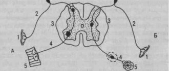
The sympathetic trunk ganglion is a cluster of perikarya of multipolar neurons with predominantly unmyelinated nerve fibers (type C), surrounded by a connective tissue capsule with septa.
Preganglionic fibers approach the perikarya of the node and form synapses, in which it acts as a mediator (acts on n-cholinergic receptors).
Some fibers pass through the nodes in transit (in particular, all splanchnic nerves). These fibers switch to postganglionic fibers in the 2nd order ganglia, which are part of the nerve plexuses (celiac plexus, renal plexus, etc.).
Postganglionic fibers are fibers that are actually directed to target tissues and, forming numerous synapses, have an adrenergic effect (acting on α1-, ά2-, β1- or β2-adrenergic receptors).
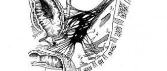
The superior cervical node is responsible for the sympathetic innervation of the craniofacial region, which gives off postganglionic fibers that, as part of the periarterial plexuses, reach the corresponding parasympathetic nodes of the head (as radix sympathica), pass through in transit and reach the corresponding organs:
- n. caroticus internus → plexus caroticus internus → n.petrosus profundus → pterygopalatine ganglion (g. pterigopalatini) → mucous membrane of the nasal cavity, palate, pharynx, lacrimal gland.
- n. caroticus internus → plexus caroticus internus → plexus ophthalmicus → ciliary node (g. ciliare) → pupillary dilator.
- nn. carotici externi → plexus caroticus externus → plexus facialis → submandibular and sublingual nodes → submandibular and sublingual glands.
- nn. carotici externi → plexus caroticus externus → plexus meningeus medius → ear node (g. oticum) → parotid gland.
Also, dendrites of sensitive pseudounipolar neurons, which are located in the spinal ganglia and are responsible for visceral sensitivity, transit through the nodes of the sympathetic trunk.
Research methods
Conventional wedge methods (inspection, palpation, percussion) make it possible to establish changes in skin color, asymmetry, swelling, infiltration. These changes can occur during inflammatory-purulent processes of the retroperitoneal tissue, tumors of the 3rd section, as well as diseases of organs located in the 3rd section. Methods for studying these organs - see separate articles, for example. Aorta, Duodenum, Ureters, Adrenal Glands, Pancreas.
Roentgenol, the study includes non-contrast methods - plain radiography and fluoroscopy of the abdominal and thoracic cavities; contrasting of the abdominal organs - contrast study of the stomach and intestines, cholegraphy (see), pneumoperitoneum (see), splenoportography (see); as well as excretory or retrograde urography (see.
), Pneumoretroperitoneum (see), etc. If there are special indications (suspicion of damage to the pancreas, blood or lymph vessels, etc.), they resort to pancreatography (see), aortography (see), selective angiography of the branches of the abdominal aorta, cavography (see), lymphography (see). To study the pelvic organs, Pneumoretroperitoneum is sometimes combined with metrosalpingography (see.
), cystography (see) and other special studies. Such double or triple contrast in combination with tomography in direct and lateral projections (see Tomography) allows you to determine the exact localization, and in some cases the nature of the pathol, process or developmental anomaly, the shape, size and contours of the kidneys and adrenal glands, the state of the perinephric tissue, which is of great value for the differential diagnosis of patol, processes in 3. p. and in the abdominal cavity.
Sources
- E. Mtui, G. Gruner, P. Dockery.
Clinical neuroanatomy and neurology according to Fitzgerald = Fitzgerald's Clinical Neuroanatomy and Neuroscience / Translation from English. edited by Yu. A. Shcherbuk and A. Yu. Shcherbuk. - Moscow: Panfilov Publishing House, 2021. - 400 p. — ISBN 978-5-91839-091-7. - S. T. Chornokulsky.
Anatomy of the vessels and nerves of the tubule (angioneurology). — Book Plus, 2016. — P. 119. - Romodanov A.P., Mosiychuk N.M., Kholopchenko E.I.
Atlas of topical diagnosis of diseases of the nervous system. - 2nd ed. - Kyiv: Vishcha School, 1987. - 231 p.