Symptoms
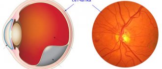
The symptoms of all forms of acute meningitis are very similar, regardless of etiology. The diagnosis of meningitis is established on the basis of a combination of three syndromes: • general infectious; • meningeal (meningeal); • inflammatory changes in the cerebrospinal fluid. The presence of one of them does not allow reliably diagnosing meningitis. For example, meningeal symptoms can be caused by irritation of the membranes without inflammation (meningism). An increase in the number of cells in the cerebrospinal fluid may be due to a reaction of the membranes to a tumor or bleeding. The diagnosis is clarified on the basis of a visual examination of the cerebrospinal fluid, as well as bacteriological, virological and other methods for diagnosing infectious diseases, taking into account the epidemiological situation and the characteristics of the clinical picture. General infectious symptoms include chills, fever, usually increased temperature, inflammatory changes in the peripheral blood (leukocytosis, increased ESR, etc.), sometimes skin rashes. The heart rate may be slow early on, but as the disease progresses, tachycardia appears. Breathing becomes faster and its rhythm is disrupted. Meningeal syndrome includes headache, nausea, vomiting, general hyperesthesia of the skin, photophobia, meningeal posture, rigidity of the neck muscles, Kernig's symptoms, Brudzinski's symptoms, zygomatic ankylosing spondylitis and the initial symptom is headache, which increases in intensity. It is caused by irritation of pain receptors in the meninges and their vessels due to the inflammatory process, the action of a toxin and irritation of baroreceptors as a result of increased intracranial pressure. The headache is intense and has a bursting, tearing character. It can be diffuse or localized more in the frontal and occipital regions, radiating to the neck and along the spine, sometimes spreading to the limbs. Already at an early stage, nausea and vomiting may be observed, not associated with food intake, occurring against the background of increased headaches. Children often, and less often adults, develop seizures. Psychomotor agitation, delirium and hallucinations are possible, but as the disease progresses, drowsiness and stupor develop, which can then progress to coma. Meningeal symptoms are manifested by reflex muscle tension due to irritation of the meninges. The most common symptoms are neck stiffness and Kernig's sign. In severe cases of meningitis, the head is thrown back, the stomach is retracted, the anterior abdominal wall is tense, the legs are brought to the stomach, and opisthotonus is detected (meningeal posture of the patient). Trismus, zygomatic ankylosing spondylitis (local pain when tapping along the zygomatic arch), pain in the eyeballs when pressing and eye movements, skin hyperesthesia, increased sensitivity to noise, loud conversation, odors, Brudzinski's symptom (upper and lower) are often observed. Patients prefer to lie motionless with their eyes closed in a darkened room. In infants, tension and protrusion of the fontanel are observed, a symptom of Lesage’s “suspension”. Venous hyperemia and papilledema may be detected in the fundus. In severe cases of the disease, the pupils are usually dilated, and sometimes there is strabismus and diplopia. Difficulty in swallowing, paresis and paralysis of the limbs with muscle hypotonia, Babinski's sign, incoordination of movements and tremor indicate damage not only to the membranes, but also to the substance of the brain, which is observed in the final stage of the disease. Control of the pelvic sphincters is impaired late, but severe mental disorders can contribute to the development of urinary retention or incontinence. Lumbar puncture should be performed in all patients with signs of meningeal irritation. With meningitis, cerebrospinal fluid pressure is often increased. Low pressure occurs when there is obstruction of the cerebrospinal fluid pathways, usually in the area of the base of the skull. In old age, meningitis usually occurs atypically: headaches are minor or absent, Kernig and Brudzinski symptoms may not be present; Trembling of the limbs and head, psychomotor agitation or apathy, and drowsiness are often observed.
Meningitis G02
Meningitis in other infectious and parasitic diseases is formed when:
anthrax (A22.8+);- gonococcal (A54.8+);
- leptospirosis (A27.-+);
- listeriosis (A32.1+);
- Lyme disease (A69.2+);
- meningococcal (A39.0+);
- neurosyphilis (A52.1+);
- salmonellosis (A02.2+);
- syphilis:
- congenital (A50.4+);
- secondary (A51.4+).
- tuberculosis (A17);
- typhoid fever (A01.0+).
Excluded: meningoencephalitis and meningomyelitis due to bacterial diseases classified elsewhere (G05.0*).
G02.0
Meningitis in viral diseases. Excluded: meningoencephalitis and meningomyelitis in infectious and parasitic diseases classified elsewhere (G05-G05.2*).
G02.1
Meningitis due to mycoses. Included :
- candidiasis (B37.5+);
- for coccidioidomycosis (B38.4+);
- cryptococcal (B45.1+).
G02.8
ICD-10 meningitis in other specified infectious and parasitic diseases classified in other headings.
Important! Meningitis in other specified infectious and parasitic diseases is a health disorder belonging to the group of inflammatory diseases of the central nervous system.
Inflammation caused by:
- African trypanosomiasis (B56.-+);
- Chagas disease (B57.4+).
Treatment
A characteristic feature of purulent meningitis is the causative agent of a bacterial nature. This could be virtually any bacteria that has entered the soft tissue of the brain. What they have in common is the principle of massive antibiotic therapy, with broad-spectrum drugs, until the specific type of pathogen and its sensitivity to antibiotics are determined. The use of drugs that destroy the bacterial population eliminates the very cause of the disease and, accordingly, leads to recovery. When determining the type of pathogen, specific antibiotic therapy is carried out with the most effective drug against this bacterium.
ICD-10 code
Serous meningitis is most often caused by viruses. However, inflammation can begin due to bacterial or fungal infection of the meninges. Due to the fact that serous meningitis can be caused by various pathogenic factors, it does not have a precise classification according to ICD-10 and is classified as “other viral meningitis”.
The disease is listed under code A87.8, where A87 is a classification of viral brain lesions, and the number 8 means viral inflammation of the brain, provoked by the action of other viruses not included in the classifier.
If the inflammation is caused by a bacterial lesion, it is classified as G00.8. This labeling describes purulent meningitis (class G00), provoked by other bacteria (this is indicated by the number 8 in the code).
Prevention
Preventive measures include measures aimed at strengthening the immune system (vitaminized nutrition, exposure to fresh air, hardening, physical education), timely treatment of infections, and elimination of chronic infectious foci in the body. Correct treatment of wounds, elimination of liquorrhea, and prophylactic use of antibiotics can prevent post-traumatic meningoencephalitis. Post-vaccination meningoencephalitis can be prevented through careful selection of the vaccinated population.
Forecast
Timely initiation of etiotropic therapy increases the chances of recovery, but the outcome of the disease depends on the etiology, course, age of the patient, and the state of his immune system. Fulminant meningoencephalitis has the highest mortality rate. Most surviving patients experience residual phenomena: paresis, speech impairment, chronic intracranial hypertension, epilepsy, psychoorganic syndrome. In young children, meningoencephalitis provokes mental retardation.
Diagnostics
Meningoencephalitis is diagnosed based on clinical manifestations, including severe headache, fever, vomiting, disturbances of consciousness and others. In addition, a number of symptoms are checked, including:
- Kernig - the patient cannot straighten the leg at the knee if it is bent at the hip joint; Brudzinsky - when a lying person’s head is tilted towards the sternum (upper symptom) or pressure is applied to the lower abdomen (middle symptom), his legs bend; Herman - when the patient’s neck is flexed, he extends his big toes; Mondonesi – when pressure is applied to the eyeballs, severe pain occurs.
In addition to general symptoms, meningoencephalitis in children of the first year is manifested by persistent bulging of a large fontanelle. During the diagnostic process, a Lessage test is performed in newborns: the child is taken by the armpits, supporting his head, and lifted. If there is pathology, its legs are fixed in a bent state.
The key diagnostic point is lumbar puncture - the collection of fluid from the spinal cord, carried out by puncture of tissue in the lumbar region. The appearance and composition of the sample, examined using the PCR method, make it possible to determine the presence of pathology and its nature. Meningoencephalitis is indicated by an increased amount of protein, high blood pressure, decreased glucose, cellular impurities, and so on.
In addition, an MRI or CT scan of the brain is performed, as well as a comprehensive examination of the patient to identify the primary foci of infection: x-ray of the lungs, nasopharyngeal smear, urine culture.
Treatment of meningoencephalitis is carried out inpatient conditions in an infectious diseases hospital. The patient is prescribed bed rest, good nutrition and careful care. Treatment tactics are determined by the form of the disease.
Purulent bacterial meningoencephalitis requires antibiotics. Depending on the detected sensitivity of the microflora, penicillins, cephalosporins, carbapenems or other drugs are prescribed. Medicines are administered intravenously over 7-10 days. For the amoebic form of the disease, antibiotics and antifungal drugs are used.
Viral meningoencephalitis is treated with gamma globulins and interferon inducers administered intramuscularly or intravenously. The duration of therapy is 10-14 days. In severe cases, such as herpetic meningoencephalitis in children, ribonuclease and corticosteroids may be prescribed.
Regardless of the etiology of the disease, the following are used:
- detoxification solutions (reopoliglucin), administered intravenously, which normalize blood composition and accelerate the removal of toxins; antihistamines (diphenhydramine, tavegil, suprastin); nootropic and neuroprotective substances to restore the functioning of the central nervous system; vitamins and antioxidants to strengthen the immune system; drugs that improve blood microcirculation; sedatives; anticonvulsants; anticholinesterase drugs and so on.
Since in most cases, after meningoencephalitis in adults and children, negative consequences are observed, patients need rehabilitation measures, which include physiotherapy and sanitary treatment.
The prognosis for meningoencephalitis is unfavorable: the percentage of deaths and severe complications is high. The course of the disease is determined by the prevalence of the pathological process, the timeliness of therapy and the age of the patient. Children and elderly people suffer from the disease very hard. The most unfavorable prognosis for meningoencephalitis in premature infants is 80% mortality when combined with other congenital defects.
Frequent consequences of meningoencephalitis in adults and children:
- paresis; hearing loss; intracranial hypertension; blurred vision; decreased intelligence; developmental delay; epileptic seizures; coma and so on.
In some cases, the disease goes away without consequences. But the patient who has undergone it must be observed by a neurologist.
Classification
The classification involves identifying forms of pathology depending on the type of infectious agent that provoked the inflammatory process in brain tissue. Bacterial meningoencephalitis develops against the background of infection with bacteria, most often pneumococci, meningococci, listeria, staphylococci, streptococci. Encephalitic meningitis occurs as a result of penetration of fungi, viruses, and protozoan microorganisms (Toxoplasma, amoeba) into the medulla.
Infectious agents enter the brain structures more often through the bloodstream. Mixed infections are distinguished, characterized by the combined participation in the pathogenesis of several types of pathogens, for example, bacteria and viruses, bacteria and bacteria, viruses and viruses. In these cases, diagnosis becomes difficult, the clinical picture unfolds atypically in comparison with the classical forms - monoinfections.
In accordance with the classification, primary and secondary forms are distinguished. The primary form develops as an independent disease. An example is tuberculous or meningococcal meningoencephalitis. Secondary forms arise as a consequence of a primary inflammatory disease or a process occurring in other organs.
The etiologically undifferentiated form is diagnosed based on the clinical picture and the results of an analysis of the cerebrospinal fluid, in which infectious agents and other characteristic changes are detected. Depending on the nature of the course, forms are distinguished - chronic, acute, subacute, fulminant (hypertoxic).


