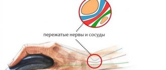
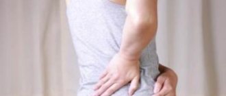
The piriformis muscle is an internal pelvic muscle that connects the pelvic bones. When it becomes inflamed, there is compression of the blood vessels, pinching of the sciatic nerve, as well as the nerve endings of the sacral spine. Because of this, a person experiences severe pain in the buttocks, which can radiate to the thigh, lower leg, or groin. This phenomenon is called piriformis syndrome.
Piriformis syndrome affects people of all ages, men and women, young and old. It is useful to know about its symptoms and treatment for everyone who plays sports, does physical labor, or leads an active lifestyle.
Reasons for the development of the syndrome
The causes of inflammation of the piriformis muscle can be different. As a rule, the following factors play a negative role:
- Overwork of the piriformis muscle, excessive physical activity;
- Hypothermia of the body;
- Constant stress, overwork;
- Finding the body in an awkward position;
- Lack of microelements in the pelvic bones (due to unbalanced nutrition or metabolic disorders);
- Pelvic injury, sprain or hematoma;
- Tumor in the roots of the spinal cord;
- Incorrectly given injection;
- Advanced stage of osteochondrosis of the sacral or lumbar spine.
Characteristic symptoms
The syndrome never occurs hidden. He announces himself immediately, giving the patient no rest.
The main symptom is a sharp pain in the pelvic area, which subsides when sitting or lying down when spreading the legs, and intensifies when crossing legs or standing up. The nature of the pain is dull, aching, sometimes shooting, jerking. A characteristic weather dependence appears: when the weather changes, the pain intensifies. Often an increase in pain occurs in a warm room at night.
If the sciatic nerve is pinched, the pain can spread to the buttocks, inner thighs and even the entire leg, down to the toes. Lameness, numbness, and loss of sensation appear.
Inflammation of the muscle can spread to the pelvic organs, in particular the bladder, which is manifested by tingling and local pain, and difficulty urinating.
The muscle and nerve tissues in the pelvic area lack blood supply. With ischemia of the sciatic nerve, pressing pain occurs with chills, burning, and a feeling of severe numbness. Warmth, changes in weather, stressful situations, and feeling the lower leg cause increased pain.
Compression of blood vessels, including the inferior gluteal artery, causes pale skin, spasms, and lameness. In a quiet position, the pain usually goes away. It is difficult for the patient to walk; he needs to relax the limb (sit or lie down) so that he can then get up and move around.
You cannot let the disease take its course. In addition to a lot of unpleasant sensations, it can cause complications. You should seek medical help.
Anatomical, topographical and biomechanical features of the piriformis muscle
The piriformis muscle (m. piriformis) is shaped like a flat isosceles triangle. It begins at the anterior edge of the upper sacrum, passes through the sciatic foramen and attaches to the inner surface of the greater trochanter of the femur. The sciatic nerve runs between the piriformis muscle and the sacrospinous ligament (see picture). The piriformis muscle does not occupy the entire sciatic foramen; it forms the upper and lower cleft. The superior fissure is occupied by the superior gluteal artery and nerve. The inferior gluteal artery and the sciatic nerve are located in the inferior fissure. In 90% of cases, the neurovascular bundle passes under the piriformis muscle. In 10% of cases, when moving to the gluteal region, it pierces the piriformis muscle. The piriformis muscle is innervated by branches of the sacral plexus from the spinal roots S1 and S2. The blood supply comes from the superior and inferior gluteal arteries.
Functionally, the piriformis muscle is designed to abduct the hip and rotate it outward. It simultaneously extends and abducts the hip, and also rotates it in a sharp flexor-abduction posture.
This muscle is necessary to “anchor” the femoral head, similar to the function of the supraspinatus muscle in relation to the head of the humerus. It keeps the hip from rapidly internally rotating during the first stages of running and walking.
Also, with its help, an oblique force is created on the sacrum and, due to the lower part of the muscle, a “shearing” force is provided for the sacroiliac joint - it pulls its side of the base of the sacrum forward, and its apex back.
This muscle promotes rocking (antinutation) of the sacrum. If the nutating muscles pull the sacrum forward, the piriformis muscle pulls its lower sections back towards the posterior sections of the innominate bones.
Spasm and hypertonicity of the piriformis muscle leads to slight stretching of its antagonists - the hip adductors. However, they simultaneously rotate the thigh outward and are in this regard a synergist with the piriformis muscle. The gluteus medius partially internally rotates the thigh, and it also abducts the thigh without being a complete antagonist of the piriformis muscle. Thus, with respect to hip abduction function, all gluteal muscles are agonists of the piriformis muscle, and all adductors are antagonists. More complex muscle complexes carry out rotational movements.
Diagnostic methods
Pain in the pelvic area is caused by many other diseases: hernias of the lower parts of the spine, inflammatory processes in the pelvis, osteochondrosis, arthritis of localization adjacent to the pelvis. Therefore, to make a diagnosis, it is important to differentiate pathologies by highlighting the real cause of pain.
The first involves manual examination of the patient in various body positions. The doctor will ask you to cross your legs, spread them under resistance, and bend your leg in a lying position. In difficult cases, a rectal or vaginal examination is used, the purpose of which is to palpate the piriformis muscle. The criterion for determining the disease is the tension of the inflamed muscle.
One of the frequently used diagnostic methods is the novocaine test. An injection of a drug from a number of novocaines is injected into the piriformis muscle. With piriformis syndrome, the pain will disappear or be significantly reduced.
The piriformis muscle is covered by the bulky muscles of the buttocks, so studying it using hardware methods is difficult and not very informative. Some exception is the echographic diagnosis of the sciatic nerve. Sonography is done on both sides to compare the results. On the affected side, there is increased blood flow and thickening of the nervous tissue. MRI and CT are also performed for diagnostic purposes, but their data are more relevant to the diagnosis of tumors. It is important to check whether there are tumors in the pelvic area. If there is a suspicion of pinching of the nerve endings of the lumbar or sacral regions, an MRI is done with a contrast agent injected into the buttock muscle.
Treatment of this pathology is long-term, it is carried out comprehensively in several directions at once.
Medication
A 10-day course of anti-inflammatory non-steroidal drugs is the basis of therapeutic treatment. They can be prescribed in the form of tablets or intramuscular injections (2 injections every 6 hours daily). Ketan drugs and other medications in this group have proven themselves well.
In case of severe pain, analgesics, antispasmodics, and muscle relaxants are prescribed. The disease that became the root cause of the syndrome is being treated.
Since the syndrome tends to worsen seasonally, courses of treatment are repeated several times and carried out twice a year.
Blockade
To relieve tension and spasm of the piriformis muscle, novocaine blockade is used. 10 ml of novocaine (dose for an adult) is injected using a syringe to a depth of 6-8 cm into the space between the piriformis muscle and the sacral root.
Surgical
The surgical method is used only in the most severe cases. This is usually a complication in the form of foot paresis. It occurs due to the strong pressure of the altered piriformis muscle on the nerve. The essence of the operation is to excise the piriformis muscle and release the sciatic nerve.
Massage
In the early stages of the disease, therapeutic massage can be effective. The session begins with kneading the paravertebral area with a transition to the lumbosacral region. The buttock on the sore side is warmed up, then the back of the leg. The session lasts 15-20 minutes. Massage should be performed under the supervision of a physician. A course of 12 procedures is recommended, repeated every month.
Physiotherapy
Physiotherapeutic procedures are useful and effective as additional methods of treatment. Typically, doctors send patients for therapy using low current devices:
- Electrophoresis;
- Treatment with dynamic currents;
- UHF;
- Phonophoresis;
- Amplipulse;
- Vacuum massage.
A laser treatment method has also appeared, but it is not yet very widespread.
Reflex methods
Reflexology includes several treatment methods that have come down to us from ancient times and are successfully used for muscle diseases:
- Acupressure massage of active points of the body;
- Acupuncture;
- Blockade;
- Laser treatment of points;
- Warming up (cauterization) of the points;
- Point-linear massaging;
- Treatment through points on the auricle.
Whether this or that method is suitable for a particular patient can only be decided by a practitioner. The choice of technique may differ depending on the stage of the disease, the general condition of the body, and existing diseases.
The basis of reflexology is the ancient Eastern teaching about active points on the body through which energy circulates.
Treatment
Conservative treatment
Conservative treatment for FMS includes pharmacologic agents (nonsteroidal anti-inflammatory drugs (NSAIDs), muscle relaxants, and medications for neuropathic pain), physical therapy, lifestyle changes, and psychotherapy.
Injections of local anesthetics, steroids, and botulinum toxin type A into the piriformis muscle can serve for both diagnosis and treatment. The practitioner should be familiar with variations in anatomy and the limitations of techniques based on anatomical landmarks. Recently, the ultrasound-guided injection method has been used. This technique has been shown to have both diagnostic and therapeutic value in the treatment of BMS.
FMS often becomes chronic, so pharmacological treatment is recommended for a short period of time.
Surgical intervention
Surgical interventions should be considered only when nonsurgical treatment has failed and symptoms become intractable and disabling. Classic indications for surgical treatment include abscesses, neoplasms, hematomas, and painful compression of the sciatic nerve vessels caused by gluteal varices.
Some authors have reported that surgical release of the piriformis muscle by cutting the tendon to free the sciatic nerve from compression results in immediate pain relief.
Sometimes the obturator internus muscle should be considered as a possible cause of pain in the area of innervation of the sciatic nerve. However, the diagnosis of obturator internus syndrome can only be made by ruling out other possible causes of gluteal pain, which is similar to how FMS is diagnosed. Surgical release of the obturator internus muscle can lead to both short-term and long-term pain relief in patients with retroacetabular pain syndrome and should be considered if conservative treatment has failed.
Postoperative management of the patient consists of partial weight reduction through the use of crutches for 2 weeks and selection of exercises. The above surgical approach has shown promising short-term results.
Treatment algorithm for retroacetabular pain syndrome:
Physical therapy
Although there are few recently published controlled studies that critically examine the effectiveness of noninvasive treatments, there are a number of treatment options for BMS that are well established in clinical practice.
Noninvasive treatments include physical therapy and lifestyle changes. According to Tonley et al., the most commonly reported physical therapy interventions include soft tissue mobilization, piriformis stretching, cryotherapy (hot packs or cold spray), and manual treatment of the lumbosacral spine.
In addition, Tonley describes an alternative approach to the treatment of BMS. Treatment focused on functional "Therapeutic Hip Exercises" exercises aimed at strengthening the hip extensors, abductors, and external rotators and correcting abnormal movement patterns. Despite the positive results (complete elimination of pain in the lower back, cessation of pain in the buttock and thigh), further research is required, since this protocol was tested in only one patient.
In order not to miss anything interesting, subscribe to our Telegram channel.
To achieve a 60-70% improvement, the patient usually undergoes 2-3 treatments per week for 2-3 months.
- Start with ultrasound treatment: 2.0-2.5 W/cm2 for 10-14 minutes. Apply the ultrasound gel using wide strokes in a longitudinal direction along the piriformis muscle from the connecting tendon to the lateral edge of the greater sciatic foramen. The patient should be in the contralateral lateral decubitus position and in the FAIR position (flexion, adduction, internal rotation).
- Before stretching the piriformis muscle, treat the same area with hot compresses or a cold spray for 10 minutes. Using hot and cold before stretching is very helpful in reducing pain.
- After this, move on to stretching the piriformis muscle, which can be performed in a variety of ways. It is important not to push down, but to direct the pressure along the surface (tangentially) to the ipsilateral shoulder (Fischman et al. (2002), level of evidence A2). When pressing downward, the sciatic nerve is compressed along the tendinous edge of the superior gemellus muscle. Another way to stretch the muscle is the FAIR position. The patient is in the supine position with the hip flexed, adducted and internally rotated. The patient then moves the foot on the affected side across and over the knee of the unaffected leg. The specialist can increase the stretch by using muscle-energetic techniques.
- After stretching the piriformis muscle, you can perform myofascial release of the lumbar muscles, as well as do Mackenzie exercises.
- Because FMS can occur when a tight piriformis muscle is forced to do the work of other large muscles (such as the gluteus maximus or gluteus medius), an alternative approach to treating FMS using a strengthening program for the hip muscles (especially weak gluteal muscles) with relearning of the movement may help. in pain relief.
Your therapist can also give you some tips to avoid worsening your symptoms.
- Avoid sitting for long periods of time; Standing and walking are required every 20 minutes.
- Make frequent stops as you move to stand and stretch.
- Prevent injury to the gluteal region and avoid further aggravating actions.
- Daily stretching is recommended to prevent recurrence of BMS.
Home exercises
The patient can also do some exercises at home.
- Rolling from side to side with bending and straightening of the knees, lying on one side of the body.
- Rotate from side to side while standing with relaxed arms for 1 minute every few hours.
- “Riding a bicycle” while lying on your back.
- Knee curls - up to 6 repetitions every few hours.
- Taking a warm bath.
Physiotherapy
Exercise therapy helps the patient fight the disease, giving the opportunity to alternate muscle relaxation with light loads. Exercises relieve tension and spasms. With the help of regularly performed gymnastics, it is possible to strengthen muscles and also avoid their atrophy. To avoid excessive stress, it is better to hold off on other workouts and going to the gym until recovery.
The following set of exercises is performed strictly in the order listed. You can do exercises 1 to 3 times a day.
- Lie on your back, bend your knees, bring your knees together and spread them several times. Then shine the light on your knees again until they touch. Push one knee with the other for several seconds: first the left with the right, then vice versa.
- Lie on your stomach with your shoulders pressed to the floor. Straighten one leg, bend the other at the knee. Using the palm of the hand opposite to the bent leg, press the knee to the floor through the other leg. You need to hold this position for as long as possible, at least 30 seconds. The same thing is repeated with the change of legs.
- Lie on your back. Bend your knees and keep them suspended. Place the sore leg on top of the healthy one (as in the “lotus” position, only lying down). Grasp the thigh of the supporting leg with your hands and pull it towards you. The exercise helps stretch the piriformis muscle, making it less prone to spasms.
- Performed with an assistant. Sit on the mat with your feet wide apart, knees bent and brought together. Leaning on the couch with one hand, stretch the other forward and begin to rise. When the elbow is fully straightened, the assistant takes the patient by the free hand and puts him on his feet (the knees open).
- For this exercise you need an expander or a tight stretch cord (tape). Attach one end of the expander to any stationary object (support), and place the other on the foot on the sore side. Then stand sideways to the support and with effort, overcoming the resistance of the expander, move your leg to the side as far as possible without bending it at the knee. Slowly return the leg to its place, holding back the compression force of the expander.
Traditional methods
Remedies recommended by traditional medicine help enhance the effect of treatment with traditional methods. As a rule, these are various warming procedures in the form of compresses.
- Compress of black radish and horseradish.
Grind a small black radish tuber and horseradish root in a blender. Add 1 tbsp. l. vinegar and salt. Leave it in a dark place for a week, then apply it to the sore spot for 20 minutes through several layers of gauze.
- Rubbing with alcohol tinctures and pepper.
Mix triple cologne with tinctures of hawthorn, valerian and hot pepper, as well as 10 crushed aspirin tablets. Leave for a week in a dark place. Rub the sore spot. Then wrap it in a plastic bag or compressor paper, and wrap a warm scarf or scarf on top.
Beeswax compress. Lubricate the sore spot with burdock oil, then apply melted (heated in a water bath) beeswax in several layers. Keep until the wax cools. For convenience, wax can be applied with a brush.
Possible complications
In advanced cases, piriformis syndrome is life-threatening, not to mention the pain syndrome that torments the patient. This is an inflammatory process in the body, and like any inflammation, it is fraught with complications. The disease is prolonged and weakens the immune system of the entire body. Pain can spread to the entire limb, causing muscle paresis, as well as to the pelvic floor area. Piriformis syndrome can cause inflammation of the pelvic organs and disrupt the function of the rectum and urinary system.
Diagnosis of piriformis syndrome
Magnetic resonance imaging is an additional way to diagnose the disease
The diagnosis is established after examining the patient, drawing up a complete clinical picture, clinical tests and instrumental research methods. To confirm or refute the presence of piriformis muscle syndrome, the following manipulations are performed:
- The piriformis muscle is palpated under the gluteal soft tissue. Sometimes a transrectal examination is required to determine its condition.
- The area of attachment of the muscle to the sacroiliac joint and along the inner region of the greater trochanter is palpated. If it spasms, pain will be felt at these points.
- When tapping the gluteal area, pain appears along the sciatic nerve.
- The patient lies on his healthy side and raises the opposite knee. If pain occurs during such manipulation, it indicates a spasm (Beatty's symptom).
- The patient is asked to lean forward without bending his knees, then the doctor presses on the buttock in the area of the projection of the exit of the sciatic nerve from the infrapiriform foramen (Mirkin test).
The doctor may suggest, as an additional measure for diagnosing piriformis muscle syndrome, to inject novocaine into the thickness of the muscle. Relief of pain is a sign that it was spasmed.
EMG, MRI, and radiography can be used as additional diagnostic methods, but clinical tests are the main tool for making a diagnosis. Below we discuss how to treat piriformis syndrome.