Descending pathways
All descending tracts of the spinal cord with their detailed characteristics and course of movement are clearly demonstrated in Table No. 2.
| No. | Descending Path View | Characteristics | |
| 1 | Lateral corticospinal, also called lateral corticospinal or main crossed pyramidal. | This pathway includes a considerable proportion of fibers of the pyramidal system. The lateral tract is localized in the lateral funiculus. As they travel, the fibers gradually become thinner. Lateral fibers conduct signals that cause conscious actions in humans. | Lateral fibers conduct signals that cause conscious actions in humans. |
| 2 | Anterior corticospinal, otherwise called corticospinal, and also straight or uncrossed pyramidal. | This path lies in the anterior spinal cord. Like the lateral pyramidal tract, the direct pyramidal tract includes cellular axons of the motor hemisphere, although they are located ipsilaterally. Initially, these axons descend towards their “own” segment. After this, as part of the anterior spinal commissure, they are transported to the opposite side, ending in the mononeurons of the anterior horn. | |
| 3 | Red-spinal or rubrospinal. | Beginning in the red nucleus of the spinal cord, this tract subsequently descends to the motor nerve cells of the anterior horns. This pathway is responsible for transmitting unconscious motor signals. | |
| 4 | Tectospinal, otherwise called tectospinal. | It is localized in the anterior funiculus near the anterior pyramidal tract. This tract starts on the roof of the midbrain. The mononeurons of the anterior horns are its final point. The tectospinal tract provides reflex protective actions in response to visual and auditory stimuli. | |
| 5 | vestibulospinal, otherwise called vestibulospinal. | This path is localized in the anterior spinal cord. The vestibular nuclei of the pons are its beginning, and the anterior spinal horns are its end. The balance of the human body is ensured precisely through the transmission of impulses from the vestibulospinal tract. | |
| 6 | Reticulospinal or reticulospinal. | This pathway ensures the transmission of excitatory signals from the reticular formation to the spinal nerve cells. |
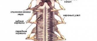
To understand the neurophysiology of the human spinal cord pathways, you will need to briefly familiarize yourself with the structure of the spine. In its structure, the spinal cord is a bit like a cylinder, covered with muscle tissue on all sides. The pathways control the functioning of the internal organs, as well as all organ systems and functions performed by the body. Injuries, various injuries, and other ailments of the spinal cord can somehow reduce conductivity. By the way, conduction may even stop completely due to the death of neurons. Complete loss of conduction of spinal signals is characterized by paralysis, manifested in a complete lack of sensation in the limbs. This is very fraught with problems with internal organs that are responsible for damage to the communication of nerve cells. Thus, injuries and other ailments of the lower spinal cord are often characterized by urinary incontinence and even spontaneous defecation.
Drug treatment will consist of prescribing medications to prevent the death of brain cells, as well as to further increase blood flow to the damaged spinal areas. Electrical impulses may be prescribed as an additional treatment to stimulate the functioning of neurons and also help maintain muscle tone.
Also, if necessary, the attending physician may prescribe the use of the following folk remedies.
Apitherapy
- Apitherapy. Bee stings effectively restore the conductivity of efferent tracts. Thus, the poisons of these insects, penetrating into damaged areas, provide them with additional blood flow. If the cause of spinal pathology is radiculitis, a growing hernia and other similar ailments, apitherapy will be an excellent addition to traditional treatment.
- Herbal medicine. Medications are prescribed to normalize blood circulation and improve metabolism.
- Hirudotherapy. Thanks to treatment with leeches, it becomes possible to eliminate congestion - inevitable attributes of vertebral pathologies.
Features of the structure of descending conductors
The descending pathways of the brain and spinal cord include several ligaments, including:
- pyramidal;
- rubrospinal;
- vestibulo-spinal;
- reticulospinal;
- rear longitudinal.
All of the above elements are motor pathways of the spinal cord, which are components of the nerve cords in the descending direction.
The so-called pyramidal tract begins from huge cells of the same name located in the upper layer of the cerebral hemisphere, mainly in the area of the central gyrus. The pathway of the anterior cord of the spinal cord is also located here - this important element of the system is directed downward and passes through several sections of the posterior femoral capsule. At the point of intersection of the medulla oblongata and the spinal cord, an incomplete decussation can be found, forming a straight pyramidal fasciculus.
In the tegmentum of the midbrain there is a conducting rubro-spinal tract. It starts from red kernels. Upon exiting, its fibers intersect and pass into the spinal cord through the varoli and medulla oblongata. The rubrospinal tract allows impulses to be transmitted from the cerebellum and subcortical ganglia.
The white matter tracts of the spinal cord begin in Deiters' nucleus. Located in the brain stem, the vestibulospinal tract continues in the spinal tract and ends in its anterior horns. The passage of impulses from the vestibular apparatus to the motor neuron of the peripheral system depends on this conductor.
In the cells of the reticular formation of the hindbrain, the reticulospinal tract begins, which in the white matter of the spinal cord is scattered in separate bundles mainly from the side and in front. In fact, this is the main connecting element between the reflex brain center and the musculoskeletal system.
The posterior longitudinal ligament is also involved in connecting motor structures to the brain stem. The work of the oculomotor nuclei and the vestibular apparatus as a whole depends on it. The posterior longitudinal fasciculus is located in the cervical spine.
How the sense of touch is born
The fibers that provide sensitivity take different paths. For example, from proprioceptors the pathways go to the cerebellum and cortex. They send a signal to this area about the condition of the joints, tendons, and muscles.
This path is made up of axons of sensory type neurons. The afferent neuron processes the received signal and, using an axon, conducts it to the thalamus. After processing in the thalamus, information about the motor system is sent to the postcentral cortex. Here the formation of sensations occurs about how tense the muscles are, in what position the limbs are, at what angle the joints are bent, whether there is vibration, passive movements.
The thin bundle also contains fibers that are associated with skin receptors. They conduct a signal that generates information about tactile sensitivity during vibration, pressure, and touch.
The axons of the second interneurons form other sensory pathways. The area where the cell bodies of these neurons are located is the dorsal horn (spinal cord). In their segments, these axons create a cross, then they go on the opposite side to the thalamus.
This pathway contains fibers that provide temperature and pain sensitivity. Also here are fibers that are involved in tactile sensitivity. Neurons located in the spinal cord receive information from brain structures.
Extrapyramidal neurons participate in the formation of the rubrospinal, reticulospinal, vestibulospinal, and tectospinal tracts. Nerve efferent impulses pass along all of these paths. They are responsible for maintaining muscle tone, performing various involuntary movements, and posture. Acquired or innate reflexes participate in these processes. In these pathways, the conditions for performing all voluntary movements controlled by the cerebral cortex are formed.
The spinal cord conducts all signals that come from the centers of the ANS to the neurons that make up the sympathetic nervous system. These neurons are located in the lateral horns of the spinal cord.
Also involved in the process are neurons from the parasympathetic nervous system, which are also localized in the spinal cord (sacral region). These pathways are entrusted with the function of maintaining the tone of the sympathetic nervous system.
Conductors of deep sensory
The structure of the nerve ligaments, acting in the ascending direction, is multi-component. These spinal cord pathways are formed by several elements:
- Burdach's bundle and Gaulle's bundle (represent pathways of deep sensitivity located on the posterior side of the spinal column);
- spinothalamic bundle (located on the side of the spinal column);
- Govers' bundle and Flexig's bundle (cerebellar tracts located on the sides of the column).
Inside the intervertebral nodes there are neuron cells with a deep degree of sensitivity. The processes, localized in peripheral areas, end in the most suitable muscle tissues, tendons, osteochondral fibers and their receptors.
In turn, the central processes of the cells, located behind, are directed towards the spinal cord. Conducting deep sensitivity, the posterior nerve roots do not go deep into the gray matter, forming only the posterior spinal columns.
Where such fibers enter the spinal cord, they are divided into short and long. Next, the pathways of the spinal cord and brain are sent to the hemispheres, where their radical redistribution occurs. The main part of them remains in the areas of the anterior and posterior central gyri, as well as in the region of the crown.
It follows that these pathways conduct sensitivity, thanks to which a person can feel how his muscular-articular apparatus works, feel any vibration movement or tactile touch. The Gaulle bundle, located right in the center of the spinal cord, distributes sensation from the lower torso. Burdach's bundle is located higher and serves as a conductor of sensitivity of the upper extremities and the corresponding part of the body.
How to find out about the pain threshold and temperature differences
To determine the level of pain, doctors use the pricking method. In the most unexpected places for the patient, the doctor applies several light injections with a pin. The patient's eyes should be closed, because He shouldn't see what's happening.
The temperature sensitivity threshold is easy to determine. In a normal state, a person experiences different sensations at temperatures, the difference of which was about 1-2°. To identify a pathological defect in the form of impaired skin sensitivity, doctors use a special device - a thermoesthesiometer. If it is not there, you can test for warm and hot water.
Trigeminal nerve
The feeling of pain is one of the most important for our life. Let's understand how the process of signal transmission through the trigeminal nerve occurs.
Where the motor fibers of the corticospinal tract intersect, the spinal nucleus of one of the largest nerves, the trigeminal, passes to the cervical spine. Through the region of the medulla oblongata, axons of sensory neurons descend to its neurons. It is from them that a signal about pain in the teeth, jaws, and oral cavity is sent to the nucleus. Signals from the face, eyes, and orbits pass through the trigeminal nerve.
The trigeminal nerve is extremely important for receiving tactile sensations from the facial area and sensing temperature. If it is damaged, the person begins to suffer from severe pain, which constantly returns. The trigeminal nerve is very large, it consists of many afferent fibers and a nucleus.
Consequences of spinal cord injury
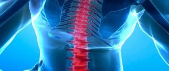
Pathological changes in conduction function can lead to disruption of the body's functionality, pain, urinary incontinence, etc. As a result of various types of injuries, spinal diseases and developmental defects, the conductivity of nerve receptors may decrease or completely cease.
If impulse conduction is disrupted, paresis of the lower extremities occurs
Complete disruption of impulse conduction may be accompanied by paralysis and loss of sensation in the limbs. In addition, there are disturbances in the functioning of internal organs, for the functionality of which damaged neurons are responsible. For example, with lesions of the lower spinal cord, spontaneous defecation is possible.
Depending on the severity of damage to the spinal nerves after injury or as a result of disease, the following manifestations are possible:
We also recommend: Cervical myelopathy
- development of congestive pneumonia;
- formation of bedsores and trophic ulcers;
- urinary tract infections;
- Spastic syndrome (pathological contraction of paralyzed muscles), accompanied by pain, stiffness of the limb and the formation of contractures;
- septic blood poisoning;
- disturbance of behavioral reactions (disorientation, fearfulness, inhibited reaction);
- a psychological change manifested by sharp fluctuations in mood, depression, causeless crying (laughter), insomnia, etc.
Impairment of conductivity and reflex activity is observed immediately after the detection of a degenerative pathological change. In this case, necrosis of nerve cells occurs, which leads to accelerated progression of the disease, requiring immediate treatment. The consequences of this condition are determined by the severity of the negative symptoms and which cells were damaged.
Consequences of spinal cord diseases
Thus, the spinal cord pathways are vital connecting elements that provide a person with the ability to move and feel. The neurophysiology of these pathways is associated with the structural features of the spine. It is known that the structure of the spinal cord, surrounded by muscle fibers, has a cylindrical shape. Within the substances of the spinal cord, associative and motor reflex pathways control the functionality of all body systems.
If spinal cord disease, mechanical damage, or developmental defects occur, conduction between the two main centers can be significantly reduced. Disorders of the pathways threaten a person with a complete cessation of motor activity and loss of sensory perception.
The main reason for the lack of impulse conduction is the death of nerve endings. The most complex degree of conduction disturbance between the brain and spinal cord is paralysis and lack of sensation in the limbs. Then problems may occur in the functioning of internal organs connected to the brain by damaged neural ligaments. For example, disorders in the lower part of the spinal trunk result in uncontrollable urination and defecation processes.
Ascending afferent pathways originating in the brainstem
The medial lemniscus, trigeminal lemniscus, ascending tract of the auditory analyzer, optic radiation, and thalamic radiation begin in the brain stem.
1. The medial lemniscus as a continuation of the thin and wedge-shaped fascicles was described earlier.
2. The trigeminal loop, lemniscus trigeminalis, is formed by processes of nerve cells that make up the sensory nuclei of the trigeminal nerve (V pair), facial nerve (VII pair), glossopharyngeal nerve (IX pair) and vagus nerve (X pair).
The axons of afferent neurons located in the trigeminal ganglion approach the sensory nuclei of the trigeminal nerve. The common sensory nucleus of the other three nerves - the nucleus of the solitary tract - is approached by axons of afferent neurons located in the genu node (VII pair) and in the upper and lower nodes of the IX and X pairs of nerves. The bodies of the first neurons are localized in the listed nodes, and the bodies of the second neurons of the path along which impulses from the head receptors are transmitted are located in the sensitive nuclei.
The fibers of the trigeminal lemniscus pass to the opposite side (some of the fibers follow on their side) and reach the thalamus, where they end in its nuclei.
The nerve cells of the thalamus are the bodies of the third neurons of the ascending tracts of the cranial nerves, the axons of which, as part of the central thalamic radiates, through the internal capsule are directed to the cerebral cortex (postcentral gyrus).
3. The ascending path of the auditory analyzer has, as its first neurons, cells located in the ganglion of the cochlear part of the vestibulocochlear nerve. The axons of these cells approach the cells of the anterior and posterior cochlear nuclei (second neurons). The processes of the second neurons, moving to the opposite side, form a trapezoidal body, and then take an ascending direction and are called the lateral loop, lemniscus lateralis. These fibers end on the bodies of the third neurons of the auditory pathway, located in the lateral geniculate body. The processes of the third neurons form the auditory radiation, radiatio acustica, which runs from the medial geniculate body through the posterior limb of the internal capsule to the middle part of the superior temporal gyrus.
4. Visual radiance, radiatio optica (see Fig.), connects the subcortical centers of vision with the cortex of the calcarine sulcus.
The optic radiance includes two systems of ascending fibers:
- the geniculate-cortical optic tract, which originates from the cells of the lateral geniculate body;
- the cushion-cortical tract, starting from the cells of the nucleus located in the cushion of the thalamus; in humans it is poorly developed.
The set of these fibers is designated as posterior thalamic radiation, radiationes thalamicae posteriores.
Ascending to the cerebral cortex, both systems pass through the posterior limb of the internal capsule.
5. Thalamic radiations, radiationes thalamicae (see Fig.), are formed by processes of thalamic cells and constitute the final sections of the ascending tracts of the cortical direction.
The thalamic radiations include:
- anterior thalamic radiations, radiationes thalamicae anteriores, are radial fibers of the white matter of the cerebral hemispheres. They begin from the superior medial nucleus of the thalamus and are directed through the anterior limb of the internal capsule to the cortex of the lateral and inferior surfaces of the frontal lobe. Some of the fibers of the anterior thalamic radiates connect the anterior group of thalamic nuclei with the cortex of the medial surface of the frontal lobes and the anterior part of the cingulate gyrus;
- central thalamic radiations, radiationes thalamicae centrales, are radial fibers connecting the ventrolateral group of thalamic nuclei with the cortex of the pre- and postcentral gyrus, as well as with the adjacent sections of the cortex of the frontal and parietal lobes. They pass as part of the posterior limb of the internal capsule;
- the lower peduncle of the thalamus, pedunculus thalami inferior, contains radial fibers connecting the thalamic cushion and medial geniculate bodies with areas of the temporal choir;
- posterior thalamic radiates (see earlier).
The essence of the spinal conduction mission
Pathways are special neural fibers that transmit signals of a certain kind to various brain centers. In medical practice, it is customary to differentiate three groups of the above fibers.
Spinal tracts
- Associative. They are intended to connect gray matter cells from dissimilar segments to form, directly near the gray matter, special bundles of their own (meaning anterior, lateral, posterior).
- Commissural. The function of these fibers is to connect the gray matter from both hemispheres, as well as similar and equidistant nerve centers of both halves of the brain to correlate and coordinate their work.
- Projection. These fibers connect the overlying and underlying brain regions. They are responsible for projecting pictures of the surrounding world onto the cerebral cortex, like on a scoreboard or television screen.
Projection fibers differ depending on the direction of the impulses sent to the ascending and descending pathways. The following three groups of ascending pathways are responsible for the delivery of signals to the brain that appear as a result of the influence of various environmental factors and phenomena on the human body.
Exteroceptive - deliver impulses from two types of receptors.
- Impulses delivered by exteroceptors. This refers to temperature, tactile and pain signals.
- Impulses of the senses: the ability to see, hear, smell and taste.
- Proprioceptive - responsible for impulses coming from the organs of movement and muscles.
- Interoceptive - intended to conduct impulses sent by internal organs.
Along descending pathways, signals pass from the subcortical centers and the cortex itself to the nuclei of the brain, as well as to the motor nuclei of the spinal horns located in front. The descending tracts include several fiber systems.
To prevent and treat JOINT DISEASES, our regular reader uses the increasingly popular NON-SURGERY treatment method recommended by leading German and Israeli orthopedists. After carefully reviewing it, we decided to offer it to your attention.
- The corticospinal cord is responsible for the mission of movement.
- The tectospinal tract, otherwise called the tectospinal tract, is a projection of the descending nervous system.
- The vestibular-spinal cord is responsible for the proper coherence in the work of the vestibular apparatus.
- The reticular-spinal cord, otherwise called the reticular-spinal tract, ensures the proper level of muscle tissue tone.
In addition, the pathways of the brain and spinal cord are also differentiated according to the tasks performed.
- Motor pathways responsible for the reflex response. Their task is to transmit “pointers” from the brain to the spinal cord and further to the muscles. Thanks to the coordinated work of these paths, the proper level of coordination of movement is ensured.
- Sensory pathways help in recognizing pain, temperature and its changes, and tactile sensations.
Nerve fibers are the guarantors of the inextricable relationship between the brain and the spinal cord, and through it, with all organ systems. The rapid transmission of appropriate signals ensures the consistency of all body movements, eliminating significant efforts made by the person himself. The pathways form bundles of nerve cells.
Types of conductive paths by direction
The ascending pathways of the spinal cord recognize urges received from various life-supporting organs of a person, with their subsequent provision to the “center”.
Descending pathways send “instructions” immediately to certain internal organs, various glands, and muscles. Signals and impulses in this case are transmitted through the spinal neural connection.
Fast and accurate data transfer is ensured thanks to the double course of the spinal tracks.
System of ascending and descending conductors
The ascending pathways of the spinal cord fulfill the human need for vision, hearing, motor functions and their contact with important systems of the body. The receptors for these connections are located in the space between the hypothalamus and the first segments of the spinal column. The ascending tracts of the spinal cord are capable of receiving and sending further impulses coming from the surface of the upper layers of the epidermis and mucous membranes, life support organs.
In turn, the descending pathways of the spinal cord include the following elements in their system:
- The neuron is pyramidal (originates in the cerebral cortex, then rushes down, bypassing the brain stem; each of its bundles is located on the spinal horns).
- The neuron is central (it is a motor neuron, connecting the anterior horns and the cerebral cortex with reflex roots; along with the axons, the chain also includes elements of the peripheral nervous system).
- Spinocerebellar fibers (conductors of the lower extremities and the spinal cord, including the sphenoid and thin ligaments).
It is quite difficult for an ordinary person who does not specialize in neurosurgery to understand the system represented by the complex pathways of the spinal cord. The anatomy of this department is truly an intricate structure consisting of neural impulse transmissions. But it is thanks to it that the human body exists as a single whole. Due to the double direction along which the spinal cord conductive pathways operate, instant transmission of impulses is ensured, which carry information from the controlled organs.
Localization of paths as they move
The ascending and descending tracts connect the spinal horns with the cerebral cortex. The spinal tracts are nerve bundles and tissues that pass through the corresponding parts of the brain. In this case, impulses can be transmitted only in one direction. The location of the spinal tracts is clearly demonstrated by the diagram in the video above.
Ascending spinal tracts and their characteristics
The bodies of the first nerve cells, acting as transmitters of various types of spinal sensitivity, lie in the corresponding brain nodes. The cellular axons of these nodes enter the spinal cord. Among them there are a couple of groups.
The medial group moves towards the posterior cord. At this point, each existing fiber is divided into a pair of branches. They are called ascending and descending. A certain number of the above branches, when moving up and down, form bundles in various spinal segments and points.
The lateral group moves to the edge, and then to the posterior column of gray matter to contact the cells of the posterior genus.
The ascending tracts of the spinal cord, otherwise called centrifugal or afferent, with their characteristics and direction of movement are described in detail in Table No. 1.
| No. | View of the ascending path | Characteristics | |
| 1 | Posterior spinocerebellar | The task of this direct cerebellar pathway is to conduct impulses to the cerebellum from muscle receptors. The spinal ganglion is the home of the first neurons. The refuge of the second neurons is the entire surface of the spinal cord in the thoracic nucleus. These neurons move towards the outside. Having reached the posterolateral spinal cord, they turn upward and follow close to the lateral spinal cord. Then they go to the cortex of the cerebellar vermis. | |
| 2 | Anterior spinocerebellar | This tract is also designed to carry impulses to the cerebellum from muscle receptors. The spinal ganglion is the home of the first neurons. And the medial nucleus of the intermediate region is the habitat of the bodies of the second neurons. Their fibers are sent to the lateral cords of both sides. Having reached the anterior outer sections of the cords, the fibers will be located above the posterior spinocerebellar tract. Turning upward, crossing the bridge and crossing, the fibers reach the cerebellar vermis, which completes this path. | |
| 3 | Spino-olive | Let this ascending conduction begin in the cells of the dorsal horns. After crossing, the axons of these cells move upward along the spinal surface. The final destination of the spino-olive tract is, accordingly, the olive nuclei. Through the above tract, data from muscle and skin receptors enters the brain. | |
| 4 | Anterior spinothalamic | Responsible for transmitting signals regarding tactile sensitivity. | The spinal ganglia are the area where the cell bodies of the first neurons are located. The path of the second neurons runs to the opposite side towards the cords. The fibers of these pathways bypass the medulla oblongata, pons and cerebral peduncles, subsequently reaching the thalamus. The third neurons lie precisely in the thalamus, following directly to the cerebral cortex. |
| 5 | Lateral spinothalamic | Carries out signals regarding temperature and pain sensations. | |
| 6 | Spinoreticular | The elements of this tract are fibers from both spinothalamic tracts. | These two pathways run through the lateral spinal cords, ending in the plate of the mesencephalic roof. |
| 7 | Dorsal-tegmental | ||
| 8 | Thin Bun | This bundle transmits “instructions” directed by the lower parts of the human torso along with its lower limbs below the 4th thoracic segment. Having reached the medulla oblongata, the bundle begins to contact its own nuclear cells. | The muscles provide “instructions” to both bundles. The first neurons of the above tracks lie in certain spinal ganglia. They move to the nuclei of the medulla oblongata. The two tubercles are the second neurons of the corresponding bundles. Their axons reach the opposite side when moving. There they form a sensory chiasm, and then move to the thalamus, already being an integral part of the medial loop. The fibers of these bundles come into direct contact with thalamic cells. The processes of these neurons are sent directly to the brain. |
| 9 | Wedge-shaped bundle | It is formed from fibers that initiate movement in the cells of the spinal ganglia and end in the sphenoid tubercle. |