Myopathy
(Greek mys, my[os] muscle + pathos suffering, disease) - a group of hereditary muscle diseases, the main clinical manifestations of which are muscle weakness, atrophy, decreased muscle tone, decreased or absent tendon reflexes, changes in the bioelectrical activity of muscles.
The first clinical and morphological descriptions of diseases of this group belong to G. Duchenne, J. Cruvelier (1853), V. Erb (1883-1884), V. K. Roth (1876, 1895). The pathology occurs in all countries of the world. The frequency of various forms is 2-6 per 100,000 population.
Classification
Issues of classification of Myopathy are being developed in various directions. Myopathies are classified according to the type of inheritance: autosomal recessive, autosomal dominant, recessive and dominant, X-linked. Depending on the time of appearance of the first symptoms and the nature of the course, M. is divided into congenital non-progressive M. and progressive muscular dystrophy (early childhood, childhood, youth and late forms). Progressive muscular dystrophy is also divided into forms depending on the predominant localization of the myodystrophic process (for example, Landouzi-Dejerine-Scapulohumeral-facial myodystrophy, Erb-Roth coxohumeral myopathy, bulbar-ophthalmoplegic M., distal M.), the nature of the spread of the process (ascending and descending options). M. with pronounced pseudohypertrophy of muscles (pseudohypertrophic Duchenne Myopathy, benign pseudohypertrophic Becker Myopathy, etc.) are classified into a separate group.
Causes of the disease
The disease, depending on the type, has different causes.
Inflammatory forms develop against the background of autoimmune reactions - a disruption of the immune system, due to which the body “attacks” its own tissues and organs. Some inflammatory forms are associated with systemic lupus erythematosus, rheumatoid arthritis, and polyarteritis nodosa4.
Infectious causes can be viruses, bacteria and other pathogens, for example:
- Trichinella;
- Toxoplasma;
- HIV infection;
- Coxsackie viruses;
- Influenza viruses;
- Bacteria of the genus Borrelia are the causative agents of Lyme disease;
- Staphylococcus aureus.
Drug-induced myopathies can develop while taking a number of drugs:
- Glucocorticosteroids;
- Lovastatin, other statins - drugs that lower cholesterol levels;
- Colchicine is an anti-gout drug;
- Amiodarone is a drug used for cardiac arrhythmias.
Etiology and pathogenesis
The causes of diseases are genetically determined defects in the metabolism of muscle tissue or the structure of muscle cells. Since the primary molecular defect is not known for any form of Myopathy, the pathogenesis has not been studied in detail. Apparently, it is not unambiguous for all forms of M. It is known, however, that in all diseases of this group, increased breakdown of muscle proteins, prevailing over their accelerated but defective synthesis, changes in blood vessels, impaired permeability of their walls, are more or less pronounced. disturbances in the structure and permeability of cell membranes, a shift in cation exchange, changes in connective tissue, etc. It is assumed that in certain forms the primary molecular defect is not localized in the muscle tissue itself (but, for example, in the nervous tissue) and serves only as a trigger factor for these changes. It is also possible that in certain forms of M. the effect of the mutant gene is more generalized and spreads to muscle, nervous and other tissues.
Pathological anatomy
Morphological changes in Myopathy are characterized by increasing atrophy of skeletal muscles, which decrease in volume and become dense, brown in color due to the proliferation of connective tissue or, on the contrary, increase in volume due to fatty tissue.
Rice. 1. Microscopic specimen of a muscle (cross section) for Duchenne myopathy in the stage of partial preservation of motor function: disorderly arrangement of muscle fibers of different sizes - atrophied (1), normal diameter (2), single hypertrophied (3); dystrophic changes in some muscle fibers (4); proliferation of connective tissue in the endomysium (5); hematoxylin-eosin; X 200. Fig. 2. Microscopic specimen of muscle (longitudinal section) for Duchenne myopathy in the immobility stage: single atrophied muscle fibers (1) among fibrous and adipose tissue; lymphoid-histiocytic infiltration (2); hematoxylin-eosin; X 200.
In various forms of muscular dystrophies (pseudohypertrophic, humeroscapulofacial, coxohumeral, ophthalmoplegic, bulbar-ophthalmoplegic), mainly the same type of histol changes are determined (Fig. 1): a decrease in the number of muscle fibers in the bundles, a sharp diffuse difference in the size of the surviving fibers with a predominance among atrophied ones, hyaline and vacuolar dystrophy in some muscle fibers, discoid and coagulative necrosis of individual fibers, splitting of hypertrophied fibers, proliferation of connective and adipose tissue in the endo- and perimysium. In some muscle fibers, sarcoplasmic bodies, sarcoplasmic masses, and ring-shaped myofibrils are found. Occasionally, regenerating fibers with round juicy nuclei and sarcoplasm rich in ribonucleoproteins are found. Muscle spindles remain unchanged for a long time. In some cases, perivascular lymphoid-histiocytic infiltrates are detected. As movement disorders increase, there is a gradual decrease in the number of muscle fibers and a leveling of their diameter due to a sharp decrease in the number and caliber of hypertrophied fibers. These changes develop most rapidly in Duchenne myopathy, a malignant variant of pseudohypertrophic myopathy. The benign variant, Becker myopathy, is characterized by a combination of pronounced processes of lipomatosis and sclerosis with a sharp hypertrophy of some muscle fibers. The latter feature is associated with long-term compensation of motor disorders in such patients. In the advanced stage of M., small islands of atrophied muscle fibers are identified against the background of severe sclerosis and lipomatosis of the endo- and perimysium (Fig. 2), while histologically it is not possible to differentiate M. from neuromuscular atrophy. In probands, muscle biopsy reveals single atrophied muscle fibers, predominantly type I, proliferation of nuclei with their transition to the center of the fiber, and a slight increase in connective tissue in the endomysium.
The earliest ultrastructural changes in muscles in Myopathy are characterized by thickening and splitting of Z-lines with subsequent destruction of muscle fiber myofibrils. Thanks to phase contrast and electron microscopy, defects in the membrane systems of muscle fibers were identified. Histoenzyme chemically marks a weakening of the reciprocal relationships between glycolytic and oxidative enzymes in muscle fibers of various types.
No changes were found in the central and peripheral nervous system. Characterized by cardiosclerosis.
Clinical picture
Rice.
3. A patient with myopathy with severe pseudohypertrophy of the calf muscles. Rice. 4. A patient with myopathy with a symptom of pterygoid scapulae: sharp protrusion of both scapulae. Rice. 5. A patient with myopathy with a symptom of “loose shoulder girdles”: lifting the patient under the armpits leads to raising of the shoulder girdles, there is no resistance from the muscles of the shoulder girdle. Rice. 6. A patient with myopathy with the “staircase” symptom: a, b, c - a series of sequential movements that the patient is forced to resort to when changing from a horizontal to a vertical position. The leading symptoms of diseases of this group are increased fatigue and muscle weakness, symmetrical muscle atrophy, decreased or absent tendon reflexes. In certain forms of the disease, pseudohypertrophy is observed (Fig. 3), when the volume of the affected muscles is increased, although their strength is reduced in the same way as with atrophy. When the myodystrophic process is localized in the facial area, the patients' facial expressions become poor. Hypomimia results in a characteristic facial expression—the “myopathic face.” A consequence of atrophy of the orbicularis oris muscle is a “transverse smile.” The lips are thickened and slightly turned outward - “tapir lips.” There are no wrinkles on the forehead - a symptom of a “polished forehead”. Damage to the striated muscles of the eyes leads to partial or complete ophthalmoplegia, ptosis, exophthalmos, lagophthalmos. Damage to the muscles of the soft palate, pharynx and larynx is manifested by impaired swallowing and phonation. Symptoms of damage to the muscles of the shoulder girdle are a limitation in the range of active movements in the proximal parts of the arms, the lag of the shoulder blades from the body is a symptom of “wing-shaped shoulder blades” (Fig. 4), the absence of resistance of the muscles of the shoulder girdle when lifting the patient by the armpits is a symptom of “loose shoulder girdles” (Fig. 5.); the patient's shoulders rise up, and the head seems to fall between them. Atrophy of the long muscles of the back and pelvic girdle is manifested by a violation of posture and gait: hyperlordosis of the spine is pronounced, the head is thrown back somewhat, the body sways rhythmically when walking - “duck gait”. Difficulty climbing stairs or getting up from a sitting position. In order to assume a vertical position, the patient is forced to resort to using his hands, leaning on neighboring objects or his own hips - standing up with a “ladder” (symptom of “ladder” - Fig. 6, a, b, c). With atrophy of the oblique abdominal muscles, the symptom of a “wasp waist” is observed. Gait disturbances such as “steppage” or “cock gait” are typical for the localization of the myodystrophic process in the muscles of the lower leg and foot. Muscle damage leads to limited joint mobility up to the formation of contractures. Pulmonary heart failure, which usually occurs in the late stage of the disease, is a consequence of the myodystrophic process in the myocardium and respiratory muscles. An electromyographic study reveals a decrease in the amplitude of oscillations, a high frequency of polyphasic potentials, and a shortening of the time of individual oscillations.
Clinical and morphological characteristics of some of the most common forms
Along with the general clinical and morphological signs characteristic of myopathies, individual forms have distinctive clinical-morphological signs; establishing the form of non-progressive congenital Myopathy is possible only on the basis of a detailed histological examination of the muscles.
Erb-Roth myopathy is a progressive muscular dystrophy, proximal, autosomal recessive. Its histological feature is the formation of giant muscle fibers followed by their splitting, as a result of which groups of small fibers are formed. Boys get sick somewhat more often than girls. Depending on the time of appearance of the first symptoms, early childhood, childhood and youth forms are distinguished. Characterized by damage to the smooth muscles of the intestine, the development of cardiopulmonary failure, contractures of large joints with relatively long-term preservation of the muscles of the distal limbs and face. The form of the disease that begins with damage to the muscles of the pelvic girdle with an ascending type of damage is known in the literature under the name Leiden-Mobius myopathy (see Leiden dystrophy).
Duchenne myopathy is a progressive muscular dystrophy, early childhood, proximal, pseudohypertrophic, recessive, X-linked. Boys are sick. A similar disease that occurs in girls is considered a pseudohypertrophic autosomal recessive form. A characteristic feature of pathomorphol. changes in the muscle already at the early stages of the process are the proliferation of adipose and connective tissue and their replacement by muscle, which explains the presence of pseudohypertrophy. The disease is characterized by the onset of muscle weakness and atrophy before the age of 3 years, a malignant course and the presence of pseudohypertrophy, especially in the calf muscles. Patients may have neuroendocrine disorders such as obesity, hyperhidrosis, etc.
A common symptom of the disease is mental retardation. The myocardium is affected early, there may be dysfunction of the diaphragm and smooth muscles of the gastrointestinal tract. tract and bladder. The activity of muscle enzymes is significantly increased in blood plasma and urine.
Becker myopathy is a progressive muscular dystrophy, late, benign, proximal, pseudohypertrophic, recessive, X-linked. The disease manifests itself at the age of 20-30 years and progresses slowly; endocrine disorders, myocardial damage and decreased intelligence are not typical.
Landouzy-Dejerine myopathy is a progressive muscular dystrophy of the scapulohumeral-facial, juvenile, slowly progressive, autosomal dominant. Histologically, in contrast to other progressive muscular dystrophies, an increase in mean fiber diameter and the appearance of small fibers characteristic of denervation are usually found.
The first symptoms of the disease usually appear at the age of 12-20 years, and long-term preservation of motor functions is typical. Depending on the predominant damage to certain muscle groups and the sequence of spread of patol, the process distinguishes the following forms of the disease: facial-scapular-humeral, facial-scapular-humeral-peroneal, facial-scapular-humeral-gluteofemoral, facial-scapular-shoulder -gluteal-femoral-peroneal and facial-scapular-shoulder-peroneal-gluteal - femoral. A variant of the disease with a later onset (27-30 years), in which the peroneal muscles are initially affected, and then, to a lesser extent, the muscles of the shoulder girdle and face, is called the scapular-peroneal form of Davidenkov’s myopathy.
Gowers-Welander myopathy (Nevin myopathy) is a progressive muscular dystrophy, distal, late, autosomal dominant. Early histol, changes resemble changes in myotonic dystrophy (see Myotonia), but as the disease progresses, the myodystrophic process becomes predominant. Symptoms of damage to the muscles of the distal extremities appear between the ages of 30 and 60 years, and the extensors are almost exclusively affected; the spread of patol, the process, as a rule, is descending, but if the leg muscles are first affected, then the gait disturbance quickly progresses. The myocardium is not constantly affected.
Kilo-Nevin myopathy is a progressive muscular dystrophy, ophthalmoplegic, juvenile, autosomal dominant. A distinctive histol, a feature of ophthalmoplegic M., in particular the bulbar-ophthalmoplegic form, is the presence in the muscle fibers of a large number of so-called. bordered vacuoles, which look like holes from a composter, surrounded by material that turns bright red with trichrome dye. The first symptoms of the disease in the form of bilateral ptosis appear before the age of 20 years. As the disease progresses, the muscles of the eyeball are affected, resulting in complete external ophthalmoplegia. In 25% of cases, the process spreads to the orbicularis oculi muscle, other facial muscles, muscles of the pharynx and larynx (bulbar-ophthalmoplegic form of M.) and muscles of the shoulder girdle. In the late stage of the disease, the muscles of the shoulder and pelvic girdle may also be affected. Cases of the disease with a later onset and a more benign course have also been described.
Non-progressive congenital myopathy is a group of diseases characterized by congenital disorders of the structure or metabolism of muscle tissue, diffuse muscle damage, lack of progression or a very slowly progressive course; in rare cases, the symptoms of the disease may become less pronounced with age.
Among the diseases of this group, based on histol, several forms are distinguished.
1. Disease of the central core, histologically characterized by the presence of central areas in the muscle fibers, which are stained with eosin more intensely than the rest of the sarcoplasm, devoid of glycogen and enzymatic activity. Electron microscopically, the rod is represented by amorphous and irregularly located fragments of myofilaments and Z-lines; mitochondria and sarcoplasmic reticulum are absent.
Rice. 7. Electron diffraction pattern of a muscle with nemaline myopathy: dark elongated (thread-like) structures are visible in the subsarcolemmal space of the muscle fiber (indicated by arrows); x 5600.
2. Nemaline Myopathy, characterized by the presence of part of the muscle fibers of thread-like structures in the subsarcolemmal zones (Fig. 7). Histochemically, ribonucleoproteins and high tyrosinase activity were detected in them.
Rice. 8. Microscopic specimen of a muscle (cross section) for myotubular myopathy: many nuclei (indicated by arrows) are located in the center of the muscle fibers; hematoxylin-eosin staining; x 200.
3. Myotubular (centronuclear) myopathy, characterized by the fact that 30-85% of muscle fibers contain centrally located nuclei, around which there are no myofibrils (Fig. 8); In general, such fibers resemble fetal muscle myotubules, which has led to the assumption that the disease is based on a lack of maturation and differentiation of muscle tissue.
4. Congenital disproportion of fiber types, characterized by a slight decrease in the diameter of fibers of type I histol and an increase in the diameter of fibers of type II histol.
5. Mitochondrial myopathies are a group of congenital slowly progressive diseases in which the main changes in muscle tissue are associated with mitochondrial abnormalities. Among the diseases of this group are: 1) pleoconial M., characterized by the fact that the mitochondria of muscle fibers contain multiple cristae and sometimes round osmiophilic inclusions; 2) megaconial M., characterized by the presence in the muscle fibers of accumulations of enlarged, bizarrely shaped mitochondria, which may contain unusual inclusions; 3) mitochondrial M. with hypermetabolism, characterized by the presence in the perinuclear areas of the muscle cell of giant mitochondria, which can be of three types: Type 1 - with a tubular type of cristae, Type 2 - with opaque bodies, Type 3 - consisting of dense concentric strands. Histochemically, high activity of redox enzymes is detected in the muscles.
What is myopathy - symptoms
They have not been identified to this day. Only one thing is clear: the disease occurs as a result of metabolic disorders of genetic origin. Other assumptions regarding the mechanism of myopathy have not been confirmed.
When the question of myopathy arises, what is it and what are the symptoms, visit an ophthalmologist. If signs of this disease appear, and there are cases of a similar diagnosis in family members of patients, then the likelihood of a history of myopathy increases.
In other situations, a rather lengthy examination is carried out, the purpose of which is to establish an accurate diagnosis and, accordingly, to deny such pathologies as disorders of the central nervous system that accompany amyotrophy (syringomyelia, chronic poliomyelitis, encephalitis, amyotrophic lateral sclerosis).
Additional difficulties arise due to manifestations that can be signs of many diseases (endogenous, exogenous). Therefore, it is possible to determine myopathy in the following diseases:
- glycogenosis;
- amyloidosis (primary);
- myoglobinuria;
- polymyositis.
Often the course of the disease is not indicated by symptoms at all, but this is only until vision begins to rapidly decline. The approach to such patients should be individual, because they realize that they are sick only in the later stages of the pathology. A biopsy can help identify this diagnosis earlier.
Unfortunately, myopathy can be malignant, in which case its consequences will be much more serious. Bad habits are a catalyst for the progression of the disease; for example, alcoholism usually leads to paresis of the lower extremities of a proximal nature. This condition is sometimes called alcoholic myopathy.
In general, to diagnose the cause of the malaise in such situations, in addition to a biopsy, the use of electromyography and laboratory testing of blood (its enzymes) will be required. The clinic has the capabilities to carry out all necessary procedures.
Diagnosis
The diagnosis is made on the basis of the clinical picture and histological examination of muscle tissue (for non-progressive congenital myopathy, the diagnosis is mainly based on the histological data of muscle biopsy). Diseases from M.'s group should be differentiated from spinal and neural amyotrophies (see), myositis (see), dermatomyositis (see), congenital muscle atrophies (see Muscular atrophy), consequences of poliomyelitis (see), Lowenthal's congenital myosclerosis, which is characterized by congenital symmetrical stiffness of joints due to sclerosis of muscles and skin. Clinically similar diseases from the group of glycogenoses and M., caused by impaired lipid metabolism with known primary molecular defects, should also be distinguished from M. Among the first, the most common and studied is McArdle's disease (see Glycogenosis). Myopathies caused by lipid accumulation include hereditary carnitine deficiency, hereditary carnitine palmitoyltransferase deficiency, hereditary pyruvate decarboxylase deficiency, and neutral lipid storage disease. Diseases of this group are manifested by muscle weakness and (or) myoglobinuria, muscle atrophy, anorexia, malnutrition, possible liver enlargement and signs of heart and kidney failure. Pathomorphologically, they reveal dystrophic changes in striated muscles, in the myocardium and the deposition of lipids in them, necrosis and degeneration of the renal tubules, accumulation of fats in them, fatty infiltration of the liver. To diagnose these diseases, it is necessary to study enzyme activity. The material for research can be skin fibroblasts, muscles, and blood lymphocytes.
1.General information
If a medical nosological term ends with “-pathy”, in different cases this may mean a diagnosis that needs clarification, a collective and too general, etiologically unclear diagnosis. Myopathy (Greek lit. “muscle disease”) is a group of diseases, congenital or acquired, the general characteristics of which are disorders of neuromuscular metabolism, degenerative-dystrophic processes and, accordingly, progressive functional failure of muscles, especially skeletal ones.
Epidemiological data regarding myopathies are contradictory and, apparently, incomplete. In any case, this group is one of the rare diseases, and even the most common of them, Duchenne muscular dystrophy, occurs no more often than 14 people per 100,000 population. Mostly men suffer.
A must read! Help with treatment and hospitalization!
Treatment
Treatment of all forms of Myopathy is pathogenetic and should be aimed at improving the trophism of muscle tissue and the conduction of impulses along the nerve fiber and through the myoneural synapse. To improve muscle trophism, complexes of amino acids (glutamic acid, leucine, methionine), ATP, anabolic steroids, potassium orotate, anticholinesterase drugs (proserine, galantamine, oxazil, nivalin, kalimin), B vitamins are prescribed. Repeated transfusions of donor blood are recommended, electrical stimulation of muscles, proserin-electrophoresis. In order to improve local blood circulation, oxygen barotherapy, ultrasound, massage, thermal procedures, and balneotherapy are used. Treatment is carried out continuously in courses, drugs are prescribed taking into account the patient’s age, form of the disease, severity of the wedge, manifestations of M.
Forecast
The prognosis is determined by the speed of progression of dystrophic changes in the skeletal muscles, as well as by the age at which the disease begins. In the most malignant form of Duchenne myopathy, complete immobility of patients develops already in childhood. The lethal outcome may be caused by an increase in pulmonary and heart failure, hypoventilation and hypostatic pneumonia. In late forms, beginning after 20–30 years, the course is relatively benign; patients can remain able to work for a long time. Myotubular, nemaline myopathies, “central core disease,” mitochondrial myopathies are usually characterized by slow progression and can become stationary with age.
Bibliography:
Gausmanova-Petrusevich I. Muscular diseases, trans. from Polish, Warsaw, 1971; Kopyeva T.N. and Potomskaya L. 3. Features of skeletal muscle metabolism in progressive dystrophy of the Duchenne form, Arch. pathol. , t. 36, v. 2, p. 35, 1974, bibliogr.; Kryzhanovsky G. N., Pozdnyakov O. M. and Polgar A. A. Pathology of the synaptic apparatus of the muscle, M., 1974; Myopathies, ed. S. Bozhinova et al., trans. from Bulgarian, Sofia, 1977, bibliogr.; Hereditary diseases of the neuromuscular system, ed. L. O. Badalyan, M., 1974; Adams R. Diseases of muscle, NY, 1975; Bethlem J. Muscle pathology, Amsterdam -L., 1970; Dubowitz Y. a. Brooke MH Muscle biopsy, a modern approach, L., 1973; Mair WGP a. Tome FMS Atlas of the ultrastructure of diseased human muscle, Edinburgh - L., 1972; New developments in electromyography and clinical neurophysiology, ed. by JE Desmedt, v. 1-3, Basel, 1973.
L. O. Badalyan, T. N. Kopieva (pat. an.).
How to diagnose myopathy?
To establish a diagnosis, a clinical picture alone is not enough, but the appearance of characteristic signs of the disease should be the reason for a thorough examination of the patient.
If congenital myopathy is suspected, molecular genetic research methods are necessary. Without them, it is impossible to establish a final diagnosis.
In addition, the examination includes3:
- Biochemical blood test, which helps assess the level of enzymes, ALT, AST, CPK (creatine phosphokinase), lactate dehydrogenase, etc.;
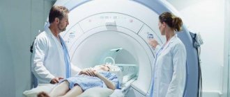
- Electromyographic, electroneuromyographic research methods - the first help to assess the potentials that arise in skeletal muscles when the muscle fiber is excited, and the second to determine the functional state of the muscles and peripheral nerves;
- Biopsy of muscle fiber, with its help it is possible to identify atrophic, destructive processes;
- Immunobiochemical, immunohistochemical studies required if autoimmune pathology is suspected.