Ultrasound of the fetal head at 1st screening
At the end of the first trimester, the expectant mother is prescribed the first ultrasound screening in utero in order to exclude such gross malformations of the fetal head as pathology of the brain, skull bones and facial skeleton.
The doctor evaluates the following fetal structures:
If everything is fine with your fetus, the doctor will write down the following in the ultrasound report:
Particular attention is paid to assessing the length of the fetal nasal bone. This is an informative criterion for early diagnosis of Down syndrome.
Examination of the skull bones already at the end of the first trimester makes it possible to identify such severe developmental abnormalities as:
- acrania;
- exencephaly;
- anencephaly;
- cranial hernia.
Anencephaly is the most common defect of the central nervous system, in which brain tissue and skull bones are completely absent.
Exencephaly - the skull bones are also absent, but there is a fragment of brain tissue.
Acrania is a developmental defect in which the fetal brain is not surrounded by the bones of the skull.
It is important to know! With these three defects, the death of the child occurs. Therefore, if they are detected at any stage of pregnancy, it is proposed to terminate it for medical reasons. In the future, the woman needs genetic counseling.
A cranial hernia is a protrusion of the meninges and brain tissue through a defect in the bones of the skull. In this case, a consultation with a neurosurgeon is required to find out whether it is possible to correct this defect with surgery after the birth of the child.
Preventive measures
To prevent the development of pathologies, a woman during the planning period should follow the principles of a healthy lifestyle, as well as take folic acid, carry out prevention and timely treatment of various chronic ailments, viral infections, etc. A medical consultation with a geneticist is mandatory.
Ventriculomegaly is a very dangerous disease that poses a direct danger to the life and health of the baby. That is why examinations and observations by a doctor should not be neglected during pregnancy. If a problem is identified early and corrected early, it increases the chances of a healthy future for your children. With the help of therapy, the consequences and complications of the disease can be eliminated.
This disease is also diagnosed in adults using CT and MRI, but deviations from the norm may not interfere with the normal functioning of the brain and central nervous system; it is only important to control these processes and constantly monitor the development of the disease.
Interpretation of ultrasound of the fetal head at 2nd screening
During the second screening, close attention is also paid to the brain and facial skeleton. Identifying pathologies of fetal development makes it possible to warn future parents about possible consequences and obtain information about the long-term prognosis.
Important indicators during examination are biparietal size (BPR), fronto-occipital size (FOR) and fetal head circumference. All these important measurements are carried out in a strictly cross-section at the level of certain anatomical structures.
The doctor evaluates the shape of the fetal head using the cephalic index (BPR/LZR ratio). Variants of the norm are:
- dolichocephalic shape (oval or oblong);
- brachycephalic form (when the skull has a rounded shape).
Important! If the fetus is found to have a lemon- or strawberry-shaped head, this is bad. It is necessary to exclude genetic diseases and concomitant malformations.
A decrease in these indicators (a small head in the fetus) is an unfavorable sign in which it is necessary to exclude microcephaly (a disease characterized by a decrease in brain mass and mental retardation). But a small head circumference does not always indicate pathology. So, for example, if all other dimensions (abdominal circumference, thigh length) are also less than normal, this will indicate intrauterine growth retardation, and not a malformation.
With an increase in the BPR and head circumference (large fetal head), they may indicate hydrocele of the brain or the presence of a cerebral hernia. If during fetometry (measurement of the fetus) all other indicators are also higher than normal, then an increase in the BPR indicates a large size of the fetus.
By the time of the second screening, all the anatomical structures of the brain have already formed and they are well visualized. Measuring the lateral ventricles of the brain is of great importance. Normally, their dimensions should not exceed 10 mm (on average 6 mm).
Note! If the lateral ventricles of the fetal brain are dilated from 10 to 15 mm on ultrasound, but the size of the head is not enlarged, this condition is called ventriculomegaly.
Chromosomal abnormalities, infectious diseases of the mother during pregnancy, and intrauterine fetal hypoxia can lead to expansion of the lateral ventricles and ventriculomegaly.
Ventriculomegaly can be:
- symmetrical (when the lateral ventricles of both hemispheres of the brain are expanded);
- asymmetric (enlargement of one of the ventricles or its horn, for example, left-sided ventriculomegaly);
- can exist in isolation from developmental defects;
- or combined with other vices.
In mild to moderate cases, careful dynamic monitoring of the size of the ventricles of the brain is necessary. In severe cases, this pathology can develop into fetal hydrocephalus (or hydrocephalus). The earlier and faster the transition from ventriculomegaly to hydrocephalus occurs, the worse the prognosis.
It can be very difficult to answer parents’ questions about how pronounced the neurological manifestations of their unborn baby will be with such a deviation and what his psychomotor development will be like. And if there is a question about terminating a pregnancy after the discovery of this pathology, you should follow the recommendations of doctors.
Hydrocephalus is another brain pathology that is detected by ultrasound. This is a condition when there is an increase in the size of the ventricles of the brain by more than 15 mm due to the accumulation of fluid (cerebrospinal fluid) in their cavities with a simultaneous increase in intracranial pressure and leading to compression or atrophy of the brain. As a rule, this pathology is characterized by an increase in the size of the fetal head.
It should be said that the most unfavorable prognosis will be when ventriculomegaly/hydrocephalus is combined with other developmental defects, chromosomal abnormalities, as well as with isolated hydrocephalus.
Hypoplasia (underdevelopment) of the cerebellar vermis can lead to disastrous consequences:
- the ability to maintain balance is lost;
- lack of muscle coordination;
- smoothness in movements is lost;
- problems appear with gait (it becomes staggering, like a drunk);
- tremors appear in the child’s limbs and head, and slow speech.
Very important for identifying this pathology is the measurement of the interhemispheric size of the cerebellum.
What is the cause of the pathology
One of the reasons for the development of ventriculomegaly in the fetus is a viral infection in early pregnancy, TORCH syndrome. The disease can be triggered by an infectious lesion of the brain during the perinatal period (prenatal).
Rarely, the cause of the pathology is neonatal sepsis - blood poisoning in an infant during the first 28 days of life.
At the Detroit Medical Research Center (USA) from 1992 to 1994. Pregnant women were monitored during the prenatal period. The scientific data obtained are presented in the publication “Mild Isolated Ventriculomegaly: Associated Anomalies and Observations” Mark W. Tomlinson, Marjorie C. Treadwel, 1997. Cytogenetic analyzes were performed on women whose fetuses had enlarged lateral ventricles by 11-15 mm. In 10-12% of cases, a positive relationship was found between the disease and chromosomal abnormalities.
Factors leading to the development of pathology:
- multifunctional immaturity of the central nervous system;
- increased reabsorption (reabsorption) of bicarbonates by the kidneys, resulting in metabolic acidosis (blood oxidation);
- disturbance of fetal hemodynamics (blood movement along the vascular bed);
- perinatal hypoxia (intrauterine oxygen deficiency);
- injuries during labor.
The listed factors lead to the disease not only in infants, but also in children and adolescents under 18 years of age. Ventriculomegaly is extremely rare in adults.
The scientific publication “Neurosurgery and Neurology of Children” by L.V. Kuznetsova, 2007, contains data from numerous clinical studies. According to their results, 50-64% of patients have stable ventriculomegaly of unknown etiology (idiopathic).
What are the lateral ventricles of the brain and what are they responsible for?
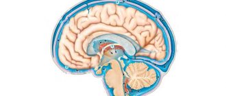
In the fetal brain there are four communicating cavities - the ventricles, which contain cerebrospinal fluid. A pair of them are symmetrical lateral, located in the thickness of the white matter. Each has an anterior, inferior and posterior horn, they are connected to the third and fourth ventricles, and through them to the spinal canal. The fluid in the ventricles protects the brain from mechanical influences and maintains stable intracranial pressure.
Each of the organs is responsible for the formation and accumulation of cerebrospinal fluid and consists of a single system of movement of cerebrospinal fluid. Liquor is necessary for stabilizing brain tissue, maintaining the correct acid-base balance, ensuring the activity of neurons. Thus, the main functions of the ventricles of the fetal brain are the production of cerebrospinal fluid and maintaining its continuous movement for active brain activity.
Diagnostics
The first signs of ventriculomegaly in the fetus during pregnancy can be detected at 17-34 weeks during a routine examination and ultrasound.
To confirm the diagnosis, karyotyping of the fetus is prescribed - studying the indicators of the set of chromosomes.
Perinatal diagnostic procedures should consist of those examinations that will help study all the anatomical systems of the fetus, especially the brain.
If dilation of the cerebral ventricles is suspected, a transverse scan of the fetal head is required to determine the size of the lateral cerebral ventricles.
Echography is also prescribed; during its implementation, the accuracy of detecting ventriculomegaly reaches 80% of cases. After birth, the child may be prescribed a CT scan or MRI - computer scans that examine the structures of the brain layer by layer.
The diagnosis is made only after evaluating all the data obtained during the examination. In approximately 9% of cases, the diagnosis gives false results.
What size should the lateral ventricles of the fetal brain normally be?
The brain structures of the fetus are visualized already during the second ultrasound (at 18–21 weeks). The doctor evaluates many indicators, including the size of the lateral ventricles of the brain and the cistern magna in the fetus. The average size of the ventricles is about 6 mm; normally, their size should not exceed 10 mm. The basic norms of fetometric parameters by week are assessed according to the table:
| Gestation period, weeks | Large cylinder head tank, mm | Head circumference, mm | Fronto-occipital size, mm | Distance from the outer to the inner contour of the crown bones (BPR), mm |
| 17 | 2,1–4,3 | 121–149 | 46–54 | 34–42 |
| 18 | 2,8–4,3 | 131–161 | 49–59 | 37–47 |
| 19 | 2,8–6 | 142–174 | 53–63 | 41–49 |
| 20 | 3–6,2 | 154–186 | 56–68 | 43–53 |
| 21 | 3,2–6,4 | 166–200 | 60–72 | 46–56 |
| 22 | 3,4–6,8 | 177–212 | 64–76 | 48–60 |
| 23 | 3,6–7,2 | 190–224 | 67–81 | 52–64 |
| 24 | 3,9–7,5 | 201–237 | 71–85 | 55–67 |
| 25 | 4,1–7,9 | 214–250 | 73–89 | 58–70 |
| 26 | 4,2–8,2 | 224–262 | 77–93 | 61–73 |
| 27 | 4,4–8,4 | 236–283 | 80–96 | 64–76 |
| 28 | 4,6–8,6 | 245–285 | 83–99 | 70–79 |
| 29 | 4,8–8,8 | 255–295 | 86–102 | 67–82 |
| 30 | 5,0–9,0 | 265–305 | 89–105 | 71–86 |
| 31 | 5,5–9,2 | 273–315 | 93–109 | 73–87 |
| 32 | 5,8–9,4 | 283–325 | 95–113 | 75–89 |
| 33 | 6,0–9,6 | 289–333 | 98–116 | 77–91 |
| 34 | 7,0–9,9 | 293–339 | 101–119 | 79–93 |
| 35 | 7,5–9,9 | 299–345 | 103–121 | 81–95 |
| 36 | 7,5–9,9 | 303–349 | 104–124 | 83–97 |
| 37 | 7,5–9,9 | 307–353 | 106–126 | 85–98 |
| 38 | 7,5–9,9 | 309–357 | 106–128 | 86–100 |
| 39 | 7,5–9,9 | 311–359 | 109–129 | 88–102 |
| 40 | 7,5–9,9 | 312–362 | 110–130 | 89–103 |
Normative and pathological parameters of BZ
Analysis of fetal brain structures is carried out in the second trimester, between 18 and 27 weeks of pregnancy. In most cases, pathological changes are detected on routine ultrasound. The diagnosis is also made at 30-33 weeks of pregnancy.
Table of the norm of the lateral ventricles (LV) in the fetus
| Fetal age | BZ width in mm |
| 18 weeks | 4,9 – 7,5 |
| 27 weeks | 5,6 – 8,7 |
| newborn | 23,5 |
| 3 months | 36,2 |
| 6-9 months | 60,8 |
| 12 months | 64,7 |
Discrepancies in parameter measurements of 0.1-0.3 mm are normal.
Degrees of ventricular dilatation (by how many mm do they increase from standard values):
- 1st (mild, insignificant) – an increase in the depth of the bodies of the 1st and 2nd basins from 5 to 8 mm, the lateral curvature is smoothed out, the outlines become more rounded, the 3rd and 4th ventricles remain unchanged;
- 2nd (moderate severity, moderate) - the body expands to 9-10 mm above the norm, the increase is uniform on all sides, the 3rd ventricle increases by 6 mm, 4 - within the anatomical norm;
- 3rd (severe, pronounced) - the depth of the bodies expands by 10-21 mm or more in all ventricles, the subarachnoid cisterns of the brain increase.
The disease is unilateral (left-sided, right-sided) or bilateral (biventriculomegaly).
What is ventriculomegaly and what types of it occurs?
If, according to the results of an ultrasound at 16–35 weeks of pregnancy, an increase in the lateral ventricles to 10–15 mm is recorded, and the size of the child’s head is within normal limits, the sonologist questions ventriculomegaly. One study is not enough to make an accurate diagnosis. Changes are assessed over time, for which at least two more ultrasounds are performed with an interval of 2–3 weeks. This pathology is caused by chromosomal abnormalities, intrauterine hypoxia, and infectious diseases that the mother suffered during pregnancy.
Ventriculomegaly can be isolated asymmetric (expansion of one ventricle or its horns without changes in the brain parenchyma), symmetric (observed in both hemispheres) or diagnosed in combination with other pathologies of fetal development. Ventricular pathology is divided into three types:
- mild - organ dilatation is 10.1–12 mm, usually detected at 20 weeks and needs to be monitored before birth;
- moderate – the size of the ventricles reaches 12–15 mm, which impairs the outflow of cerebrospinal fluid;
- pronounced - ultrasound readings exceed 15 mm, which disrupts brain function and negatively affects the vital functions of the fetus.
If the lateral ventricles are enlarged to 10.1–15 mm (deviation from the table standards by 1–5 mm), borderline ventriculomegaly is diagnosed. It may be asymptomatic up to a certain point, but in fact it indicates the emergence of a complex pathological process that gradually changes the functioning of many important organs.
What it is
Ventriculomegaly is a change characterized by an increase in the size of the ventricles in the brain. As a result of the progression of this pathology, various brain ailments arise and neurological abnormalities appear.
Important! A timely undetected pathology, in addition to a negative effect on the central nervous system, rapidly worsens the functioning of the heart and provokes the development of abnormalities in the musculoskeletal system.
There are 4 ventricles in the brain, 2 of them are located in the white matter and are called lateral. The interventricular foramina serve as peculiar connecting elements between the third ventricle (located between the visual tuberosities), which is connected to the fourth via the cerebral aqueduct. The location of the latter is the bottom of the diamond-shaped dimple. It is attached to the central canal of the spinal cord.
The ventricular system is necessary for the production of cerebrospinal fluid, which, when pathology occurs, must enter the spinal canal. When its outflow is disrupted, ventriculomegaly develops. At the same time, the ventricles expand. Normally, the size of one of them should be 1-4 mm, and with the development of diseases - 12-20 mm.
Find out what happens in the body of a pregnant woman and fetus on 1, 2, 3, 4, 5, 6, 7, 8, 9, 10, 11, 12, 13, 14, 15, 16, , 18, 19, 20, 21 , 22, 23, 24, 25, 26, 27, 28, 29, 30, 31, 32, 33, 34, 35, , 37, 38, 39, 40 weeks of pregnancy.
Treatment of pathology
When treating a pathology, the doctor sets two goals: finding and eliminating the cause of abnormal organ enlargement and neutralizing its consequences for the newborn. In case of a mild isolated form, which is not caused by chromosomal abnormalities, the expectant mother is prescribed drug therapy: taking diuretics, vitamins, injections of drugs that prevent hypoxia and placental insufficiency. Exercise therapy has a good effect (the emphasis is on the pelvic floor muscles). To prevent neurological changes in the baby's body, the expectant mother is prescribed medications to retain potassium.
In the postpartum period, infants are given several courses of massage aimed at relieving muscle tone, strengthening, and eliminating neurological symptoms. Monitoring by a neurologist is required in the first weeks, months and years of life. Severe forms of pathology require surgical treatment after the birth of the baby. In the brain, neurosurgeons install a tube that has a drainage function to maintain proper circulation of cerebrospinal fluid.
The prognosis for a child with a severe form and chromosomal abnormalities is unfavorable.


