Structure
All ventricles of the brain have special choroid plexuses. They produce liquor. The ventricles of the brain are connected to each other by the subarachnoid space. Thanks to this, the movement of cerebrospinal fluid occurs. First, from the lateral ones it penetrates into the 3rd ventricle of the brain, and then into the fourth. At the final stage of circulation, the outflow of cerebrospinal fluid into the venous sinuses occurs through granulations in the arachnoid membrane. All parts of the ventricular system communicate with each other using channels and openings.
Tumors of the third ventricle
Treatment of cancer >> Books on oncology >> N. A. Popov, “Intracranial tumors” Len. dept. Medgiz Publishing House, Leningrad, 1961 OCR Wincancer.Ru Given with some abbreviations
The clinic of tumors of the third ventricle has many common features with neoplasms of the lateral ventricles. Recognizing them is no less difficult, and the clinical picture can be very contradictory.
Tumors of the third ventricle are also characterized by the early development of hypertensive-hydrocephalic syndrome and its intermittent course with congestive optic nerves, attacks of headache with vomiting and dizziness. This is explained by the same mechanism of temporary exclusion of the ventricle from the general cerebrospinal fluid circulation system (blockage of the foramina of Monro) and the occurrence of severe hydrocephalus. But unlike tumors of the lateral ventricles, focal symptoms associated with the pathological influence of the neoplasm on the autonomic centers of the bottom and walls of the third ventricle may be more clearly presented here.
These are disturbances of thermoregulation, water-salt and fat metabolism, sexual function, sleep disorders, attacks of diencephalic epilepsy, etc. The appearance of special paroxysmal conditions, such as tonic postural seizures (Penfield and Erickson), is possible. Whether they are, by the mechanism of their origin, mesencephalic (decerebrate attacks) due to the involvement of the adjacent region of the midbrain or subcortical seizures of striatal epilepsy, they acquire a certain significance in the semiology of tumors of the third ventricle. The mentioned authors observed diencephalic vegetative attacks with phenomena of decerebranion rigidity when the subtubercular region was affected.
According to B. G. Egorov, the anamnesis of these patients is noteworthy. During the development of the disease, “peculiar twilight states are observed in the presence of very significant exacerbations of headaches; patients fall or drop objects.” These phenomena should be considered as “paroxysmal changes in tone of a cataplectoid nature.”
However, the described vegetative symptoms and paroxysmal states do not necessarily accompany tumors of the third ventricle - in this regard, much depends, obviously, on the position of the tumor inside the ventricle, the direction of growth, the nature and size of the tumor itself; Only general brain symptoms remain obligatory. Therefore, decisive for the recognition of tumors of the third ventricle is a contrast X-ray examination - ventriculography, by puncture of both lateral ventricles, with the aim of establishing the patency (blockade) of the Monroy foramina and the nature of the filling of the ventricular cavities with air.
B. G. Egorov reports on a number of patients with the localization in question, who were successfully operated on and followed for a long time. This gives him grounds to assert that “primary tumors of the ventricular system of the brain, being always well isolated, are operable. The study of the long-term consequences of their surgical treatment gives reason to believe that radical surgical treatment of this type of tumor undoubtedly has a future.”
Patient D., 19 years old, was admitted to the hospital on January 2, 1959. He complained of moderate headaches in the frontal region, sometimes nausea, foggy vision, especially in the morning, transient watering in the eyes. Ill for about a month; At school age, one night there was an attack of a sharp headache. At the beginning of December 1958 there was “flu”, low-grade fever. Objectively: General condition is good. Sharply expressed congestive papillae of the optic nerves. Slight bitemporal narrowing of the visual field to red. Visual acuity in the right eye was 0.8, in the left eye - 1.0 (later it fell steadily). Increase in the size of the blind spot in both eyes. Limitation of outward movements of the eyeballs; transient diplopia. Slight weakness of the inferior branch of the left facial nerve. Very mild membrane symptoms. Deep reflexes are uniformly reduced.
Craniography: Blurry contours of the back of the sella turcica, widening of the entrance to it, slight divergence of the sutures. Chronic purulent tonsillitis. Slight leukocytosis in the blood with a neutrophilic shift and subfebral temperature. The headache is intermittent, not intense, more often at night and in the morning, rarely with vomiting. At times, the pulse slows down (up to 48 per minute).
Biochemical research. Fasting blood sugar is 67 mg% Daily diuresis is normal. Serum proteins - 6.4% (albumin - 4.5%, globulins - 1.9%). Cholesterol: 108-168 mg%. Bilirubin - 6.4 mg% (indirect reaction). In the cerebrospinal fluid: chlorides - 585-690 mg%; sugar - 77 mg%. Quick's reaction: in 4 hours, 4.14 g of sodium benzoate was released - 89.9%.
Ventriculography through the posterior horn of the left lateral ventricle: Significant dilatation of the entire left lateral ventricle; gas did not penetrate into the third ventricle. Ventriculography through the anterior horn of the right lateral ventricle: filling mainly of the right lateral ventricle along its entire length; There is no gas in the third ventricle. Conclusion: Hydrocephalus of both lateral ventricles, without displacements or filling defects, blockade of the foramina of Monroy.
Subsequently, the patient's condition fluctuated, but, in general, gradually worsened. The state of pronounced euphoria and the weakening of criticism, which was noticeably increasing, attracted attention. Shell symptoms appeared, at times blackouts, and motor agitation. Clinical diagnosis: Tumor of the third ventricle. Due to the serious condition, decompressive trepanation was performed in the right infratemporal region. The patient's condition improved, and hypertensive symptoms decreased. The second stage of the operation with an attempt to revise the ventricles of the brain: no tumor was found. The patient underwent the surgical intervention satisfactorily, but his condition continued to deteriorate, and he died on 6/V.
Section: Tumor - glioma of the third ventricle of the brain, size 3X 1.5-2 cm, located in the anterior parts of it and blocking the Monroe foramina. It is fused with the wall of the ventricle. Severe hydrocephalus of the lateral ventricles. Ependimitis. Brain swelling. Left-sided focal pneumonia.
The main difficulty in the clinical recognition of this case was the absence of any obvious focal brain symptoms in the presence of hydrocephalic-hypertensive, especially pronounced congestive nipples, which were steadily increasing and leading to a decrease in vision. This circumstance, as well as the age of the patient, made it possible first of all to think about a cerebellar tumor. The possibility of arachnoiditis of the posterior cranial fossa was not excluded, since the appearance of the first symptoms was preceded by “flu” with an increase in temperature.
Those minor deviations that occurred in relation to metabolic functions and other autonomic disorders; (transient changes in white blood, slight transient hyperthermia, etc.) could be associated with hydrocephalus and should not necessarily be considered as primarily diencephalic. Some of these symptoms were identified later, when ventriculography data made the presence of a tumor of the third ventricle quite obvious.
It was noteworthy that, despite the pronounced congestion of the nipples, the patient very rarely vomited. This circumstance in this case could be related to the fact that the level of occlusion corresponded to the third ventricle, while hydrocephalus of the fourth ventricle was absent, therefore, there could be no irritation of the vomiting center. As is known, this symptom is often and pronounced in neoplasms of the posterior cranial fossa, especially the fourth ventricle; Apparently, this circumstance may be important in the differential diagnosis between a tumor of the third ventricle and the cerebellum. It should be remembered, however, about the importance of individual irritability of the vomiting center, as evidenced by many clinical facts.
The mental changes that developed in the patient later were apparently associated with increasing hydrocephalus of the lateral ventricles; they resembled those disorders that more often occur with tumors of the frontal lobes and are not characteristic of neoplasms of the posterior cranial fossa. Known fluctuations in the patient's condition could be retrospectively explained by the valve mechanism of blockage of the foramina of Monroy, which carries out the outflow of cerebrospinal fluid from the lateral ventricles.
Thus, the differential diagnosis between a tumor of the third ventricle and the cerebellum, i.e., determining the level of occlusion, was very difficult, as is often the case in connection with closed hydrocephalus, scarcity or even the absence of clear focal symptoms observed in both cases , the possible common origin of stem and diencephalic symptoms - primarily focal in one case and secondary in nature - in another case. Therefore, the issue could finally be resolved only with the help of ventriculography.
Indeed, a contrast X-ray study showed closure of the foramen of Monroy with good filling of the highly dilated lateral ventricles (without displacements or filling defects), which indicated in favor of a tumor of the third ventricle. The same localization could be indirectly supported by the fact that after ventriculography no focal symptoms were revealed, as is sometimes observed in cases of neoplasm of the posterior cranial fossa.
The general condition of the patient made it possible to carry out a contrast study and thereby resolve doubts; when this is impossible, one has to rely on an accurate account of clinical data, especially since electroencephalography and the method of radioactive isotopes in such cases do not give convincing results.
Tumors of the bottom and walls of the third ventricle (paraventricular) give a unique clinical picture. Neoplasms of this kind are rare and have not yet been studied enough, and their general and differential diagnosis can be very difficult.
Patient Sh., 51 years old, was admitted to the clinic on March 30, 1949, with complaints of headaches, loss of vision, and increased thirst. She got sick a month ago, when she first developed a headache, transient double vision, and her vision began to rapidly deteriorate. Soon the patient began to experience extreme thirst. Three weeks later, complete blindness was established with an unchanged fundus, bilateral anosmia, Kernig's sign on the left, and low-grade fever.
Objectively: General condition is quite satisfactory, temperature is normal, pulse 82 beats per minute, blood pressure 120/100, polydipsia. Bilateral hyposmia, blindness in both eyes, fundus without changes. The pupils are evenly dilated and do not react to light. Sharply expressed spontaneous nystagmus. Corneal reflexes are preserved, knee and Achilles reflexes are animated, slightly higher on the left; “fan symptom”, more clearly expressed on the left leg; abdominal reflexes are not evoked. No motor or sensory disorders are observed.
The cerebrospinal fluid is slightly opalescent, protein - 0.66%o, globulin reactions are positive, cytosis - 95 per 1 mm3, predominantly lymphocytes. Blood unchanged. X-ray of the skull without abnormalities.
On the 4th day after admission, the patient’s condition worsened: the temperature increased to 38.3, pulse 112 per minute; headaches intensified, excruciating thirst and vomiting arose. After another 2 days, neck rigidity, Kernig’s sign, and bilateral Babinski’s sign appeared. The fundus remained normal until the end of the patient's life. The daily amount of urine is 4 liters. Treatment with penicillin was started (a total of 9,200,000 units were administered). Repeated lumbar puncture: the fluid is slightly turbid, xanthochromic; globulin reactions are sharply positive, protein 0.99%o, formed elements 360 per 1 mm3 (neutrophils predominate). Three days later, the patient’s condition worsened even more, psychotic symptoms appeared - delusions, hallucinations. The patient died 10 days after admission.
Clinical diagnosis (presumable): Meningitis of the base of the brain (tuberculous?). Section: Glioma of the bottom of the third ventricle, growing through its bottom and forming a node at the base of the brain, sharply compressing the chiasm.
The disease in this case began with headaches and a rapid decrease in vision, which soon led to blindness (due to compression of the chiasm). Here it was easier to recognize the localization than the nature of the process. The diagnosis of meningitis seemed certain; the absence of congestive nipples and other hypertensive symptoms testified against another, the only other possible diagnosis - a tumor of the third ventricle. Correct assessment of such early symptoms as amaurosis and polydipsia, and therefore clinical diagnosis, was very difficult.
The visual field could not be examined due to amaurosis, and there was no atrophy of the optic nerves (before they even reached the eyeball). Due to the rapidly developing blindness and early onset of polydipsia, the presence of a lesion at the base of the brain was obvious, but the nature of the process, due to a number of symptoms unusual for a tumor (fever, inflammatory changes in the cerebrospinal fluid, meningeal phenomena), was mistakenly recognized. The appearance of these symptoms, apparently of hypothalamic origin, is of interest in brain tumors from a pathophysiological point of view. It is known that significant changes in the cerebrospinal fluid are observed mainly in tumors localized in the cavities or walls of the ventricles and the posterior cranial fossa. Thus, the rapid development of the disease, meningeal syndrome, changes in the cerebrospinal fluid in the absence of congestive optic nerves and other hypertensive symptoms seemed to make the diagnosis of meningitis certain, and the presence of a tumor could hardly be recognized.
Pleocytosis in the cerebrospinal fluid in brain tumors is common, but only in some cases does it reach high numbers.
Thus, I. Ya. Razdolsky observed two patients where the number of cells reached 379 and even 5314. Similar observations are given by D. G. Schaefer and E. F. Voronkina, A. B. Iozefovich, L. L. Sinegubko and S. Pekker . Large pleocytosis was described by Kaffka, Christiansen and many others. etc. See further: Tumors of the quadrigeminal area >>
Lateral divisions
Conventionally, they are called the first and second. Each lateral ventricle of the brain includes three horns and a central section. The latter is located in the parietal lobe. The anterior horn is located in the frontal, the lower - in the temporal, and the posterior - in the occipital zone. In their perimeter there is a choroid plexus, which is distributed quite unevenly. So, for example, it is absent in the posterior and anterior horns. The choroid plexus begins directly in the central zone, gradually descending into the lower horn. It is in this area that the size of the plexus reaches its maximum value. For this reason, this area is called a tangle. Asymmetry of the lateral ventricles of the brain is caused by a disturbance in the stroma of the tangles. This area is also often subject to degenerative changes. This kind of pathology is quite easily detected on ordinary radiographs and carries a special diagnostic value.
Hydrocephalus accompanied by increased intracranial pressure
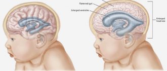
Hydrocephalus of the brain in an adult has symptoms that are not as pronounced as hydrocephalus in children. In a child, “dropsy of the brain,” accompanied by an increase in cerebrospinal fluid pressure, causes not only headaches, crying, anxiety, and impaired consciousness, but also in infancy leads to a change in the configuration of the cranium, a rapid increase in head circumference, and bulging of the fontanelle.
The average person often does not pay attention to such manifestations of pathology as headaches and sleep disturbances. All this is attributed to overwork at work and constant stress. symptoms , forces you to seek help
- headache of a bursting nature , occurring most often in the morning immediately after sleep. The rate of increase in pain depends on the rate of development of hydrocephalus;
- nausea and vomiting at the height of headache. Vomiting with hydrocephalus rarely brings relief and does not depend on food intake. Sometimes this is the first symptom of hydrocephalus, especially with tumors located in the posterior cranial fossa;
- sleep disturbance (drowsiness during the day and insomnia at night);
- persistent hiccups;
- impairment of consciousness of varying degrees (from stupor to coma);
- visual disturbances , most often manifested by double vision. This symptom develops as a consequence of compression of the abducens nerves. Paroxysmal disorders also occur in the form of limitation of visual fields, which arise due to a decrease in venous outflow from the eye and damage to the optic nerve;
- a congestive optic disc is formed , which is detected during examination of the fundus by an ophthalmologist. This symptom is characteristic only of chronic and subacute hydrocephalus, since during the development of acute “dropsy of the brain” it is often delayed;
- pyramidal insufficiency , manifested by symmetrical pathological foot signs (Babinsky's symptom, Rossolimo, etc.);
- Cushing's triad , which includes increased blood pressure accompanied by bradycardia and bradypnea (decreased breathing).
It must be remembered that the severity and speed of onset of symptoms in hydrocephalus depends on the type of disease, namely the rate of increase in intracranial pressure. With an acute increase in cerebrospinal fluid pressure, the symptoms will be pronounced, but some may be “late” (for example, changes in the fundus).
Third cavity of the system
This ventricle is located in the diencephalon. It connects the lateral sections with the fourth. As in other ventricles, the third contains choroid plexuses. They are distributed along its roof. The ventricle is filled with cerebrospinal fluid. In this department, the hypothalamic groove is of particular importance. Anatomically, it is the border between the visual thalamus and the subtubercular region. The third and fourth ventricles of the brain are connected by the aqueduct of Sylvius. This element is considered one of the important components of the midbrain.
Fourth cavity
This section is located between the pons, cerebellum and medulla oblongata. The shape of the cavity is similar to a pyramid. The floor of the ventricle is called the rhomboid fossa. This is due to the fact that anatomically it is a depression that looks like a diamond. It is lined with gray matter with a large number of tubercles and depressions. The roof of the cavity is formed by the lower and upper brain sails. It seems to hang over the hole. The choroid plexus is relatively autonomous. It includes two lateral and medial sections. The choroid plexus attaches to the lower lateral surfaces of the cavity, extending to its lateral inversions. Through the medial foramen of Magendie and the symmetrical lateral foramina of Luschka, the ventricular system communicates with the subarachnoid and subarachnoid spaces.
Changes in structure
The expansion of the ventricles of the brain negatively affects the activity of the nervous system. Their condition can be assessed using diagnostic methods. For example, a computed tomography scan reveals whether the ventricles of the brain are enlarged or not. MRI is also used for diagnostic purposes. Asymmetry of the lateral ventricles of the brain or other disorders can be caused by various reasons. Among the most popular provoking factors, experts call increased formation of cerebrospinal fluid. This phenomenon accompanies inflammation in the choroid plexus or papilloma. Asymmetry of the ventricles of the brain or changes in the size of the cavities may be a consequence of impaired outflow of cerebrospinal fluid. This happens when the holes of Luschka and Magendie become impassable due to inflammation in the membranes - meningitis. The cause of obstruction may also be metabolic reactions due to venous thrombosis or subarachnoid hemorrhage. Often, asymmetry of the ventricles of the brain is detected in the presence of space-occupying neoplasms in the cranial cavity. This could be an abscess, hematoma, cyst or tumor.
Methods for diagnosing the disease
Diagnosis of hydrocephalus consists not only in detecting its signs, but also in trying to establish what disease of the nervous system it was caused by. This usually does not cause difficulties, given modern examination methods.
The tactics of further treatment of the patient depend on the correct diagnosis. The methods used in examining adults and children differ somewhat, because in children a clear clinical picture comes to the fore: changes in the shape of the skull, depression of consciousness, convulsive seizures, and impaired psychomotor development. Therefore, an MRI or CT scan of the brain may not be needed; neurosonography will be enough. Since neuroimaging diagnostic methods require lying still, this will require the use of sedatives or light anesthesia, which is not always possible due to the child’s health condition.
So, diagnostic methods for hydrocephalus are divided into instrumental and non-instrumental.
Non-instrumental diagnostic methods
Non-instrumental diagnostics includes the following methods:
- interviewing the patient, asking for anamnesis of life and illness. If the patient is in clear consciousness, the neurologist clarifies his complaints, previous diseases and injuries to the nervous system, how quickly the symptoms developed, and what the first of them was. In case of depression or impairment of consciousness, this information is obtained from the patient’s immediate environment;
- neurological examination - allows you to identify focal changes that arise as a result of neoplasms in the brain, leading to a block of the cerebrospinal fluid pathways; signs of increased intracranial pressure (pain when pressing on the eyeballs, trigeminal points, changes in visual fields, etc.); pyramidal insufficiency and gait disturbance, changes in the shape of the skull (in older people this may occur during osteoporotic processes);
- neuropsychological testing reveals signs of dementia, affective disorders (depression, etc.);
- examination by an ophthalmologist - examination of the fundus often reveals congestive changes in the area of the optic nerve head.
Instrumental diagnosis of hydrocephalus
It is impossible to confirm the diagnosis based on complaints and physical examination, so they resort to the use of instrumental methods:
- radiography of the skull (craniography) – in adults this examination method is not very informative. With its help, the size of the skull, the condition of the sutures and bones are determined. With a long-term increase in intracranial pressure, signs of porosity and destruction of the sella turcica may be detected;
- echoencephaloscopy is an ultrasound method that allows you to identify signs of hydrocephalus and indirectly confirm the presence of a space-occupying lesion in the brain by the displacement of its midline structures;
- lumbar (spinal) puncture followed by biochemical and cytological analysis of the cerebrospinal fluid - is performed only in the absence of a space-occupying formation in the cranial cavity. With hypertensive hydrocephalus, cerebrospinal fluid flows out under pressure and extraction of 35 - 50 ml of fluid leads to a significant improvement in the patient's condition. Subsequent analysis may reveal signs of hemorrhage, increased protein levels;
- CT or MRI of the brain - changes detected using these examination methods confirm not only the presence of hydrocephalus, but also “explain” the reason for its formation. That is, in addition to the expansion of the ventricles, an increase in the size of the grooves and subarachnoid space, the following is detected: a block of the cerebrospinal fluid pathways with a space-occupying formation, damage to the meninges and choroid plexuses in the ventricles, or signs of neurodegenerative diseases are visualized.
Only a qualified neurologist can establish the correct diagnosis. When the first symptoms of the disease appear, you should immediately contact a specialist, since rapidly increasing hydrocephalus can lead to dislocation of the brain stem structures and death.
General mechanism for the development of disturbances in the activity of cavities
At the first stage, there is difficulty in the outflow of cerebral fluid into the subarachnoid space from the ventricles. This provokes expansion of the cavities. At the same time, compression of the surrounding tissue occurs. Due to the primary blockage of fluid outflow, a number of complications arise. One of the main ones is the occurrence of hydrocephalus. Patients complain of sudden headaches, nausea, and in some cases vomiting. Disorders of autonomic functions are also detected. These symptoms are caused by an acute increase in pressure inside the ventricles, which is characteristic of some pathologies of the liquor-conducting system.
Treatment of hydrocephalus of the brain in adults
The most effective treatment for hydrocephalus is shunt surgery. Although, with a compensated course of the disease, you can limit yourself to drug therapy for some time. Drugs used for “dropsy of the brain” are mainly aimed at reducing intracranial pressure by removing “excess” fluid from the body. It is also important to improve microcirculation and metabolism of brain cells.
Conservative treatment of hydrocephalus: main groups of drugs
Conservative therapy is carried out under the supervision of a doctor either in a hospital or on an outpatient basis. Since decompensation of the disease can occur suddenly and be complicated by cerebral edema.
Hydrocephalus treatment includes the following groups of drugs:
- diuretics: loop (Lasix, furosemide, hypochlorothiazide, torsemide, diacarb, acetazolamide), osmotic (mannitol) and potassium-sparing (veroshpiron, spironolactone). When using the first two groups, it is necessary to take potassium supplements (asparkam, panangin) in parallel. These drugs are ineffective in the normotensive form of the disease;
- vascular drugs (cavinton, vinpocetine, nicotinic acid);
- neuroprotectors (Ceraxon, Pharmaxon, Gliatilin, Gleacer);
- metabolic agents (actovegin, cortexin, cerebrolysin, cerebrolysate);
- anticonvulsants (carbamazepine, lamotrigine, valprocom) are used for the development of convulsive syndrome.
Drugs are prescribed and discontinued only by the attending physician, taking into account the individual characteristics of the patient.
Surgical methods to combat hydrocele
Surgery, namely shunt surgery, is the main method of treating hydrocephalus. If the disease is caused by a mass formation in the brain (cyst, tumor, aneurysm), then, if possible, it is removed.
In case of acutely developing hydrocephalus in emergency situations, a lumbar puncture with the removal of no more than 50 ml of cerebrospinal fluid can alleviate the condition, but only in cases of absence of “plus” tissue in the brain. ventricular drainage is also used , when a catheter is inserted directly into the ventricles of the brain through a burr hole in the skull. The disadvantage of this method is the high risk of developing infectious complications.
In other cases, ventriculoperitoneal, ventriculoatrial or lumboperitoneal shunting is used . When cerebrospinal fluid from the ventricles is drained through a catheter located under the skin into the abdominal cavity, into the atrium, or from the spinal canal into the abdominal cavity, respectively.
Often with this method of treatment a number of complications arise:
- infections;
- violation of shunt patency;
- subdural hematomas and hygromas;
- hemorrhages;
- epileptic seizures;
- rapid outflow of cerebrospinal fluid, which can lead to herniation of stem structures.
In recent years, an endoscopic method , which consists of forming pathways for the outflow of cerebrospinal fluid from the third ventricle into the cisterns of the brain. The advantage of such surgical intervention is less trauma, restoration of the physiological dynamics of cerebrospinal fluid, and a reduced risk of complications.


