Author
: Grachev Ilya Illarionovich
Editor
: Efremov Mikhail Mikhailovich
Publication date: 10/20/2014 Update date: 11/12/2020
The most common manifestation of serious problems with the spine is vertebrogenic dorsalgia - back pain caused by various factors that are traumatic, degenerative, neoplastic and inflammatory.
Any type of dorsalgia has serious complications, so you should not delay treatment.
See how easy it is to cure a disease in 10 sessions
Causes of the disease
Vertebral cervicalgia according to ICD-10 is classified as dorsopathies - code M54.2. The main reason for its appearance is associated with diseases of the cervical spine. Most patients are diagnosed with osteochondrosis, characterized by degenerative changes in the intervertebral discs. In addition, reasons may be:
- osteophytes in the area of intervertebral joints. They are areas of bone tissue growth that can compress neural and vascular structures;
- disc protrusion, characterized by its protrusion beyond the intervertebral space;
- infectious processes, benign and malignant tumors in the spine;
- vertebral fractures and other injuries: subluxations, dislocations;
- diseases of the vertebral arteries;
- rheumatoid arthritis, ankylosing spondylitis, etc.
These diseases lead to organic changes in the cervical spine. This creates the background for the development of pain and other symptoms. Clinical manifestations of cervicalgia occur as a result of exposure to trigger factors: acute stress, uncomfortable position of the head and neck during sleep, hypothermia, curvature of posture, excess body weight, prolonged sedentary work, heavy physical activity, etc.
Classification
Vertebrogenic cervicalgia can have various forms. The disease is most often classified depending on the nature of its course:
- acute variant, characterized by intense pain and accompanying neurological changes. Most often occurs after spinal injuries and with pathologies of the spinal cord roots. Lasts up to 7-10 days;
- Chronic cervicalgia manifests itself with moderate symptoms with mild neuralgia. Clinical signs of the disease persist for up to 2 months or more. It occurs against the background of chronic pathologies: tumor lesions, vascular diseases, osteoporosis, etc.
In addition to the nature of the course, experts take into account the causes of the disease. Based on this, spondylogenic and discogenic variants of cervicalgia are distinguished. In the first case, the disease occurs due to pathological changes in the bone tissue of the vertebrae due to osteoporosis, neoplasms or injuries. Violations lead to negative effects on the nerves and pain. With discogenic, or true cervicalgia, the pathological focus is detected in the intervertebral discs. As a rule, patients are diagnosed with osteoporosis.
Depending on the localization of pain and its spread to other parts of the spine, the following variants of the disease are distinguished:
- cervicocranialgia;
- cervicobrachialgia;
- cervicothoracolumbalgia;
- vertebrogenic cervicothoracalgia.
These options are accompanied by pain in the head, arms, lumbar and thoracic spine, respectively.
Symptoms
Did you know that...
Next fact
The main symptom of the syndrome is neck pain, which can be aching, constant throbbing or shooting.
With cervicobrachialgia, pain radiates to other parts of the body - to one or both arms, the back of the head, shoulders or shoulder blades.
The severity of this symptom increases during coughing, sneezing, and making all kinds of head movements (turning, bending).
Over time, the tension in the neck muscles is accompanied by limited movement (the patient cannot turn his head separately from his body). Prolonged intense pain causes hypotension, which gradually turns into atrophy.
Another symptom is sensory disturbances , which can occur on the right or left. The patient may complain of numbness in the limbs, a crawling sensation, and even cramps in the hands.
Diagnostics
Since cervicobrachialgia occurs against the background of other diseases, the patient’s examination should be comprehensive . Diagnosis begins with a visual examination, during which stoop, asymmetry, and any signs of deformation are revealed.
Then palpation (palpation) and tapping are carried out to determine the location of the pathological process. The doctor also evaluates sensitivity disorders and mobility of the cervical spine.
But the most important diagnostic methods to detect the root cause of the syndrome are the following instrumental studies:
- magnetic resonance imaging – designed to identify protrusions and disc herniations, as well as their localization;
- radiography performed in different planes;
- CT scan;
- biochemical tests of urine and blood to identify or exclude concomitant pathologies.
The most preferred diagnostic method is MRI , which visualizes the structures of the spinal cord and intervertebral discs. As for x-rays, they only show bone tissue.
Clinical manifestations
Acute and chronic vertebrogenic cervicalgia manifests itself with a characteristic complex of symptoms. When questioned by a doctor, the patient expresses the following complaints:
- pain in the neck area that has a burning or pulling character. Unpleasant sensations can spread to the back of the head, shoulders and back, all the way to the lower back;
- when sneezing and sudden movements of the head, the pain intensifies;
- characteristic signs of cervicalgia with muscular-tonic syndrome - compactions and pain in the muscles associated with a violation of their tone;
- numbness and tingling on the skin of the face and scalp;
- restriction of neck mobility. Patients experience difficulty turning their head or movements are completely absent, as they lead to severe pain;
- dizziness and migraine-like pain attacks;
- tinnitus, flashing “spots” before the eyes and other visual disturbances;
- nausea, general weakness and fainting;
- unsteady gait.
If these symptoms appear, you should immediately seek medical help. Attempts to treat cervicalgia on your own can lead to progression of the underlying disease and the development of complications.
Cervicalgia syndrome has different severity in patients. In the initial stages of the disease, the patient may notice mild pain in the cervical spine during sudden head movements or uncomfortable posture during sleep. Without treatment, the disease progresses, leading to increased symptoms.
Diagnostic measures
The diagnosis of vertebrogenic cervicalgia is made on the basis of a comprehensive diagnosis. In addition to collecting existing complaints and the history of the development of the disease, the examination includes the following methods:
- X-ray of the cervical spine, allowing to identify traumatic and non-traumatic disorders in the musculoskeletal system. Using an x-ray, a specialist can identify vertebral fractures, deformation of intervertebral discs, osteophytes, etc. The method is used at the initial stages of diagnosis to determine the nature of the pathology and select further examination tactics;
- computed tomography is a variant of x-ray examination that allows you to obtain a three-dimensional image of the spine and adjacent tissues. Used to identify signs of damage to the osteoarticular apparatus;
- Magnetic resonance imaging is suitable for assessing the condition of intervertebral discs, nerve roots, blood vessels and soft tissues. These formations are poorly visible on x-ray examination. MRI is used to detect tumors, inflammatory processes in the nervous system, etc.;
- Ultrasound with Dopplerography of the vertebral arteries if there is suspicion of their compression and impaired blood flow;
- If there is pain in the thoracic back, electrocardiography is performed. An ECG allows you to exclude a painful attack during angina pectoris and myocardial infarction;
- To assess the speed of transmission of nerve impulses along the nerves, electromyelography is performed. The study is indicated for patients with paresis and paralysis that may accompany cervicalgia.
Additionally, laboratory tests are carried out: clinical and biochemical blood tests, serological diagnostics to identify neuroinfections, etc. These methods make it possible to clarify the diagnosis and identify concomitant diseases. The results are always interpreted by a doctor. Attempts to make a diagnosis yourself lead to incorrect diagnosis and selection of ineffective therapy.
Necessary diagnostics
Vertebrogenic pain syndrome is treated by a neurologist or a vertebrologist who specializes in diseases of the spine. The specialist will prescribe all the necessary examinations to determine an accurate diagnosis and prescribe appropriate treatment.
First of all, a detailed survey is carried out to determine the location and nature of the pain. Next, hardware tests are prescribed.
The most informative studies are CT and MRI of painful areas of the spine. In some cases, radiography is sufficient to confirm the diagnosis.
The main purpose of the research is to exclude diseases that are similar in symptoms and have a similar clinical picture in order to prescribe effective treatment (spinal injury, cancer, metastases, neoplasms, inflammatory processes in the spinal region, etc.).
Treatment approaches
Therapy for symptoms of cervicalgia is aimed at eliminating the underlying disease and alleviating the patient’s condition. For this purpose, medicinal and non-medicinal methods are used.
Use of medications
For cervicalgia, the following groups of medications are used:
- non-steroidal anti-inflammatory drugs (Ketorolac, Nimesulide, etc.) in the form of tablets, ointments or gels. NSAIDs eliminate pain and reduce the severity of the inflammatory process. If efficiency is low, the doctor may prescribe glucocorticosteroids - Dexamethasone, etc.;
- for chronic pain syndrome, antidepressants are used (Fluoxetine, Amitriptyline). They eliminate pain and improve the patient’s quality of life;
- muscle relaxants: Tizanidine, Baclofen, Tolperisone, etc. Eliminate increased tone and muscle spasm in the cervical and thoracic region. Thanks to this, pain is reduced;
- drugs that improve blood circulation (Actovegin, Cerebrolysin);
- nootropics: Phenotropil, Piracetam and their analogues. Improve the condition of nervous tissue, including spinal roots;
- chondroprotectors - Artra, Chondroitin sulfate, etc. Inhibit degenerative processes in intervertebral discs and prevent the progression of osteochondrosis.
All medications are prescribed only by the attending physician. The drugs have indications and contraindications that should be taken into account before selecting treatment. Otherwise, it may not be effective and may lead to side effects.
Using traditional approaches, such as medicinal herbs, is not recommended. They have no proven effectiveness and safety.
Non-drug methods
When the patient’s condition is stabilized and there are no acute signs of the disease, therapy is supplemented with medicinal approaches: physical therapy, massage and physical therapy. The following physiotherapeutic procedures are used: magnetic therapy, UHF, diadynamic therapy, medicinal electrophoresis, phonophoresis and reflexology. Physiotherapy is carried out in courses of 7-10 sessions.
Therapeutic massage and exercise therapy can normalize muscle tone in the neck, strengthen the muscle corset and prevent exacerbations of the disease. These methods are used under medical supervision. After the patient has mastered the technique of performing therapeutic exercises, they can be performed at home.
All patients with vertebrogenic cervicalgia are advised to use an orthopedic pillow and mattress to properly distribute the load on the spine during sleep. In the acute period of pathology, a cervical collar is used to fix the neck in a physiological position. In case of acute pain syndrome, injections are given to the right and left of the spine with anesthetics: Novocaine or Lidocaine. Injections can eliminate pain for a long time.
Surgical interventions
Surgeries for cervicalgia are performed in the following conditions:
- aneurysm and dissection of the vertebral artery;
- progression of the pathology against the background of ineffective conservative therapy;
- compression of the nerve roots;
- spinal cord damage.
Depending on the patient's condition, various surgical interventions can be performed: laminectomy, discectomy, spinal canal decompression and foraminotomy. Laminectomy involves removing part of the vertebra that is compressing the nerve roots. In this case, part of the intervertebral disc may also be removed. During a discectomy, the disc is completely removed, after which adjacent vertebrae are tightly fixed to each other. This operation is rarely performed as it leads to disability.
When choosing decompression of the spinal canal, formations that cause compression are eliminated: tumors, osteophytes, etc. Foraminectomy is characterized by the removal of osteophytes that lead to damage to nerve roots or blood vessels. After surgery, the patient needs comprehensive rehabilitation to prevent negative consequences and disability.
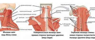
Cardialgia and abdominalgia of vertebrogenic and myofascial origin
Pain is an extremely complex phenomenon, and the search for the causes of pain does not always end with a clear somatic diagnosis. In a number of patients, the nature of thoracic and abdominal pain remains unclear, which leads to diagnostic and treatment errors, incorrect choice of treatment tactics, and even unjustified surgical interventions. Thus, in 10–30% of cases after appendectomy, histological examination does not reveal any morphological changes, while the clinical picture of acute appendicitis may be caused by vertebrogenic pseudoappendiceal Lehman syndrome, which occurs when 10–12 lower thoracic roots are affected on the right. Coronary angiography performed in patients with a clinical picture of typical angina pectoris reveals the normal state of the coronary arteries in 10–20% of these patients, and in patients with an atypical picture of angina pectoris, unchanged coronary arteries are found in 70% of cases [1, 2]. Patients with cardialgia and abdominalgia of vertebrogenic and myofascial origin, as a rule, search for the substrate of their disease for a long period of time, and the possibility of pain in connection with musculoskeletal and autonomic factors most often seems unlikely to them. Often, internists, having not found the anatomical substrate of the pain syndrome, experience difficulties in the management of such patients and refer them to a neurologist. Therefore, the neurological view of this problem is becoming increasingly relevant.
Cardialgia of vertebrogenic and myofascial origin
The formation of cardialgia against the background of spinal pathology is due to the presence of close connections between the cervical spinal motion segments (SMS) and the heart through the sympathetic formations of the cervical region with the corresponding segments of the spinal cord. There may be two circles of pathological impulses: proprioceptive - from the affected SMS (spine, transverse costal joints, cervical rib, anterior scalene muscle, scapula, chest, arm) to the projection zone of the dermatome, myotome and sclerotome; afferent - from the heart through the phrenic nerve, the spinal cord into the periarticular tissues of the cervical spine and upper shoulder girdle, with subsequent projection onto the skin into the corresponding Zakharyin-Ged zones. Painful impulses from these vicious circles reach the cerebral cortex along the spinothalamic pathway. As a result, pain associated with damage to the spine and peripheral joints of the upper limb can be projected onto the heart region, often simulating attacks of coronarogenic chronic as well as acute coronary heart disease [3, 4]. An important and common mechanism of cardialgia of non-coronary origin is irritation of the endings of the sinuvertebral nerve with a subsequent compensatory reaction in the form of spasm of certain muscle groups of the upper quadrant zone with their biomechanical overload, that is, with the formation of myofascial dysfunction with the formation of trigger points and, accordingly, the occurrence of pain. Myofascial syndromes can develop against the background of degenerative-dystrophic changes in the spine, but they can also have a different genesis (trauma, sprain, muscle strain, etc.). The main clinical forms of myofascial disorders, in which pain in the chest and heart region can occur, are the syndromes of the pectoralis major and minor muscles, and less commonly, the anterior scalene muscle syndrome [5, 6].
Cardiac syndrome of vertebrogenic and myofascial origin is characterized by muscular-tonic and dystrophic changes in the anterior chest wall with characteristic pain manifestations. Patients may complain of pain in the left half of the anterior chest wall, of a constant nature, but intensifying with sharp turns of the head, torso, abduction of arms to the sides, lifting heavy objects, severe coughing, sometimes the pain appears or intensifies when lying on the left side. Typically, patients regard such pain as cardiac pain, but note that taking nitrates does not have a positive effect. Palpation of the muscles of the anterior chest wall reveals signs of myofascial dysfunction in the form of local painful areas and compactions. Pathognomonic signs of myofascial pain are myofascial trigger points - areas of local pain in the muscle, mechanical pressure on which causes not only intense local, but also referred pain. The occurrence of pain and activation of trigger points in the pectoralis major muscle occurs when lifting heavy objects, especially in front of you, when loading the arm in an abducted position, and when staying for a long time with lowered shoulder girdles, which leads to contraction of the muscle. The pain radiates along the anterior surface of the chest wall, the medial surface of the shoulder and forearm. With myofascial dysfunction of the pectoralis minor muscle, which occurs as a result of trauma during sudden or prolonged lateral abduction of the shoulder and throwing back the arm, including during sleep, when working with outstretched and raised arms, the pain resembles angina pectoris. Pain sensations are localized along the midclavicular line at the level of the III–V ribs and radiate into the arm along the ulnar edge to the hand, accompanied by paresthesia. This is due to compression of the neurovascular bundle between the coracoid process of the scapula, the first rib and the tense pectoralis minor muscle. Pain and activation of trigger points intensify when walking with a cane, coughing, or compression of the muscle by the strap of a bag or backpack.
Costosternal syndrome (“anterior chest wall syndrome,” “costochondritis,” “costosternal chondrodynia”) is one of the most common causes of chest pain. Palpation reveals multiple areas of pain: in the left parasternal region, in the projection of the pectoral muscles and sternum. When the upper costal cartilages are affected, the pain radiates to the heart area, usually intensifying with movements of the chest. The pain can be shooting and lasting a few seconds, or dull, aching, lasting several hours or days. There is often a feeling of tension associated with pain due to muscle spasm. Palpation of the area of the costochondral joints helps to identify the source of pain if it is located in these sections. For the purpose of differential diagnosis with coronary pain, intercostal nerve blocks are used with the introduction of local anesthetics along the posterior axillary line, which bring pronounced relief to patients. Pain when pressing on the xiphoid process of the sternum (xyphodynia or xyphoidalgia) may be accompanied by pain along the anterior surface of the chest and in the epigastrium. The intensity of pain can vary from mild to high and requires the exclusion of coronary pathology or acute diseases of the abdominal organs. Pain may occur or intensify when bending forward and turning the body, and especially after eating, leading to an increase in pressure behind the xiphoid process. With deep palpation of the xiphoid process, pain can radiate to the sternum, as well as to the shoulder girdle and back. No specific radiological changes were detected in xyphoidalgia.
Diagnosis of myofascial pain is based on the results of local palpation of the muscles of the anterior chest wall (major, pectoralis minor and others), assessment of muscle function, identification of trigger points and pain intensity, palpation of parasternal points. Characteristic of cardialgia of vertebrogenic and myofascial origin are the connection of pain with the movement of the spine (flexion, extension, rotation of the neck and torso), increased pain when coughing, sneezing, straining; muscle tension and soreness on palpation. When examining patients, signs of vertebral syndrome are revealed (deformation and biomechanical disorders of the spine, limitation of movements in it, tension and soreness of the paravertebral muscles, the presence of zones of hyperesthesia or hypoesthesia). Reducing pain during therapeutic and drug blockades, “dry puncture”, manual therapy, and post-isometric relaxation is of diagnostic importance. Changes in spondylograms confirm the presence of degenerative-dystrophic changes in the spine in the patient, but it must be remembered that the detection of these signs is not yet a sufficient argument for considering the connection between pain in the heart area and the presence of degenerative changes in the spine. Only a thorough examination and cooperation of doctors of different specialties will eliminate coronary causes of cardialgia. In addition, it is important to know that with increased vertebrogenic myotonic reactions against the background of a dystrophic process in the cervical motor segment in patients with coronary atherosclerosis, so-called reflex angina is possible, that is, cardiovascular pain syndrome of mixed origin [7]. Therefore, a combination and layering of symptoms of vertebrogenic pathology and coronary heart disease is possible, which often complicates diagnosis and, accordingly, adequate therapy.
Abdominalgia of vertebrogenic and muscular nature
The nature of these abdominal pains is determined by vertebro-vegetative-visceral relationships, which is explained by the anatomical and physiological features of the structure of the somatic and autonomic nervous systems, the interaction of which occurs with the close participation of the spine. There are non-reflex and reflex options. In non-reflexive, primary nociceptive afferentation from the affected organ can destabilize the mechanisms for processing sensory signals at the entrance to the segmental apparatus. This leads to irritation of neurogenic groups of the dorsal horn of the spinal cord with excitation of sensory channels belonging to the myoderm and the development of hyperalgesic areas in the dermatome, myotome, sclerotome and splanchnotome - the so-called “vicious circle” of viscerovertebrovisceral influence is formed. The reflex variant can be of a vertebrovisceral, visceromotor and viscerosclerotomy nature. The basis for understanding reflex vertebrobdominal pain should be the fact that the presence of organic damage to the nerve trunks is not necessary; in this case, impulses are transmitted from receptors of the affected spine or other tissues [8, 9]. With this type of pain, a vicious circle of vertebrobviscerovertebral influences can form. The vertebrogenic nature of pain can be caused by degenerative changes in the spine, tuberculosis, tumor or spinal injury. In vertebrogenic diseases, referred pain in the abdominal area is characterized by simultaneous and more pronounced pain directly in the area of the vertebrae and back, local tension in the back muscles, pain upon percussion of the corresponding vertebra or its joints, and limited mobility. Moreover, when the process is localized in the lower thoracic segments, reflex muscular-tonic and compression syndromes manifest themselves in the form of unilateral or bilateral girdle pain in the abdominal area (usually in the area of one or another root), sometimes local changes in muscle tone, which have a clear connection with movements in the spine .
There are three groups of vertebrogenic visceral syndromes:
a) visceralgic syndromes - characterized by the prevalence in clinical practice of pain that is localized in the area of a specific organ; the mechanism of their occurrence is associated with irritation of the radicular structures, sympathetic ganglia, as well as a violation of the autonomic neurotrophic regulation of internal organs (for example, ischemic disorders caused by regional changes in vascular tone); b) viscerodysfunctional syndromes - manifested by dysfunction of an organ without the occurrence of pronounced organic changes in its tissues (for example, vertebrogenic gastrostasis or flatulence); this syndrome is especially characteristic of lesions of the nodes of the borderline sympathetic trunk; c) viscerodystrophic syndromes of vertebrogenic nature - lesions of internal organs arise due to a violation of the neurotrophic function of the autonomic nervous system; in essence, they constitute the initial stage of the formation of a somatic disease, which can later develop into a certain nosological form [10, 11].
Myofascial pain syndromes, accompanied by abdominal pain, are characteristic of local muscle hypertonicity in the area of the rectus, oblique, transverse abdominal muscles, iliocostal muscles of the chest, multifidus muscles and pyramidal muscles. The genesis of such pain is not only vertebrogenic causes, but long-term muscle tension, for example, in athletes, injuries to the abdominal wall, operations in this region, etc. Patients may complain of “burning in the abdomen” or “heaviness”, pain is often unilateral localized, combined with pain in the lower back and back of a permanent nature. Important characteristics of such pain are the connection with body movement, changes in intra-abdominal pressure, and limitation of movements. For myofascial pain, painful muscles and trigger points are identified. Sometimes symphysosternal Brugger syndrome develops: usually after several lumbargic episodes, at the moment of physical overexertion, pain appears in the abdominal wall, which becomes constant and intensifies with coughing, sneezing, and sudden turns of the body [12]. Radicular syndromes at the thoracic level of the spine are rare, so radicular abdominalgia is uncommon. The diagnosis of vertebro- and myogenic abdominalgia is confirmed by the good effect of nonsteroidal anti-inflammatory drugs (NSAIDs). Doctors often use the term “intercostal neuralgia” for pain in the abdomen or chest, but under the guise of this term, myofascial, radicular pain or postherpetic neuralgia most often appear. Intercostal neuralgia is not currently identified as an independent nosological form [13].
Therapy for vertebrogenic and myofascial cardialgia and abdominalgia should be aimed at the vertebral and extravertebral mechanisms of pathogenesis in accordance with the existing tactics for the treatment of vertebrogenic and myofascial syndromes [14]. Combinations of NSAIDs and muscle relaxants are of great importance in treatment. The use of muscle relaxants allows you to reduce the dose of NSAIDs and thereby reduce the possibility of unwanted effects. When choosing NSAIDs, preference should be given to drugs with greater analgesic and anti-inflammatory activity. When choosing a drug, it is important to have several dosage forms; it is more advisable to start treatment with injection forms and switch to tablets. Among muscle relaxants, you should choose drugs with the fewest possible side effects and greater therapeutic breadth. An effective method of local influence is therapeutic and drug blockades, their main goal is to block pain and eliminate its etiopathogenetic basis. The most commonly used combination of several drugs: dexamethasone, lidocaine, cyanocobalamin. It is possible to use local therapy with drugs with chondroprotective effects, for example, chondroitin and glucosamine sulfate. This allows you to reduce the need for NSAIDs and improve the metabolism of cartilage tissue; in addition, these drugs have some analgesic effect. Non-drug methods of therapy are also used: physiotherapy, manual therapy, post-isometric relaxation, “dry punctures”, exercise therapy, acupuncture, transcutaneous electrical stimulation, electromagnetic therapy, etc. One of the promising directions for correcting the muscular-tonic component is the use of biofeedback methods. Modern and promising methods of non-drug therapy include kinesio taping. This method appeared in the mid-1970s. and is based on the use of special elastic cotton tapes coated with hypoallergenic acrylic glue for fastening to the patient’s skin. With the help of kinesio taping, you can normalize muscle tone using muscle techniques by influencing superficial and deep tissue proprioception. These techniques can be used to correct pathobiomechanics, indirectly affecting muscle tone [15]. Lymphatic drainage kinesiotaping techniques can locally improve microcirculation and tissue perfusion. Analgesic and anti-inflammatory effects appear a few minutes after application of the tape; the patient feels a decrease in pain, warmth in the area where the tape is applied, and an increase in range of motion. The use of various techniques for applying kinesio tape provides a unique opportunity to correct the biomechanics of each patient, taking into account his individual characteristics, which contributes to a more effective regression of pain manifestations.
Patients with cardialgia and abdominalgia of vertebrogenic and myofascial origin, having a pathology “at the intersection of specialties,” present a considerable diagnostic and therapeutic difficulty for the practicing physician. That is why cooperation and collegiality between doctors of various specialties is important in deciding on the final diagnosis and choosing the optimal treatment tactics.
Literature
- Moldovanu I.V. Abdominalgic syndrome. In the book: Autonomic disorders / Ed. AM Wayne. M., 1998.
- Danilov A. B. Cardialgia and abdominalgia. Pain syndromes in neurological practice / Ed. Veina A. M. M.: Medpress-inform, 2001. 284–292.
- Zaslavsky E. S. Painful muscular-tonic and muscular-dystrophic syndromes (etiology, pathogenesis, clinical picture, treatment). Diss. ... MD Novokuznetsk, 1980. 252 p.
- Travell D. G., Simons D. G. Myofascial pain. T. 1. M.: Medicine, 1989. 255 p.
- Ivanichev G. A. Myofascial pain. Kazan, 2007. 392 p.
- Esin R. G., Erpert D. A. Myogenic pain syndrome. In the book: Pain. Principles of therapy, pain in manual medicine / Ed. R. G. Esina. Kazan: Offset Company, 2008. pp. 120–131.
- Yaroshevsky A. A., Morozova O. G. Musculoskeletal pain in the chest area as an interdisciplinary problem // News of medicine and pharmacy, 2012. No. 405. pp. 34–40.
- Popelyansky Ya. Yu. Orthopedic neurology. Vertebroneurology. A guide for doctors. Kazan, 1997. T. 1. 554 p.
- Danilov A. B. Abdominalgic syndrome. In: Pain syndromes in neurological practice / Ed. A. M. Veina. MEDpress-inform, 2001. 368 p.
- Merzenyuk O. S. Reflex vertebrobvisceral syndromes: new approaches in manual therapy: dis. ...Dr. med. Sci. Krasnodar, 2001. 306 p.
- Vasilyeva L. F. Manual diagnosis and therapy of dysfunction of internal organs. Novokuznetsk, 2002. 243 p.
- Rybak V. A., Gordeeva I. E., Barulin A. E. Abdominalgia syndrome in neurology // Medicine Bulletin. 2013. No. 1. pp. 13–20.
- Golubev V. L. Pain syndromes in neurological practice. 3rd ed. 2010. 336 p.
- Podchufarova E. V., Yakhno N. N. Pain in the back and limbs. In the book: Diseases of the nervous system: A guide for doctors / Ed. N. N. Yakhno. M., 2005. T. 2. pp. 306–331.
- Barulin A. E., Kalinchenko B. M., Puchkov A. E., Ansarov Kh. Sh., Babushkin Ya. E. Kinesiotaping in the treatment of pain syndromes // Volgograd Journal of Medical Scientific Research. 2015. No. 4. pp. 29–31.
T. L. Vizilo1, Doctor of Medical Sciences, Professor A. D. Vizilo A. G. Chechenin, Doctor of Medical Sciences, Professor E. A. Polukarova, Candidate of Medical Sciences
Federal State Budgetary Educational Institution of Further Professional Education RMAPO Ministry of Health of the Russian Federation, Moscow
1 Contact information
Patient prognosis
With timely seeking medical help and complex therapy, the prognosis is favorable. Eliminating the causes of the underlying disease allows you to get rid of the symptoms of cervicalgia and prevent its relapses in the future. If the patient has not consulted a doctor for a long time and is self-medicating, complications may develop: muscle paresis and paralysis, impaired skin sensitivity, arterial hypertension, etc.
Cervicalgia with severe pain syndrome, which occurs against the background of severe damage to the spine and nerve structures, is the reason for deferment from the army. Patients are also not allowed to engage in professional activities involving heavy lifting and prolonged standing.