Correction of agnosia
Agnosia is rarely independent and is more often accompanied by serious illnesses or brain damage. A complete examination and thorough diagnosis help to identify the causes of this or that type of agnosia, only after this individual symptomatic drug therapy is selected.
- complete recovery is possible if the underlying disease is cured;
- long-term remission supported by prophylactic doses of drugs;
- adaptation of a person with lifelong agnosia: various activities aimed at socialization and assimilation of lost skills in partial form through the development of new neural connections.
Diagnostics
When you first detect one of the main signs of agnosia, you should make an appointment at the diagnostic office. Some of them work for free with government support.
To correctly diagnose the disease, the following procedures are performed:
- Survey. Sometimes the severity of the disease can be established already at this stage.
- Neurological examination. The doctor finds movement disorders characteristic of a specific illness.
- Consultation with a psychiatrist. Helps rule out the possibility that the patient needs treatment for a mental illness.
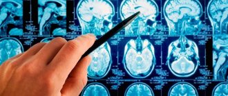
- Tomography. Research using special technical means detects tumors, degenerative processes in the brain, and other signs.
Agnosia is just a syndrome that can be detected at different stages of disease progression. Research will help determine the nature of the pathology and select effective methods for its elimination.
General information
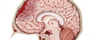
Gnosis translated from Greek means “knowledge.” It is a higher nervous function that ensures recognition of objects, phenomena, and one’s own body. Agnosia is a complex concept that includes all violations of the gnostic function. Gnosis disorders often accompany degenerative processes of the central nervous system and are observed in many organic brain lesions resulting from injuries, strokes, infectious and tumor diseases.
Classic agnosia is rarely diagnosed in young children, since their higher nervous activity is in the developmental stage and the differentiation of cortical centers is not complete. Gnosis disorders most often occur in children over 7 years of age and in adults. Women and men get sick equally often.
Agnosia
Impaired visual recognition (visual anosia)
11>
Gnostic disorders (agnosia) are a violation of the recognition of stimuli (objects of the surrounding world) related to one or another modality.
Visual agnosia
occur with damage to the secondary projection zones of the cortex (18 and 19 Brodmann areas), localized in the parieto-occipital region of the cerebral cortex. With visual agnosia, there is a disorder of perception and recognition of previously familiar objects while consciousness and intelligence are preserved. Mental pathology is described by the phrase: “It sees, but does not understand.” Elementary visual functions in gnostic disorders are preserved (visual acuity, visual fields, color perception are not impaired). Children with visual agnosia see objects, touch them, hear sounds, but cannot understand what they mean. Adult patients lose the ability to recognize even familiar stimuli perceived by the senses.
In the clinic of local brain lesions, different types of visual gnosis disorders are distinguished, which indicates a high functional differentiation of the visual cortex. The specific designation for visual agnosia depends on whether it refers to an object, space, movement, color, or symbol. Accordingly, the following types of visual agnosia are distinguished:
– object agnosia;
– agnosia for colors;
– agnosia for faces;
– finger agnosia.
Visual agnosia was first described by G. Munch in 1881, observing a dog with an affected occipital region, in which elementary visual functions were preserved (she saw surrounding objects = did not bump into them, but she “did not understand” their meaning). The term “agnosia” was proposed by S. Freud.
The following types of visual agnosia have diagnostic significance in neuropsychological practice: object, facial, optical-spatial, color, letter, color, simultaneous and symbolic.
Object visual agnosia is characterized by a violation of the recognition of individual objects and images or a violation of the integrity of the perception of an object with the possible recognition of its individual features or parts. It is based on defects in recognizing the shape and contours of an object. Most often, this disorder occurs with bilateral lesions of the temporo-occipital regions of the brain, but it can also be caused by unilateral lesions of the right or left hemispheres. Bilateral lesions cause severe disturbances in objective visual gnosis; patients do not recognize even simple images of everyday objects and confuse similar images.
The impossibility of visual identification of an object can manifest itself as a listing of individual fragments of an object or its image (fragmentation), or the isolation of only individual features of an object that are insufficient for its complete identification. For example, recognizing the image of “glasses” as a “bicycle”, since there are two circles connected by crossbars; identification of a “key” as a “knife” or “spoon” based on the selected features “metal” and “long”.
Object agnosia can have varying degrees of severity - from maximum (difficulty in recognizing real objects) to minimal (difficulty recognizing contour images in noisy conditions or when superimposed on each other). As a rule, the presence of extensive object agnosia indicates bilateral damage to the occipital region. Thus, with severe damage, the perception of real objects is impaired: the patient does not recognize previously familiar objects or can only describe their individual features. To facilitate identification, he tries to feel the object, and if it is food, then taste it. Often, to identify an object, the patient names randomly selected features by brute force and thus tries to “recognize” the object. A less severe gnostic visual disorder is manifested by the fact that the patient recognizes real objects, but does not recognize schematic images, inverted or superimposed drawings.
In mild cases, only the time of tachistoscopic recognition increases when a stimulus is presented in a dosed manner (normally, the time sufficient to recognize a stimulus is 0.01 sec; with visual gnostic disorders, the recognition time can increase to 1 sec or more).
With unilateral lesions of the occipital region of the brain, differences in the structure of visual object agnosia can be seen. Damage to the left hemisphere is manifested to a greater extent by a violation of the perception of objects by the type of listing of individual details. The pathological process in the right hemisphere leads to the virtual absence of the act of identification. In this case, the patient can evaluate the visually presented object according to its significant characteristics, answering the researcher’s questions about the relationship of this object to “living - inanimate”, “dangerous - non-dangerous”, “warm - cold”, “big - small”, etc.
Diagnosis of subject visual gnosis[5].
1. Recognition of depicted objects (methodology of A. R. Luria). 16 pictures are presented, which depict externally similar objects, for example, a snake - a belt, a cap - a plate, a table lamp - a mushroom, etc.
2. Recognition of objects with missing features (unfinished pictures).
3. Poppelreiter's test - identification of superimposed images.
4. Recognition of disguised (hidden) figures.
5. Folding pictures from parts.
6. Correlating an object with a form.
Facial agnosia (prosopagnosia) often develops with right-sided lesions of the secondary and tertiary projection areas of the visual cortex. Often prosopagnosia manifests itself as an independent gnostic defect. Face agnosia is a selective gnostic disorder, manifested in difficulties in recognizing familiar faces. In some cases, with a gross manifestation of the defect, patients do not recognize their loved ones, photographs from a family album, cannot imagine or describe a familiar face, evaluate people by random signs (moles, hairstyles, etc.), as well as by voice, gestures. In rare cases, patients with such disorders find it difficult to evaluate facial expressions expressing a particular emotion, and also see distorted grimaces. Agnosia on faces
caused by a lesion in the temporo-parietal-occipital regions of the right hemisphere.
| Prosopognosia is characterized by difficulty or inability to distinguish familiar faces |
Manifestations of prosopagnosia vary in severity. Thus, a mild degree is characterized by difficulty distinguishing familiar faces and recognizing facial expressions. In severe cases, the patient does not recognize previously familiar people, does not identify their gender, age, and uses auxiliary signs for identification: voice, gestures, gait, etc. In some cases, he does not even recognize himself in the mirror. Violations of moderate severity are characterized by difficulty in identification from a photograph or schematic image. The patient cannot combine the front and profile.
There are apperceptive and associative facial agnosia. Apperceptive
characterized by a violation of gender and age identification.
With associative
prosopognosia, the analysis and synthesis of perceptual information is available (the patient recognizes gender, age, determines the degree of similarity of faces), but associative connections are impaired. Thus, the patient cannot provide any information about the person he sees, while the emotional reaction to this person is preserved.
Diagnosis of facial gnosis [H4] [6] .
1. Recognition of familiar faces. Presentation of photographs of outstanding domestic writers.
2. Identification of unfamiliar persons. The subject is presented one by one with standards - three images of faces unfamiliar to him - and is asked, looking at them, to find identical ones in a set of 20 images. If completed successfully, the task becomes more complicated: the photograph is presented for 10 seconds, and the search is carried out from memory.
2. Emotional identification of portraits. The patient is presented with images of faces (photographs or diagrammatic images) that reflect certain emotional states: joy, pleasure, surprise, fear, anger, grief, contempt, pride - and is asked to identify them.
Optical-spatial agnosia is characterized by the fact that the patient, while retaining the recognition of individual objects, loses the ability to navigate in a familiar space. With less severe lesions, orientation in real space is preserved, but there is a loss of the ability to distinguish between “right and left,” impaired orientation in geographic maps, in the position of clock hands, and loss of the ability to mentally rotate an object by 90–180°.
Optical-spatial agnosia occurs when there is predominant damage to the superior parietal and parieto-occipital parts of the cortex of the left or right hemispheres of the brain, due to which the complex interaction of several analyzer systems (visual, auditory, tactile, vestibular) occurs. Optical-spatial agnosia is especially severe in cases of symmetrical bilateral lesions.
Features of the functioning of the visual areas of the brain make it possible to recognize such spatial characteristics of visual images as size, distance, direction, and relative position of objects. These characteristics of objects are learned in ontogenesis due to associative connections between various modalities: tactile, auditory and visual. Such disorders vary in form depending on the location of the lesion and, above all, on the side of the brain in which it is located. During the period of mastering the corresponding types of activity, the cause of such disorders can be not only lesions, but also various dysfunctions in the maturation of brain structures.
Left hemisphere lesions or dysfunctions lead to disturbances in spatial orientation activity. They are characterized by insufficient discrete-logical analysis of optical-spatial objects. In these cases the following are violated:
1) schematic ideas about the spatial relationships of objects of reality (inability to rotate a figure in space, orientate oneself in a geographical map, a clock, spatial games, etc.);
2) various types of constructive activities, drawing;
3) body diagrams (autotopagnosia);
4) naming and understanding words denoting spatial relationships: prepositions with spatial meaning on, in, under, above
etc., adverbs such as
far, on the side, below
, etc., on the basis of which agrammatism, characteristic of patients with semantic aphasia, often arises;
5) identification and naming of fingers (finger agnosia);
6) writing and reading (based on a defect in analytical-synthetic, simultaneous actions for sound-letter analysis of the composition of a word).
Right hemisphere lesions with optical-spatial agnosia and dysfunction are observed much more often than left hemisphere lesions. They cause a lack of holistic perception of the spatial situation:
1) simultaneous agnosia, in which there is an inability to assess the meaning of a plot picture due to the fragmentation of the perception of the spatial situation, although recognition of individual objects, as a rule, remains intact;
2) impaired recognition of a familiar spatial situation, inability to reproduce it from memory;
3) a violation of the body diagram (autotopagnosia), when orientation in the location of body parts is difficult because they are perceived as distorted in size, disproportionate.
The listed situations are especially pronounced on the contralateral (opposite to the lesion on the left half of the body).
As a consequence of a violation of the body diagram and weakening of visual control, difficulties arise in constructing movement in space, i.e. apraktoagnosia,
consisting in the disintegration of established everyday skills, for example, dressing (apraxia of dressing), the ability to draw, perform professional actions, etc.
The most striking and characteristic symptom of optical-spatial disorders that occur when the right hemisphere of the brain is damaged is unilateral spatial agnosia. With it, the phenomenon of ignoring the left half of space, as well as visual, auditory, and tactile stimuli emanating from the left half of space, appears.
| Optical-spatial agnosia is characterized by the patient losing the ability to navigate in a familiar space |
Diagnostics of optical-spatial gnosis [H5] [7] .
1. Draw a floor plan.
The study can begin with asking the patient to orient himself in the real space in which he is currently located (draw a plan of the department, tell how to get from this office to the exit; indicate which of the visible objects is closer or further).
2. Determination of parts of the world: the experimenter places a dot on the picture. The subject is asked to determine the direction of the world.
3. "Blind compasses." The figure shows only one part of the world, and not in a typical place. Task: identify the direction indicated by the arrow.
4. Line orientation test by A. Benton. The subject needs to be shown in the drawings which of the set of lines corresponds to the lines exhibited as stimulus material (they need to be correlated by angles of inclination). Instead of recognition, the option of sketching lines is possible.
5. Drawing figures
The experiment usually begins with asking the subject to draw a “house” and a “cube” on his own. In case of inadequacy of the drawing, it is proposed to copy the same object from the sample.
6. Drawing “House – tree – man.” When drawing in patients with optical-spatial agnosia, the overall scheme of the drawing may disintegrate, the spatial arrangement of parts of the drawing may be disturbed, and proportions may be disturbed.
7. Test with rotation of figures.
The subject is offered a form with rows of drawn squares, inside each of which there are one or two graphic marks located in a certain way in the space of the square. The leftmost square is a sample, in relation to which you need to mentally rotate the subsequent squares 90° clockwise and then select the one that fully matches the sample.
8. Braid cubes. The subject is offered a series of increasingly complex patterns of two-color patterns that must be laid out from the upper faces of the cubes. The cubes have two opposite faces painted red, two – white, and two more – diagonally divided into white and red triangles.
Agnosia for colors
or
color agnosia
, is caused by damage or dysfunction of the temporo-occipital regions of both the left, dominant, and right, subdominant hemispheres. Color perception is preserved, i.e. the patient distinguishes colors, names them correctly, but has difficulty correlating the color with a specific object: he cannot remember what color an orange, carrot is, or cannot name objects of a certain specific color. In addition, there is evidence that when the right hemisphere is damaged, difficulties may arise in differentiating mixed colors (brown, purple, orange, pastel colors). Color agnosia of the dominant type, like other types of visual agnosia, is characterized by a violation of abstractness and generalization in perception. Patients with color agnosia cannot select shades of color into a single color scheme - a selective disorder accompanied by forgetting the names of colors.
| With color agnosia, the patient experiences difficulty relating a color to a specific object. |
11>
Date added: 2017-05-02; views: 9058; ORDER A WORK WRITING
Find out more:
Treatment of agnosia
The development of agnosia is facilitated by brain damage that occurs as a result of the disease. An important nuance is that in right-handed people the disease develops when the left hemisphere is damaged, in left-handed people - vice versa. The main pathological changes that provoke the development of agnosia are:
- Alzheimer's disease.
- Circulatory problems, such as stroke.
- Encephalopathy.
- Tumor diseases of the brain.
- Traumatic brain injuries.
- Parkinson's disease.
In medicine, there is no single protocol for the treatment of agnosia, since it depends on the root cause of the disease, its type and degree of neglect. In parallel with the treatment of the main illness, work is carried out with a psychiatrist, speech therapist, and neuropsychologist. This is necessary in order to help a person adapt to life in the presence of such pathologies.
Agnosia is an unpleasant and dangerous condition that can threaten health and interfere with a normal, fulfilling life. If you seek help in a timely manner, you can get rid of symptoms and improve your condition. It is strictly forbidden to self-medicate, use traditional medicine or other non-traditional methods without first consulting a specialist.
This disease is quite multifaceted and therefore, before the doctor prescribes a specific treatment, he must organize a large number of examinations required to make an accurate diagnosis.
In order to recognize the causes of the pathology and select the correct treatment, a physical examination is performed, which helps to identify primary deviations in the functioning of certain types of sensitivity, because they can complicate the subsequent assessment of the patient’s health.
Scientists have repeatedly studied agnosia and currently there are quite conflicting opinions about how to organize its treatment, because each individual case requires an individual approach depending on the causes of the disease.
Experts have not developed a general treatment, but to achieve compensation for the pathology, the help of a speech therapist, as well as a specialist who decides on occupational therapy, is necessary.
The degree of recovery of the patient after the disease depends on the location of the lesions, their size and also on the age of the patient. As practice shows, recovery takes about three months, but treatment in general can last up to one year. Regardless of this, more attention is paid to the treatment of the underlying disease that provoked the formation of agnosia.
To date, there is no effective treatment for agnosia. We are talking about damage or lesions of the brain, so the main methods and manipulations are aimed at restoring these departments:
- Drugs are prescribed that improve blood circulation in the brain. Blood pressure is controlled.
- Surgeries are performed to remove tumors, ruptures, etc. from the brain. Without surgery, pills will not help in this case.
- Medicines that help restore neuropsychological functions.
The patient is constantly under consultation with a neuropsychologist.
Many doctors perceive this disease as ordinary amnesia. The patient simply needs to be re-taught the skills that were lost. If a person suffers from visual agnosia, then he is again taught shapes and colors, the relationship of objects in space, etc. If auditory agnosia has developed, then the person is taught sounds.
We are talking about damage that is difficult to restore with modern medicine. However, in some cases, such manipulations are effective and help patients adapt to life. The exception is somatoagnosia, which requires constant medical supervision.
If agnosia is a consequence of a mental illness, then treatment is aimed at eliminating this disease. Since brain diseases are not always completely cured, the restoration of its parts also becomes incomplete.
If agnosia is the result of abuse of toxic substances, then it is recommended to protect the patient from alcohol, poison, drugs and other substances. The body is cleansed of these substances, as well as taking medications that improve brain function.
go to top
Characterized by a disorder in determining the parameters of space. Here are the types of this violation:
- Agnosia of depth. A person cannot correctly localize objects in three spatial coordinates. Especially in depth. Reason: disturbances in the parieto-occipital region (middle sections, as a rule).
- Stereoscopic pathology. Inability to see a three-dimensional image, in other words. Reason: disruption of the left hemisphere.
- Spatial one-sided pathology. A person simply does not perceive what is happening on the left side of him. Cause: damage to the parietal lobe, contralateral side of the prolapse.
- Violation of topographic orientation. With this pathology, a person retains his memory, but loses the ability to navigate. He can get lost in his yard, get lost in the city where he has lived all his life. Cause: damage to the parieto-occipital region.
Having one of the pathologies is not just inconvenient, but dangerous. If, for example, a person is diagnosed with optical-spatial visual agnosia, the signs of which were discussed above, then he can easily get lost even in his own apartment. Therefore, the disease must be dealt with. But how?
Agnosia is a violation of various types of perception (tactile, auditory and visual) while maintaining consciousness and sensitivity. This condition is pathological and occurs due to damage to the cortex and subcortical structures of the brain.
Why is this happening? What causes agnosia? What types of pathology are there? What symptoms indicate its presence? And most importantly, how to deal with it? Well, this and much more will be discussed now.
Agnosia is a pathological condition characterized by a violation of various types of perception: visual, auditory, gustatory, olfactory, tactile. At the same time, the functions of the sensory system responsible for perceiving signals from the environment and internal environment remain normal. It occurs against the background of damage to the cortex and subcortical structures of the brain after injury, stroke, infections, degenerative diseases, and tumor processes.
Mental development disorders
30.11.2017
3.7 thousand
2.5 thousand
7 min.
Agnosia is a violation of the recognition of objects and stimuli of various modalities (visual, auditory, tactile and others).
This deviation occurs in children and adults due to the presence of certain pathological conditions of the brain. Treatment is carried out taking into account the underlying disease and the type of agnosia.
Correction of the patient's condition
Treatment is prescribed by a specialist after a thorough examination and based on the results obtained. Self-therapy in this situation is not effective and is contraindicated; it can lead to irreversible consequences of brain destruction.
To eliminate the disease, it is necessary to eliminate the underlying pathology that provoked this deviation.
In most cases, treatment lasts no more than three months - sufficient time to recover. But there are exceptions when therapy is carried out for six months to a year. A positive result depends on the patient’s age group, severity and nature of the lesion.
After the provoking cause has been eliminated, it is necessary to make some adjustments that are aimed at eliminating the disease and the disturbed state of the body. Therefore, experts recommend:
- attend speech therapy classes;
- psychotherapeutic sessions;
- study with teachers;
- spend more time on occupational therapy.
In case of agnosia, it is recommended to constantly monitor blood pressure and, if necessary, take medications that improve blood flow and nutrition to the brain (nootropics and antiplatelet agents).
If a patient has been diagnosed with a brain tumor, then surgery to remove the tumor is mandatory.
Anticholinesterase drugs are prescribed to improve neuropsychological function. At the same time, classes are held with a neuropsychologist.
Pathogenesis
The cerebral cortex has three main groups of associative fields that provide multi-level analysis of information entering the brain. Primary fields are connected to peripheral receptors and receive afferent impulses coming from them. Secondary association areas of the cortex are responsible for analyzing and summarizing information coming from primary fields.
Next, the information is transferred to tertiary fields, where higher synthesis and development of behavioral tasks are carried out. Dysfunction of secondary fields leads to disruption of this chain, which is clinically manifested by the loss of the ability to recognize external stimuli and perceive holistic images. In this case, the function of the analyzers (auditory, visual, etc.) is not impaired.
Why does agnosia occur?
Cognition disorders are a symptom of many diseases of the nervous system, accompanied by damage to the gray and nearby white matter of the brain, namely the secondary projection-associative zones. Damaging factors disrupt the normal relationships between neurons in these areas.
The most common causes of agnosia are the following pathologies:
- volumetric formations of the brain (cyst, tumor, vessel aneurysm, arteriovenous malformation, abscess). Disruption of the normal functioning of neurons occurs due to the growth of “plus” tissue, which puts pressure on healthy cells and leads to their destruction;
- circulatory disorders in the brain (ischemic or hemorrhagic stroke, subarachnoid hemorrhage, chronic encephalopathy, etc.), which results in the death of neurons in associative fields;
- traumatic brain injuries (most often brain contusions and injuries accompanied by the development of intrathecal hematomas) - damage to neurons occurs either at the time of injury or when post-traumatic complications occur;
- neuroinfections (bacterial, viral, fungal or prion etiology), which lead to the development of encephalitis. In this case, focal or diffuse damage to the cerebral cortex occurs;
- autoimmune processes (multiple sclerosis, post-vaccination encephalitis, etc.) - also lead to damage to cortical neurons and subcortical structures;
- neurodegenerative pathologies (Alzheimer's disease, Pick's disease, Parkinson's disease, Huntington's disease, etc.) - atrophic processes destroy the normal connections of neurons in the projection-associative fields.
Identification of agnosias
Despite the fact that agnosia is not a common pathology, its diagnosis should be carried out comprehensively. Most often, gnostic dysfunction is found in adults. However, cases of identifying symptoms of agnosia in a child are not uncommon (at a younger age, we are talking about a delay in the formation of gnosis centers; in puberty, true agnostic disorders can be diagnosed).
A patient with suspected cognitive impairment should be evaluated by a neurologist to identify focal neurological deficits. The presence of additional symptoms can help carry out topical diagnosis and identify the area of brain damage. The symptoms of true cognitive impairment and pseudoagnosia are similar.
To clarify the type of agnosia, a number of neuropsychological tests are carried out. It includes specially developed materials that allow the assessment of higher cortical functions in general and their individual manifestations in particular.
To assess the state of visual gnosis, the patient is asked to look at images of objects, people, animals, plants, and color schemes. Some pictures may be shaded or covered with a curved line (so-called noisy pictures). Additionally, the patient is asked to look at images of parts of an object; with their help, simultaneous agnosia can be identified.
When checking for the presence of pathology of acoustic gnostic functions, the patient is asked to close his eyes and the most common sounds are played (most often they clap their hands, let them listen to a ticking alarm clock, rattle keys).
To identify astereognosis, the doctor gives the patient an object, which he must feel with his eyes closed, and then determine what it is. Violations of the body diagram are established by interviewing the patient.
To clarify the degree of formation of gnostic functions in children, there are similar neuropsychological materials adapted for a child of a certain age.
Establishing diagnosis
This neurological disorder is not a common disease. Most often diagnosed before the age of 18.
Occurs as a result of many precipitating factors that affect each patient differently. In most cases, a comprehensive neurological examination is necessary to make a diagnosis.
The following research methods are used in diagnosis:
- magnetic resonance imaging;
- CT scan;
- neuropsychological and physical examinations.
Be sure to pay attention to symptoms and complaints, how long ago they appeared, and what could have provoked them. The specialist asks the patient to identify objects using different senses.
The neurological examination consists of the following:
- assessment of mental functions;
- assessment of visual functions;
- assessment of auditory functions.
A neuropsychological examination consists of the following: assessment of the patient’s condition using questionnaires and conversations. If necessary, the patient is referred for consultation to a psychiatrist.
Article on the topic: Signs of vitamin deficiency in children - how does a lack of vitamins C, A, D, K, E and group B manifest themselves?
Classification
Depending on the area of damage in clinical neurology, agnosia is classified into the following main groups:
- Visual – lack of recognition of objects and images while maintaining visual function. Develops with pathology of the occipital, posterior parietal parts of the cortex.
- Auditory - loss of the ability to recognize sounds and phonemes, and perceive speech. Occurs when the cortex of the superior temporal gyrus is damaged.
- Sensitive - impaired perception of one's own body and recognition of tactile sensations. Caused by dysfunction of the secondary fields of the parietal regions.
- Olfactory - a disorder of smell recognition. It is observed with damage to the mediobasal areas of the temporal lobe.
- Gustatory - the inability to identify taste sensations while maintaining the ability to perceive them. Associated with pathology of the same areas as olfactory agnosia.
Prognosis and prevention
It is impossible to prevent the occurrence of agnosia with one hundred percent probability. However, it is known that maintaining physical fitness and regularly training the mind by memorizing poetry or other similar materials, as well as performing thinking exercises, helps maintain the mental health of the body into old age.
If you don’t want to give up and are ready to really, and not in words, fight for your full and happy life, you may be interested in this article .
Diagnosis of the disease
The diagnosis of agnosia is made based on medical history (trauma, stroke, tumor) and the clinical picture of the disease. Special tests are also performed to determine the type of agnosia.
The patient is asked to identify simple objects using various senses. If the doctor suspects denial of half of space, then he asks the patient to identify paralyzed parts of his body, or objects in different parts of space.
Conducting a neuropsychological examination helps determine the presence of more complex types of agnosia.
Brain imaging methods (MRI or CT) are also used to establish the nature of central lesions (hemorrhage, infarction, massive intracranial process) and to identify areas of cortical atrophy.
To identify primary disorders of certain types of sensitivity, a physical examination is performed.
The examination is aimed at identifying agnosia and searching for its cause. Determining the clinical form of agnosia allows us to establish the localization of the pathological process in the brain. The main diagnostic methods are:
- Interview with the patient and his relatives. The purpose is to establish complaints, the onset of the disease, its connection with injury, infection, and cerebral hemodynamic disorders.
- Neurological examination. During the study of the neurological and mental status, along with agnosia, the neurologist identifies signs of intracranial hypertension, focal neurological deficits (paresis, sensory disorders, cranial nerve disorders, pathological reflexes, changes in the cognitive sphere) characteristic of the underlying disease.
- Consultation with a psychiatrist. Necessary to exclude mental disorders. Includes a pathopsychological examination and a study of personality structure.
- Tomographic studies. CT, MSCT, and MRI of the brain make it possible to visualize degenerative processes, tumors, inflammatory foci, areas of stroke, and traumatic injury.
Agnosia is only a syndrome; a syndromic diagnosis can occur at the initial stage of diagnosis. The result of the above studies should be the establishment of a complete diagnosis of the underlying disease, the clinical picture of which includes a disorder of gnosis.
To diagnose the disease, it is extremely important to consult a specialist - a neurologist. To make an accurate diagnosis, a number of diagnostic techniques and neurological studies are performed. The main goal of all research is to establish the root cause, the main disease that provoked brain damage.
Diagnostic methods used to establish a diagnosis:
- A thorough examination of the patient, collecting anamnesis and clarifying the presence of hereditary diseases. Most often, the study reveals tumor diseases, injuries, consequences of stroke and other diseases.
- Additionally, consultation with highly specialized specialists is carried out to exclude other causes of symptoms. As a rule, the help of an ophthalmologist, oncologist, cardiologist, psychiatrist and others is required.
- Carrying out various tests that help determine the degree of perception impairment in order to differentiate the disease from other possible ones.
- Carrying out computed tomography and magnetic tomography. Using these procedures, damaged areas of the brain can be identified.
Auditory agnosia occurs as a result of damage to the auditory analyzer. If the temporal part of the left hemisphere has been damaged, then a disorder of phonemic hearing occurs, characterized by a loss of the ability to distinguish speech sounds, which can lead to a disorder of speech itself in the form of sensory aphasia.
If the right hemisphere is damaged, the patient ceases to recognize absolutely all sounds and noises. If the anterior parts of the brain are affected, then all processes proceed with the preservation of the auditory and visual systems, but with a violation of the general perception and concept of the situation. Most often, this type of auditory agnosia is observed in mental illnesses.
Arrhythmia of auditory agnosia is characterized by the inability to understand and reproduce a specific rhythm. Pathology manifests itself when the right temple is affected.
A separate type of auditory agnosia can be identified as a process that manifests itself as a violation of understanding the intonation of other people’s speech. It also occurs with right temporal lesions.
Agnosia is a pathological condition that is formed as a result of damage to the cortex in the brain, as well as nearby subcortical structures. If the brain damage is asymmetrical, then unilateral agnosia may appear.
Facial agnosia usually develops due to damage to the right hemisphere of the brain, usually in the posterior or middle regions. The strength of the pathology manifestation varies, ranging from impaired recognition and memory of faces during experimental tests to the inability to recognize one’s own reflection in the mirror. Selective impairment of face memory may also develop.
The face, as a visual object, has a certain specificity compared to objects. The possibility of impaired perception of faces is confirmed by data on the difficulties of playing chess in people with damage to the right hemisphere of the brain.
In addition, the perception of a human face contains a certain individuality of the perceiver, who captures something of his own in the facial features, completely subjective even when examining portraits of famous personalities. The specificity of the person in question lies in its uniqueness, which expresses the individuality of the sample in relation to the original perceived by a person.
A special form of this pathology includes a violation of a person’s recognition of his own body. This process often occurs when a large part of the right hemisphere of the brain is affected. In this case, a person ceases to recognize his own limbs and may experience the presence of several arms or legs.
Tests and diagnostics
To diagnose agnosia, various types of neuropsychological tests are usually used:
- for example, Head's test, during which the patient needs to repeat the correct position of the hands, pose, gesture, or their mirror image;
- tests to determine the location and number of touches to the hands and face;
- Poppelreiter's test - for combining and highlighting individual 3, 4, 5 contours; in patients with visual agnosia, difficulties arise in their identification.
Tachistoscopic studies can also be carried out to determine the speed and accuracy of image perception and the level of attention shown at the same time. If other types of agnosia are suspected, patients may be asked to recognize objects by sound, shape, touch, name numbers and letters in various types of writing, and reproduce rhythm.
Symptoms of agnosia
The basic symptom of the condition is the inability to recognize perceived sensations while maintaining the ability to feel them. Simply put, the patient does not understand what he sees, hears, feels. Differentiated agnosia is often noted, caused by loss of function of a particular affected part of the cortex. Total agnosia accompanies pathological processes diffusely spreading in cerebral tissues.
Visual agnosia is manifested by confusion of objects, the inability to name the object in question, copy it, draw it from memory or a started drawing. When depicting an object, the patient draws only its parts. The visual form has many options: color, selective agnosia of faces (prosopagnosia), apperceptive - recognition of the characteristics of an object (shape, color, size is preserved), associative - the patient is able to describe the entire object, but cannot name it, simultaneous - inability to recognize a plot from several objects while maintaining recognition of each object separately, visual-spatial - a violation of the gnosis of the relative position of objects. A disorder in recognizing letters and symbols leads to dyslexia, dysgraphia, and loss of the ability to do arithmetic calculations.
Auditory agnosia with damage to the dominant hemisphere leads to partial or complete misunderstanding of speech (sensory aphasia). The patient perceives phonemes as meaningless noise. The condition is accompanied by compensatory verbosity with repetitions, insertion of random sounds and syllables. When writing, omissions and rearrangements may be noted. Reading saved. Damage to the subdominant hemisphere can lead to loss of musical hearing, the ability to recognize previously familiar sounds (the sound of rain, dog barking), and to understand the intonation features of speech.
Sensitive agnosia is characterized by a disorder of gnosis of stimuli perceived by pain, temperature, tactile, and proprioceptive receptors. Includes asteriognosis - the inability to identify an object by touch, spatial agnosia - impaired orientation in a familiar area, a hospital ward, one’s own apartment, somatognosis - a disorder in the sense of one’s own body (proportionality, size, presence of its individual parts). Common forms of somatognosia are finger agnosia - the patient is unable to name fingers or show the finger indicated by the doctor; autotopagnosia - the feeling of the absence of a separate part of the body; hemisomatoagnosia - the feeling of only half of one’s body; anosognosia - unawareness of the presence of a disease or a separate symptom (paresis, hearing loss, visual impairment).
Causes of agnosia
Agnostic deviations develop against the background of various pathological changes in the cerebral cortex during the process of ontogenesis, the existence of the organism from the embryonic state to death. In medicine, there are different causes of the disease; depending on the nature of agnosia, a diagnosis is made and treatment is prescribed.
Disorders of the circulatory system in the brain
The death of neurons containing information collected by the senses leads to its loss and reduction in cognitive function. This disease affects people who have experienced an ischemic or hemorrhagic stroke - spontaneous hemorrhage as a result of the destruction of blood vessels.
This form of the disease appears suddenly; depending on the location of the effusion, the occipital, temporal or frontal region is damaged. Timely localization of the site will increase the likelihood of future cure.
Hemorrhagic stroke, which led to an accelerated course of agnosia, is a consequence of the voluntary destruction of blood vessels due to high blood pressure. Symptoms increase quickly; when they are detected, it is necessary to accurately determine the location and cause of the pathology, and carry out treatment with possible surgical intervention.
Chronic cerebral ischemia
Progressive degradation of the cerebral circulatory system leads to the appearance of various types of dementia, including serious disorders. A secondary consequence of the disease can be an almost complete cessation of higher mental activity, in which the patient lives like a plant, unable to satisfy minimal needs on his own.
Description of symptoms:
- Disorders of various types of memory - the patient becomes forgetful, cannot remember the simplest things. To ensure that important information is always at hand, the patient is forced to create tips similar to school cheat sheets.
- Decreased stamina. A person always looks tired, he loses interest in the world around him and becomes apathetic, avoiding unnecessary movements.
- Pain and noise in the head, this manifestation intensifies over time.
- Poor orientation in the surrounding space.
- Sleep disturbance.
- Tearfulness and hot temper, replacing each other.
- Loss of ability to walk.
- Disorder of gnostic functions.
- Inability to focus on one object or express emotions.
And also against the background of loss of normal perception of reality, periodic dizziness and impaired swallowing function appear.
Brain tumors
With the development of cancer, loss of various cognitive functions is possible. Tumors disrupt the normal functioning of the organ, and depending on the location of the tumor, various consequences can occur. Neoplasms destroy neurons; when they grow rapidly, agnosia can progress rapidly in a short period of time.
Disturbances in the processes of recognition and touch during oncogenesis and the development of cancer pathologies are accompanied by:
- Left-sided or right-sided (depending on location) headaches, appearing most often in the morning or evening. The sensations may intensify from rotating the head and tensing the abdominal muscles. Painkillers do not work, but taking an upright position can relieve pain.
- Slowing brain activity. The auditory-speech function is impaired if the tumor has affected the lower parietal part of the brain, apraktoagnosia is observed - loss of the ability to perform habitual, long-learned actions.
- Involuntary muscle contractions in the legs or arms with dullness of tactile function. The next stage in the development of cancer is periodic loss of consciousness.
- Hallucinations, impaired coordination, unreasonable sensation of temperature - the patient thinks that the room is cold or hot.
Further damage to the brain by the tumor leads to irritability, hormonal imbalances and agnostic disorders - the patient suffers from the inability to write anything or remember the purpose of objects.
Traumatic brain injury
In pathopsychology, several disorders are identified that are a direct consequence of physical damage to the brain or cortical lobes. Neuronal destruction is observed immediately at the time of injury or as a consequence of post-traumatic processes.
The likelihood of restoration to a normal state depends on the severity of the injury and the presence/absence of secondary pathologies. If the damage is irreversible, the symptoms do not worsen. The patient, depending on the location of the injury, may lose certain cognitive abilities.
Encephalitis
A dangerous disease characterized by acute inflammation of brain tissue. It can be a consequence of a tick or other insect bite, acute flu, or complications after vaccination. There are several signs that, together with gnostic disorders, indicate the development of encephalitis:
- increase in temperature, sudden or gradual;
- characteristic fever starting at temperatures above 39 degrees;
- severe headache accompanied by speech impairment - it is difficult for a person to understand the semantic structure of sentences and put his wishes and thoughts into words;
- nausea and vomiting, after which there is no relief;
- seizures and development of epilepsy;
- disorders of consciousness in the form of drowsiness, lethargy; if unilateral inflammation spreads to the second hemisphere, the patient may fall into a coma;
- paresis of the limbs - gait disturbance, feet drooping when lifting the leg, a person cannot get up from a sitting position or keeps his head constantly tilted forward.
If such signs are detected, accompanied by a violation of verbal or taste function, you should contact a neurologist.
Degenerative diseases of the central nervous system
Pick's disease, Huntington's chorea and Alzheimer's disease - these diseases lead to serious damage to brain function.
More than 9,000 people have gotten rid of their psychological problems using this technique.
Each of them can be recognized by its characteristic features:
- Pick's disease. Progressive brain damage leading to complete cessation of higher mental activity.
- Alzheimer's disease. The first symptom is a tremor or tic of the left or right hand, accompanied by a gradual breakdown of short-term memory and dysfunction of the musculoskeletal system.
For these and other diseases, agnosia is a secondary manifestation that occurs due to irreversible destruction of nerve cells and brain tissue. Orientation in space and recognition of objects are preserved, but sometimes there is a loss of amodal meaning - the world gradually ceases to be recognizable to humans.
Autotopagnosia
And this is the second type of disorder related to somatoagnosia. A person’s presence can be recognized by his ignoring half of his body. And many patients simply do not recognize some of its individual parts or incorrectly assess their position in space.
This type of pathology includes:
- Hemisomatoagnosia. A person ignores half of the body, but its functions are partially preserved. The patient simply does not use them. He may “forget” that he has a left arm, leg, etc.
- Somatoparagnosia. The patient perceives the affected part of the body as foreign. He may seriously believe that there is someone else next to him, and it is he who owns his second leg, arm, etc. In severe cases, people feel the “separateness” of the body, as if it were sawn into two parts.
- Somatic allosthesia. The patient feels an increase in the number of his limbs. He may think that he has two or three left hands, for example.
- Autotopagnosia of posture. A person is not able to determine exactly what position his body parts are in. He really doesn't know whether his hand is down or up, whether he's sitting or lying down.
- Disorientation. With this pathology, the patient does not know such concepts as “right” and “left”. This is usually caused by damage to the left parietal lobe.
- Finger agnosia. Specific pathology. If it is present, a person is not able to show on his hand the same finger that the doctor shows on his hand.
Apractoagnosia is also a disorder of this type. This is a special case. People with this disorder have difficulty performing spatially oriented movements. It is difficult for them to make the bed (they place the bedspread crosswise rather than lengthwise), find their way into a room or ward, get their foot in a pant leg, or put on a T-shirt with the right side.
As in any other case, when diagnosing somatoagnosia, neuropsychological methods, samples and tests are used.
Treatment of pathology
Treatment for agnosia depends on the disease causing the disorder. Therapy includes conservative, neurosurgical and rehabilitation measures.
Neurosurgical treatment is indicated in cases of volumetric processes in the brain, massive hemorrhages and some types of traumatic brain injuries. But after this, drug therapy and rehabilitation are required.
Conservative treatment consists of the use of the following groups of drugs:
- vascular drugs (pentoxifylline, cytoflavin, nicotinic acid, vinpocetine) - acting on the microvasculature, improve perfusion in the brain;
- nootropics (piracetam, lucetam, thiocetam, picamilon) – regulate energy metabolism in neurons;
- metabolites (actovegin, cortexin, cerebrolysate) - normalize the nutrition of brain cells;
- neuroprotectors (ceraxon, pharmacoxon, gleacer, gliatilin, noocholine) – have a protective effect on neurons, help improve interneuron connections;
- antioxidants (mexipridol, neurox, mexiprim, mexicor) – limit the effect of free radicals on brain cells;
- vitamins (especially B vitamins: combilipen, compligamma B, milgamma) - contribute to the restoration of neurons, their axons, and improve synaptic transmission of impulses.
For encephalitis, according to the etiofactor, treatment is carried out with antibiotics, antiviral or antifungal agents.
The rehabilitation part of therapy includes:
- work with psychologists to understand and accept one’s defect, various types of psychotherapy (art therapy, animal therapy, cognitive behavioral therapy);
- occupational therapy – used to improve the patient’s functioning, promotes more complete socialization;
- classes with a speech therapist - most often needed for auditory and visual agnosia, when dyslexia and dysgraphia occur.
High-quality treatment of agnosia often requires a multidisciplinary approach and the participation of several highly specialized doctors (neurologist, neurosurgeon, psychiatrist, rehabilitation specialist, speech therapist, etc.).
Recovery and restoration of gnostic functions depends on the pathological process that caused agnosia, the age of the patient, and the timeliness of diagnosis and treatment of the disease. Young patients usually return to normal life after 3 to 9 months of intensive treatment and rehabilitation. Degenerative processes in the brain, unfortunately, cannot be reversed; Currently existing drugs only stop the progression of the disease.
Prevention of agnosia involves taking good care of your own health and consulting a doctor when the first signs of damage to the nervous system appear.
Types of visual agnosia
This classification is based on a violation of the perception of various types of objects or their signs. Each form of agnosia is associated with damage to certain brain structures. Clinical psychologists correlate disorders of visual gnosis with syndromes of lesions in the occipital and temporo-parietal regions of the brain [1].
It manifests itself in the inability to recognize and identify information that comes through the visual analyzer. There are the following types of agnosia of this type:
- Subject. The person does not recognize objects. He can describe some of their signs, but he is not able to say what exactly he sees. Cause: damage to the convexital surface of the left occipital region.
- Prosopagnosia. The person does not recognize the face. If his friend stands in front of him, he will distinguish his eyes, lips, nose, cheeks, hair, but will not understand who it is. In especially severe cases, people do not even recognize themselves in the mirror. Cause: damage to the lower occipital region of the right hemisphere.
- Colored. A person cannot select the same shades and determine how this or that color relates to any object. For example, he doesn’t understand that an orange is orange. Cause: damage to the occipital region of the left dominant hemisphere.
- Optical. A person cannot imagine an object and give it a characteristic (describe texture, shape, size, shade, etc.). He is asked to describe a cucumber, and he understands what exactly they are talking about, but he cannot talk about this vegetable. Cause: bilateral damage to the occipital-parietal region.
- Simultaneous. A person is capable of perceiving only one semantic unit. He sees only one object, regardless of its size. Cause: narrowing of the visual field, damage to the anterior part of the dominant occipital lobe.
- Optical-motor. A person cannot direct his gaze in the required direction or focus on a given object. It is even difficult for him to read, since the transition from one word to another is a real problem. Cause: bilateral damage to the occipital-parietal region.
With any visual agnosia, as can already be understood, the symptoms are very specific. A person suffering from this pathology has to do a lot of work to analyze what he sees.
Treatment
After a thorough diagnosis, it will be possible to determine methods to combat the disease.
Note!
Traditional medicine and other similar approaches are powerless against agnosia. If a certain person promises you a cure as a “servant of God”, offers dubious potions with alkaloids or occult rituals, be sure that you are dealing with scammers. You need to get treatment from professionals.
Conservative methods include:
- Use of vascular or thrombolytic drugs. Many of them can be purchased at pharmacies only with a prescription. They dilate blood vessels in the brain and change the tendency to form blood clots to normal levels.
- Use of vasoactive compounds. They increase the blood supply to tissues that have undergone the development of ischemic processes. Requires careful use.
- Consumption of neurometabolites, antioxidants. Glycine, pyritinol and other analogues increase resistance to hypoxia - oxygen starvation.
- Use of anticholinesterase compounds. Medicines stabilize cognitive functions.
- Encephalitis therapy. Provides for the implementation of measures aimed at curing encephalitis.
- Psychotherapy. Allows you to get rid of alcohol addiction, which can lead to the development of agnosia, and maintains the moral state of the patient.
- Speech therapy. An experienced teacher will help you cope with the manifestations of auditory agnosia.
Occupational therapy also helps to overcome the disease. By choosing an activity that is feasible for the patient, you can speed up recovery and restore his faith in himself.
Anosognosia
This is one of the types of somatoagnosia - a pathology in which a person does not recognize parts of his own body. This type of disease includes the following manifestations:
- Anosognosia hemiplegia. The person does not realize that he has unilateral paresis or paralysis, or denies it.
- Anosognosia of blindness. The patient denies or is unaware that he is not sighted. He perceives confabulatory images as real.
- Anosognosia aphasia. With this disorder, a person does not notice his speech errors at all, even if he speaks as unintelligibly as possible.
In simple terms, the term “anosognosia” refers to a lack of awareness of one’s illnesses and illnesses. A person not only denies them out of principle - he really sincerely believes that he is healthy.
What is typical for a person in such a state?
This neurological disease can only be diagnosed by a specialist based on the following symptoms:
- a broken landmark on the ground, the patient does not understand the map;
- the patient denies that he has abnormalities and defects, although there is a violation;
- the patient is not interested in the defect;
- the patient ceases to recognize objects by touch and sounds;
- the patient does not perceive his body;
- the ability to recognize faces, even close people, is impaired;
- the patient is unable to recognize complex images and also ignores much of what he sees in space.


