Paresis of the abducens and oculomotor nerve
Optic nerve palsy is a neurological disease that limits the movement of the muscles of the visual organs.
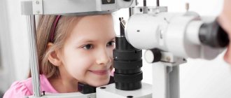
Normally, the muscles of the visual organs are innervated by three pairs of nerves. If one or more nerves are damaged, muscle function will be impaired. The damage to each nerve has its own distinctive features, which facilitates the diagnosis of the disease. But damage to several nerves at once makes it difficult to make a correct diagnosis; it requires time and a thorough examination. Paresis of the abducens and oculomotor nerves should be observed by a specialist. It is very important to see a doctor immediately after the first symptoms appear. In this case, there is a greater chance of getting rid of the disease as quickly as possible. At the Yusupov Hospital you can undergo high-quality diagnostics and treatment of optic nerve palsy. The hospital has neurology and rehabilitation departments where such diseases are successfully treated.
What is abducens nerve palsy?
Paresis of the abducens nerve of the left eye and the right eye occurs with the same frequency. In most cases, the disease affects one side, rarely both. Paresis of the abducens nerve can be suspected by characteristic features: the patient has difficulty turning his eye towards the affected nerve. Paresis of the abducens nerve impairs the function of the rectus lateralis muscle, and the patient cannot fully avert the eye to the side. The patient experiences diplopia when looking straight ahead, which intensifies when the eye is turned towards the affected side. Other symptoms of abducens nerve palsy include:
- forced position of the head (impaired vision leads to an attempt to adapt to the changes that have arisen, which provokes the occurrence of involuntary positions);
- uneven gait (also associated with visual impairment);
- loss of orientation;
- dizziness.
Why does abducens ophthalmic nerve paresis occur?
Paresis of the abducens ophthalmic nerve is a consequence of any disease in the head, central nervous system, or other organs and systems. Paresis of the abducens ophthalmic nerve can be caused by:
- infectious and inflammatory diseases of the brain (encephalitis, meningitis);
- infectious and inflammatory diseases such as syphilis, diphtheria, influenza, etc.;
- severe intoxication (alcohol, drugs, chemicals);
- botulism;
- stroke;
- heart attack in the head area;
- otolaryngological diseases;
- tumors in the brain;
- increased intracranial pressure;
- diabetes mellitus (in which the functioning and structure of blood vessels is disrupted);
- multiple sclerosis.
Causes
The causes of paralytic strabismus are usually caused by damage to:
- Nuclei of the motor nerves of the eye in the human brainstem.
- Trunks of the motor nerves of the eye;
- Muscles that provide movement of the eyeball.
- Tumors of the orbit of the eye.
- Injuries to the eyeball.
- Traumatic brain injury.
- Neuroinfections.
- Myasthenia.
- Multiple sclerosis, etc.
The disease can be congenital or acquired.
How does oculomotor nerve palsy manifest?
The most negatively affecting the functioning of the visual organs is paresis of the oculomotor nerve. Symptoms of the disease will be pronounced and will allow the doctor to suspect this pathology. The oculomotor nerve performs a very important function in eye movement. It provides the work of the superior, inferior and medial rectus muscles, the inferior oblique muscle, and the muscle responsible for raising the upper eyelid. The oculomotor nerve innervates the sphincter of the pupil, ensuring its response to light (constriction and dilation). Therefore, if the oculomotor nerve is damaged, performing many eye movements becomes impossible.
Patients experience double vision, the pupil does not respond to light, ptosis develops, difficulty opening and closing the eye, and difficulty moving the eye.
Rarely, only the oculomotor nerve is affected. Usually the condition is accompanied by disruption of the abducens, trigeminal and lateral nerves. The pathology occurs against the background of diabetes mellitus, arterial hypertension, brain cancer, microinfarctions of the head vessels, and strokes.
Paresis of the oculomotor nerve and abducens nerve: treatment in Moscow
The main method of treatment for paresis of the oculomotor and abducens nerve is the elimination of the disease that caused it. The Yusupov Hospital provides comprehensive treatment for this pathology, which helps eliminate the underlying disease and its consequences. Before prescribing therapy, the patient undergoes a thorough examination, which will help identify the underlying disease and the extent of nerve damage. At the Yusupov Hospital, diagnostics are performed using the latest high-precision equipment, which allows us to determine the cause of the disease even in the most difficult cases. After making a diagnosis and determining the condition of the patient’s body, the doctor draws up the most optimal treatment strategy.
Complex treatment of paresis of the oculomotor and abducens nerve will include drug therapy (drugs are selected depending on the type of underlying disease) and rehabilitation. The course of physiotherapy and rehabilitation is carried out in a specialized center at the Yusupov Hospital, where experienced specialists work in the field of restoring lost functions. Without a course of rehabilitation, paresis of the oculomotor and abducens nerves may resolve within 2-3 months after getting rid of the underlying disease. A rehabilitation course at the Yusupov Hospital allows you to speed up the process of restoring lost functions, contributes to the effective elimination of the consequences of the disease, the patient’s speedy recovery and return to a full life.
On topic: What did you eat for muscles?
You can make an appointment with neurologists, rehabilitation specialists, physiotherapists and other clinic specialists, get information about the work of the neurology clinic, rehabilitation, or clarify other questions of interest by calling the Yusupov Hospital.
Source
Treatment of convergent strabismus in the MGK
At the Moscow Eye Clinic, patients with convergent strabismus are provided with a wide range of diagnostic and therapeutic procedures. Diagnostics are performed by qualified specialists with unique clinical experience. When prescribing treatment, a strictly individual approach is used. The clinic has an extensive methodological and hardware base for the treatment of such pathologies. These techniques are suitable for both children and adults:
- Infrared laser therapy (exposure to infrared radiation on the organ of vision at close range), the procedures of which make it possible to improve the hemodynamics of eye tissue and relieve spasm of accommodation.
- Laser therapy sessions designed to improve spatial vision and accommodation functions.
- Exercises on the device “Synoptophore Sinf-1”. This device is the main one for stimulating vision, necessary for strabismus and other types of binocular vision disorders. It allows you to carry out sets of special exercises aimed at merging images, which makes it possible to train vision impaired by strabismus. In addition, such training significantly increases visual reserves and improves eye mobility.
- Treatment with the Spekl-M device, using two types of laser, which is indicated for clouding of optical media, amblyopia and strabismus of various origins.
- Especially for young patients with strabismus, MGK offers training with the Rainbow BZR-1 kit. This kit, in a playful way, helps restore central fixation and other functions of binocular vision.
Doctor of Medical Sciences Svetlana Gavrilovna Chernysheva deals with the problems of all types of strabismus at the Moscow Eye Clinic. Svetlana Gavrilovna is an ophthalmic surgeon, professor, the best specialist in Moscow in the field of diagnosing binocular oculomotor pathology in adults and children, as well as its functional and surgical treatment. Proficient in various methods of surgical treatment of all types of strabismus.
You can find out the cost of a particular procedure or make an appointment at the Moscow Eye Clinic by calling 8 8 (499) 322-36-36 (daily from 9:00 to 21:00) or using the online registration form.
Yakovleva Yulia Valerievna
Causes of paresis of the eye muscles in children
A person is able to move the eyeball thanks to 3 cranial nerve pathways. The main role in this process is played by the oculomotor nerve, which goes under number 3. It is responsible for the movements of the eyelid and eye, as well as its reaction to exposure to light. The abducens nerve controls the abduction of the eyeball, and the trochlear nerve turns it outward and downward. These nerve pathways are numbered 6 and 4. Any disturbances in the innervation of the extraocular muscles affect the ability to see fully. Among such disorders, paresis of the eye muscles in children can be identified, since, in addition to the main causes that are characteristic of adults, they often exhibit congenital anomalies. Treatment is usually carried out using a whole range of procedures; in especially severe cases, surgery is used.
Causes and symptoms
Abducens nerve palsy manifests itself as isolated paralysis. In this case, a person cannot fully avert his eyes and a double image of one object appears (diplopia). This phenomenon occurs due to a violation of the innervation of the lateral muscle, for which the abducens nerve is responsible. Similar symptoms are characteristic of diseases of the orbit, so you should undergo detailed diagnostics to make a diagnosis.
The abducens nerve is damaged due to the following factors:
- Aneurysm;
- Damage to the carotid artery;
- Traumatic brain injuries;
- Infectious diseases;
- Oncological diseases;
- Microinfarctions and strokes;
- Pathologies of the nervous system;
- Multiple sclerosis.
The abducens nerve in children is also injured due to the above factors. However, other reasons are also common for children:
- Gradenigo syndrome;
- Mobius syndrome;
- Duane's syndrome.
Damage to the trochlear nerve causes partial paralysis of the eye and the person has a double image in an oblique or vertical plane. This symptom intensifies when the eye is lowered down, so people suffering from this pathology often walk with their heads tilted to the healthy side to reduce the manifestation of diplopia. During diagnosis, myasthenia gravis (autoimmune pathology of nerve and muscle tissue) and diseases of the orbit should be excluded.
Damage to the trochlear nerve occurs in virtually the same way as to the abducens nerve, but in this case the main cause is trauma and micro-strokes. Oncological pathologies rarely affect this nerve pathway.
Oculomotor nerve palsy usually occurs in conjunction with disruptions of the facial, abducens, and trochlear nerve pathways. A separate form of pathology appears extremely rarely. This nerve is damaged primarily due to an aneurysm. It arises on the posterior communicating artery and gradually compresses the nervous tissue.
The nerve can be damaged by a growing tumor, as well as manifestations of stroke and multiple sclerosis. In most cases, such factors affect the nucleus of the nerve tract and the posterior longitudinal fasciculus. Sometimes neuropathy of the oculomotor nerve, caused by the above reasons, manifests itself in the form of bilateral drooping eyelids (ptosis). In more rare cases, paresis of the superior rectus muscle of the eye is observed. It is localized on the reverse side of the main site of damage.
According to statistics, the oculomotor nerve is often damaged due to a microinfarction. It can occur due to vascular pathologies, such as diabetes and hypertension. Such diseases usually do not immediately lead to disruptions in cerebral circulation and they should be predominantly in an advanced state. Neuritis of this nerve does not affect the reaction of the pupil to light, but in rare cases it is slightly weakened. A microinfarction occurs near the cavernous sinus or in the area of the interpeduncular fossa. It takes about 3 months for the oculomotor nerve to recover after suffering a disorder.
You should consult a doctor if you notice several symptoms characteristic of paresis of the eye muscles, especially when it comes to children. Among the common manifestations of optic neuropathy, the most basic can be identified:
- Diplopia;
- drooping eyelid;
- Strabismus;
- Decreased pupil reaction to light;
- Inability to turn the eyeball inward;
- Loss of the ability to quickly look at objects located at different distances from each other;
- Protrusion of the eye.
On the subject: How to lose weight if you have a lot of muscles
Accommodation
In pediatric ophthalmological practice, an accommodation study may be required to recognize conditions such as paralysis, paresis or spasm of accommodation, as well as to resolve the issue of rational optical vision correction for ametropia.
Determining the performance of the ciliary muscle makes it possible to identify children who have an increased risk of myopia, and in the presence of myopia, it is more correct to justify treatment tactics. Accommodation studies using the methods described below can usually be carried out in children 5-6 years of age and older. In order to judge the state of absolute accommodation, the nearest and further points of clear vision of each eye are determined. It is more convenient to conduct research using special devices - proximeters. In the absence of a proxymeter, use a regular millimeter ruler and a screen with a test object - a Landolt ring for visual acuity of 0.7-0.8 at a distance of 33 cm. The screen can be fixed to the ruler using a slider.
Determination of the nearest point of clear vision (pp) is carried out as follows. The light source is installed behind the patient, above his head. The end of the ruler with zero division is slightly rested on the outer edge of the orbit on the side of the eye being examined. The screen with the object is placed at a distance of 2-3 cm from the eye, and then gradually moved away. In this case, the screen should be in the frontal plane, and the ruler should be in a direction parallel to the visual axis. As soon as the subject is able to indicate the direction of the break in the optotype, the screen is stopped and the distance from it to the eye is measured using a ruler, i.e., the position of the nearest point of clear vision.
Usually the study is carried out 2-3 times and the average value of the indicator is calculated. To convert to diopters, you need to divide 100 by the resulting distance in centimeters. For example, if the nearest point is located at a distance of 8 cm from the eye, then its dynamic refraction in this position will be equal to 12.5 diopters. To speed up the calculation, use a table for converting linear distances into diopter values (Table 16).
Table 16. Position of the nearest point of clear vision
To determine the position of the further point of clear vision (pr), a reducing lens is used, artificially bringing this point closer to the eye. For myopia of 2.0 diopters or more, reduction is not required; for weaker myopia and emmetropic refraction, a +3.0 diopter lens is used; for farsightedness, the lens power should be 3.0 diopters greater than the degree of farsightedness.
The study under reduction conditions is carried out in the same way as when determining the nearest point of clear vision, with the only difference that the screen with the object is placed at a distance of approximately 60 cm from the eye and it is moved towards the eye, and not from the eye. The position of the reduced further point of clear vision is determined using a ruler at the moment when the subject can already indicate the direction of the break in the optotype. The position of this point is determined in diopters and the power of the refractive lens is subtracted from the resulting value, resulting in the diopter value of the true further point of clear vision. In practice, this indicator can be taken as the static refraction of the eye. The closer the pr is to the eye, the stronger the refraction.
Knowing the pp and pp in diopters, you can determine the volume of absolute accommodation using the following formula:
A = pp - pp
The volume of accommodation changes throughout the day:
the smallest volume is observed at 8-10 o'clock, the largest at 16-18 o'clock. It should be no less than the lower limit of the norm, which for children aged 6-7 years is 7.0 diopters, 8 - 10 years - 8.0 diopters, at 11-20 years old it is equal to 10.0 diopters. With accommodation paresis, the volume of absolute accommodation is significantly reduced due to the removal of the nearest point of clear vision from the eye. The more pronounced the accommodation paresis, the further this point is from the eye. With accommodation paralysis, it coincides with the position of the further point of clear vision. With a spasm of accommodation, the volume of absolute accommodation also decreases significantly, but due to the approach of the further point of clear vision to the eye. With a pronounced spasm, this point may coincide with the nearest point of clear vision.
To determine the performance of the ciliary muscle during visual work at close range, a study is carried out using an eye ergograph
or determine the reserve of relative accommodation. An ergograph developed at the Moscow Research Institute of Eye Diseases named after. Helmholtz, consists of a drum with two support posts on which guides are fixed.
A carriage connected to a writing pen moves along the guides; on it is a flashlight with a matte screen, on which an optotype is applied (Landolt ring, corresponding to visual acuity 0.7 for a distance of 33 cm). The screen is illuminated from behind by light bulbs. By changing the voltage on them, you can create different levels of illumination of the test field. The drum and carriage are driven by electric motors. The ergograph is installed on a special table with a built-in control panel. The remote control has a carriage travel switch and two toggle switches for turning on the flashlight lamp and the drum drive.
The subject's head is placed on a special stand. Using the carriage travel switch, the screen with the optotype is brought closer to his eye until the subject can no longer distinguish the cut in the Landolt ring (the ring that looked like the letter “C” becomes similar to the letter “O”). As soon as this happens, the observer changes the movement of the carriage and moves the screen away until the examinee can again distinguish the cut in the ring; then the carriage is brought closer again, moved away, etc. The frequency of movements is 19-20 per minute. The duration of the study is determined by its objectives (usually 5-10 minutes).
All screen movements are recorded on the paper drum tape. The results of the study are a curve of a sequential row of teeth. On each tooth there are two points, one of which is located on the lower, the other on the upper tip. The first corresponds to the position of the nearest point of clear vision - the maximum, but short-term tension of the ciliary muscle, the second, the so-called near point of clear vision - not the maximum, but long-term sustained tension.
There are four types of curves:
Type I (Fig. 54) - the nearest and closest point of clear vision throughout either maintain their position or approach the eye of the subject;
Type II a - wave-like ergogram with small waves and minor changes in amplitude; Type II b - wave-like ergogram with large waves and significant changes in amplitude; III a type - a stepped ergogram with a gradual “removal” of the nearest and near points of clear vision from the eye and their abrupt approach to the eye; III b type - the ergographic curve gradually “removes” from the eyes until the moment when recording becomes impossible; Type IV - atypical forms of curves that require a separate description. Type I ergograms characterize the normal performance of the ciliary muscle, the rest - an increasing weakening of the accommodative ability of the eye for near. Atypical forms of ergograms are extremely rare.
Rice. 54. Types of ergograms. Explanation in the text.
When analyzing an ergogram, it should be taken into account that its amplitude depends on the distance from the eye to which the closest and nearest points are located: the closer they are to the eye, the smaller the amplitude. In this regard, with high myopia, the teeth have a height of 1-2 mm, but when measured in diopters, their amplitude is significant (up to 1.0 diopters or more).
ergometry technique has been proposed
, allowing you to study the accommodative ability not only for near, but also for distance. This is achieved by transferring the further point of clear vision to a final distance (25-50 cm) using a reducing lens.
In order to get an idea of the performance of the ciliary muscle, you can use a simple technique for determining the reserve of relative accommodation, the results of which closely correlate with the data obtained from ergography. The relative accommodation reserve is determined in the following way. A patient wearing glasses that completely correct ametropia is asked to read text No. 4 (corresponding to visual acuity 0.7) of Sivtsev’s table for near from a distance of 33 cm.
If he can read this text, then they begin to add negative spherical lenses symmetrically to both eyes, increasing their power by 5.0 diopters. Using the strongest negative lens, with which reading is still possible, the amount of relative accommodation reserve is determined. This study can be conveniently carried out using a device for determining visual acuity at close range POZB-1. Approximate norms for the reserve of relative accommodation: at 7-10 years - 3.0 diopters, at 11-13 - 4.0 diopters, at 14-20 - 5.0 diopters.
Diagnostics
It is easiest to recognize the lesion, since this pathological process is characterized by drooping eyelids, dilated pupils and abnormal deviations of the eyes. Based on such signs, making a diagnosis will not be a problem, but often they are combined with each other in various combinations, so the doctor suspects the secondary nature of the disease. To differentiate paresis of the eye muscles from other possible ailments, the ophthalmologist will need to prescribe an examination, which consists of the following procedures:
- Fundus examination;
- Determination of visual acuity and degree of mobility of the eyeball;
- Light reflex test;
- Angiography (to identify vascular pathologies);
- Magnetic resonance imaging (checks brain tissue for abnormalities).
Sometimes a consultation with a neurologist may be required. If it was not possible to determine the cause of the pathology, then the patient should be registered with a doctor and periodically examined. To prevent the condition from getting worse, your doctor may recommend special sets of exercises and other treatment methods.
Symptoms
Paralytic strabismus is usually accompanied by the following symptoms:
- Deviation of the affected eye from the point of fixation;
- Double vision, which is eliminated by closing one of the eyes;
- Different primary and secondary angles of strabismus.
- Pathological positioning of the head (corrective gesture that helps reduce double vision, which can cause secondary torticollis).
Examination reveals deviation of one eyeball from its natural position. In this case, the patient cannot “move” the affected eye to any direction.
Course of therapy
Treatment methods for paresis of the eye muscles in children are not particularly different from those in adults. However, it must be borne in mind that most congenital anomalies are corrected through surgery. If the operation is successfully performed, the extraocular muscles are partially or completely restored. If the problem is compression of the nerve pathway, then the main task is to eliminate the cause.
After eliminating the factor influencing the development of muscle paresis, treatment is adjusted towards restoring blood flow and damaged nerve fibers. For this purpose, exercises that strengthen the oculomotor muscles are often used. They serve as the basis for the treatment of minor injuries and are a good preventive measure. In severe cases of the disease, therapeutic exercises well complement the main course of therapy.
Drug therapy for paresis may include the following:
- Vitamin complexes;
- Preparations for strengthening the extraocular muscles and restoring their innervation;
- Eye drops;
- Medicines that improve blood circulation;
- Corrective glasses and bandages.
The pathology can be treated with medications only according to the regimen prescribed by the doctor, so as not to aggravate its course and not to impair vision, especially if the child is sick. It is recommended to combine drug therapy with other methods, namely:
- Steriopictures. By watching them, the extraocular muscles are trained and blood flow improves. The nerve tissues that innervate the muscles of the eye are extremely tense during the procedure, due to this the lost innervation is restored. The procedure must be carried out under the supervision of a specialist so as not to cause complications;
- Electropheresis. This physiotherapeutic procedure is carried out with a 1.5% solution of Neuromidin. The duration of one session of electropheresis usually does not exceed 20 minutes, and it acts directly on the synapses (junctions) of the muscle and nervous tissue of the eyeball. After a course of such therapy, the patient’s severity of paresis decreases and the innervation of the eye muscles improves.
It is impossible to eliminate some causes of paresis in children, for example, congenital anomalies, without surgical intervention. Their duration and degree of risk depend on the type of operation and the factor that influenced the development of the pathology. In the case of severe damage to the optic nerves, it will not be possible to completely eliminate the problem, but there will be a chance to save the child’s vision.
Due to paresis of the extraocular muscles, many complications develop, such as strabismus, ptosis, etc. In children, this pathological process is often a consequence of congenital anomalies. It may not appear immediately, but only over time. This is why it is important to see an ophthalmologist and other doctors, especially in the first years of a child’s life.
Source
Treatment
Special exercises and physical therapy
Gymnastics for the eyes is carried out in combination with exercises and massage for other parts of the back, as well as the head and temples. It allows you to restore the muscle frame.
Gymnastics for the eyes strengthens the ciliary muscle, allowing the eye to adapt faster. Special exercises help adjust the level of curvature of the lens.
Physiotherapy is carried out after relief of the signs and causes of the disease. It allows drugs to better penetrate tissues.
Eye drops
Medicines relieve spasms. Prescribed:
- Cyclomed;
- Atropine.
Medicines dilate the pupil. Sometimes Irifrin is used, which relaxes the ciliary muscle as much as possible.
The course of treatment and dosage is determined by an ophthalmologist after a comprehensive examination of the patient’s eyes and determining the cause. Sometimes it is enough for a patient to use eye drops for 1 week to obtain a positive effect.
Treatment is carried out in combination with exercises and physiotherapy, since eye drops provide short-term results.
Surgical intervention
The operation is performed if the disease has led to myopia, which is difficult to correct with glasses or lenses and the patient constantly has to use vision correction devices.