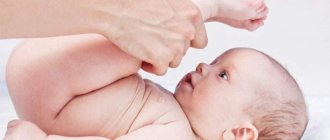
Most of the population have at least once experienced muscle twitching in different parts of the body. This symptom often frightens people, and searching for answers on the Internet is a dead end, because often such queries lead to sites describing serious diseases of the nervous system.
For example, fasciculations in polyneuropathy and ALS are characteristic manifestations indicating disturbances in the functioning of the motor neuron. Muscle twitching can also appear in people who do not have neurological diseases. Specialists at the Neurology Clinic of the Yusupov Hospital conduct high-precision examinations and create individual therapy programs for patients who experience tremors and fasciculations.
Causes
Muscle twitching may be a symptom of a pathological condition that can lead to death or a significant deterioration in quality of life:
- Oncology of the brain and spinal cord;
- Neuroinfections;
- Damage to peripheral nerves;
- Intoxication;
- Motor neuron diseases.
However, in most cases, muscle fasciculations are benign in nature and do not cause harm to life. Muscle fasciculations are uncontrolled, visible short-term muscle contractions. They are usually a sign of heavy exercise, fatigue or stress. Fasciculations can be observed on different parts of the body, including the eyelids, cheeks, lips and tongue.
Make an appointment
Source of energy for reduction
To contract a muscle, the energy of ATP hydrolysis is used, but the muscle cell has an extremely efficient system for regenerating the ATP supply, so that in a relaxed and working muscle the ATP content is approximately equal. The enzyme phosphocreatine kinase catalyzes the reaction between ADP and creatine phosphate, the products of which are ATP and creatine. Creatine phosphate contains more stored energy than ATP. Thanks to this mechanism, during a burst of activity in the muscle cell, the content of creatine phosphate drops, but the amount of the universal energy source - ATP - does not change. Mechanisms for regenerating ATP reserves may vary depending on the partial pressure of oxygen in surrounding tissues (see Anaerobic Organisms).
Muscle fibrillations and fasciculations
Fibrillation is a spontaneous contraction of a specific muscle fiber that does not lead to contraction of the entire muscle. Fibrillation is not noticeable through the skin (unlike fasciculation). At the Yusupov Hospital, neurologists using highly sensitive equipment carry out differential diagnostics between fibrillation and muscle fasciculation.
During an electromyographic study, fibrillations are detected as an asynchronous irregular short (1-5 ms) low-voltage (20-300 μV) discharge in the muscle (usually occurring up to 30 discharges in 1 s).
The reasons are:
- Trauma to the body or axon of a motor neuron;
- Primary muscle disorders (myopathy).
Muscle fasciculations on electromyography are detected as a longer discharge (from 8 to 20 ms) and a higher voltage (2-6 mV). Fasciculations occur at irregular intervals (frequency 1-50/min).
In a healthy person, fasciculations occur during the period of muscle relaxation: during, before or after sleep, at rest. With active movements, such twitching disappears, but resumes again at rest. Unlike fibrillations, they are not accompanied by muscle weakness, loss of sensitivity and atrophy in muscle tissue.
Muscle fasciculation is the cause of:
- Motor neuron diseases (ALS - amyotrophic lateral sclerosis, progressive spinal amyotrophy and others);
- Benign fasciculations;
- Painful muscle fasciculations syndrome;
- Traumatic damage or compression of the root and peripheral nerve;
- Facial myokymia (brain tumor, multiple sclerosis);
- Isaacs Syndrome (Isaacs Syndrome);
- Some forms of facial hemispasm;
- Post-paralytic contracture of facial muscles;
- Iatrogenic fasciculations.
Neuromuscular response to strength training[edit | edit code]
Source: "Training Programs"
, scientific ed.
Author:
Prof. Dr. Tudor Bompa, 2021
Muscle structure[edit | edit code]
Muscle
is a complex structure responsible for movement.
Muscles are composed of sarcomeres, which contain a specific combination of fibrillar proteins - myosin (thick filaments) and actin (thin filaments), which play an important role in muscle contractions. Thus, a sarcomere
is a contractile element of muscle fiber, consisting of myosin and actin protein filaments.
In addition, the ability of a muscle to contract and exert force depends specifically on its type, cross-sectional area, and the length and number of fibers within the muscle. Fiber number is determined by genetics and cannot be influenced by training; however, training can change other variables. For example, the number and thickness of myosin filaments increases through rigorous training with maximum strength load. Increasing the thickness of muscle filaments increases muscle size and contraction strength.
The human body is made up of different types of muscle fibers, divided into groups, and each group belongs to one motor unit. Overall, our body contains thousands of motor units containing tens of thousands of muscle fibers. Each motor unit contains hundreds or thousands of muscle fibers that remain at rest until they need to act. The motor unit controls a set of fibers and directs their actions according to the “all or nothing” law. This law means that when a motor unit is stimulated, the impulse sent to its muscle fibers either spreads completely - thus irritating the entire set of fibers - or does not spread at all.
Different motor units respond to different training loads. For example, performing a bench press at 60% RM recruits a specific set of motor units, whereas larger motor units wait for a higher load. Since the sequential recruitment of motor units depends on the load, it is necessary to develop special programs to activate and adapt the main groups of motor units and muscle fibers that play a dominant role in the chosen sport. For example, training for short distance sprints and track and field events (such as shot put) should use heavy loads to help develop the strength needed to optimize speed and explosiveness.
Muscle fibers perform different biochemical (metabolic) functions; More specifically, some are better physiologically adapted to work under anaerobic conditions, while others work better under aerobic conditions. Fibers that use oxygen to produce energy are called aerobic, type I, red, or slow fiber. Fibers that do not require oxygen are called anaerobic, type II, white, or fast. Fast-twitch muscle fibers, in turn, are divided into subtypes IIA and IIX (sometimes called IIB, although type IIB is almost never found in humans [1]).
Slow and fast fibers exist in approximately equal proportions. However, depending on their function, some muscle groups (eg, hamstrings, biceps) contain more fast-twitch fibers, while others (eg, soleus) contain more slow-twitch fibers. In Table 2.1 we compare the characteristics of fast and slow fibers.
Comparison of fast and slow fibers
| SLOW FIBERS | FAST FIBER |
| Red, type I, aerobic | White, type II, anaerobic |
| • Slowly get tired • Nerve cell is smaller - innervates from 10 to 180 muscle fibers • Develop long, sustained contractions. • Used to develop endurance • Activated during low- and high-intensity activities | • Get tired quickly • Large nerve cell - innervates 300 to 500 (or more) muscle fibers • Develop short, strong contractions • Used to develop speed and strength • Activated only during high-intensity activities |
Training can influence these characteristics. Danish scientists Andersen and Aagaard [2][3][4][5][6] in their studies show that with volume loads or lactate-inducing training, IIX fibers acquire the characteristics of IIA fibers. That is, the myosin-rich chain of these fibers becomes slower and copes with lactate activity more effectively. These changes can be reversed by reducing the training load (tapering), causing IIX fibers to return to the original characteristics of the fastest fibers[3]. Strength training also increases fiber size, which produces more force.
The contraction of a fast motor unit is faster and more powerful than the contraction of a slow motor unit. As a result, the proportion of fast-twitch fibers tends to be higher in successful speed-strength athletes, but they also fatigue more quickly. Athletes with higher slow-twitch fibers, on the other hand, tend to excel in endurance sports because they can perform low-intensity activities for longer periods of time.
Activation of muscle fibers occurs according to the principle of magnitude, also known as Henneman's principle[7], according to which motor units and muscle fibers are activated from smaller to larger. Activation always begins with slow fibers. During low- to moderate-intensity exercise, the slow-twitch fibers are activated and do most of the work. Under heavy load, the slow fibers contract first, then the fast fibers are involved in the process. During repetitions to failure with a moderate load, fast-twitch fiber motor units are gradually activated to maintain force production while previously recruited motor units become fatigued (see Figure 1).
rice. 1. Sequential activation of motor units in a set of exercises until concentric failure
There may be differences in the distribution of muscle fiber types among athletes participating in different sports. This is illustrated in Fig. 2 and 2.3, representing the total percentage of fast and slow muscle fibers in athletes in selected sports. For example, the significant difference between sprinters and marathon runners makes it clear that success in some sports is at least partially determined by the genetic composition of the athlete's muscle fibers.
rice. 2. Distribution of fiber types in men in different sports. Note the predominance of slow fibers in athletes involved in aerobic sports and the predominance of fast fibers in athletes involved in speed-strength sports
Therefore, the peak power produced by athletes also relates to the distribution of fiber types—the higher the percentage of fast-twitch fibers, the more power the athlete produces. The percentage of fast-twitch fibers also relates to speed: the faster an athlete's speed, the higher the percentage of fast-twitch fibers they have. Such people make excellent sprinters and jumpers, and such natural talent should be channeled into speed-strength sports. Trying to train them for, say, distance running is a waste of talent; in such disciplines they will have only average success, while they can become excellent sprinters, baseball players or football players (the list of speed-strength sports does not end there).
rice. 3. Distribution of fiber types in women in different sports
Benign crampy-fasciculation syndrome
The most common symptom of benign crampy-fasciculation syndrome is muscle twitching in the thighs (including fasciculations in the buttocks) or lower legs (fasciculations in the calf muscle).
People may also experience:
- Numbness;
- Convulsions;
- Itching and trembling;
- Sudden contractions or involuntary muscle spasms;
- Symptoms of anxiety (headache, shortness of breath, sensation of a lump in the throat).
Fasciculations in the legs and buttocks can last from a few seconds to several years (appearing at regular intervals, for example, two to four times a day).
When conducting research, there was no decrease in the speed of nerve excitation, as well as a decrease in sensitivity or changes in reflexes.
Most often, symptoms can be caused by excessive exercise (in the gym, lifting weights), stress and anxiety.
Video illustrations:
Video illustration of the lever hypothesisMyosin clings to the actin filament, grabs onto it, turns the tail, then unhooks and returns the tail to its previous position. Video by Kenneth Holmes. |
Video illustration of the “Roll and lock” hypothesisMyosin first looks for a suitable position on the actin filament, then weakly connects to it, then adheres tightly - in this case, the entire head turns and develops some force, and only then the tail turns. Video by Mary Reedy. |
Fasciculations in ALS
Amyotrophic lateral sclerosis (ALS) is an incurable disease, with the progression of which the patient develops muscle atrophy and weakness. Fasciculations in ALS are manifested by spontaneous, rapid and irregular muscle contractions. Fasciculations in this disease occur at the initial stage due to damage to central or peripheral motor neurons. In different forms of ALS, the frequency of fasciculations is different, but their development is accompanied by muscle weakness.
Patients who develop tongue fasciculation in ALS experience difficulty swallowing, increased salivation, and voice changes. When assessing the degree of muscle damage using electromyography, specialists identify positive fasciculations potentials, indicating chronic denervation.
Specialists at the diagnostic center of the Yusupov Hospital have the necessary skills and equipment to identify neurological diseases and their signs. Thus, during the examination, the frequency of fasciculations in ALS is determined.
Make an appointment
Physiological properties
Like all vertebrates, the human body has three most important properties of skeletal muscle fibers:
- contractility - contraction and change in tension during excitation;
- conductivity - movement of potential throughout the fiber;
- excitability is a response to a stimulus by changing the membrane potential and ionic permeability.
The muscles become excited and begin to contract from nerve impulses coming from the centers. But in artificial conditions, electrical stimulation is used. The muscle can then be stimulated directly (direct stimulation) or through the nerve innervating the muscle (indirect stimulation).
Muscle twitching due to polyneuropathy
Polyneuropathy is characterized by multiple lesions of the peripheral nerves, in which continuous fasciculations, muscle weakness, trembling of the fingers, and impaired respiratory function occur.
Fasciculations can be caused by increased excitability of nerve endings. During fasciculation, ion channels can regulate the intensity of the manifestations of involuntary twitching. As sodium channels are blocked, fasciculations decrease with medication.
Neurologists, when treating patients with induced fasciculations, use drugs registered in the Russian Federation. A complement to drug therapy for neurological diseases is physical therapy, physiotherapy and massages performed by specialists at the Yusupov Hospital Rehabilitation Clinic.
Innervation
This process in striated muscle fibers is realized through nerve fibers, namely the axons of motor neurons in the spinal cord and brain stem. One motor neuron innervates several muscle fibers. The complex with a motor neuron and innervated muscle fibers is called a neuromotor unit (NME), or motor unit (MU). The average number of fibers that one motor neuron innervates characterizes the size of the muscle MU, and the inverse value is called innervation density. The latter is large in those muscles where movements are small and “subtle” (eyes, fingers, tongue). On the contrary, its small value will be in muscles with “rough” movements (for example, the torso).
Innervation can be single or multiple. In the first case, it is realized by compact motor endings. This is usually characteristic of large motor neurons. Muscle fibers (called physical or fast muscle fibers in this case) generate action potentials (APs) that are propagated to them.
Multiple innervations occur, for example, in the external eye muscles. No action potential is generated here, since there are no electrically excitable sodium channels in the membrane. In them, depolarization spreads throughout the fiber from the synaptic endings. This is necessary in order to activate the mechanism of muscle contraction. The process here does not happen as quickly as in the first case. That's why it's called slow.
Diagnostics
A neurologist is involved in the diagnosis and treatment of fasciculations, as well as their accompanying pathologies. If you constantly experience muscle spasms-twitching, then you need to consult a doctor to find out the cause of their occurrence.
The neurology department of the Yusupov Hospital employs real specialists in their field who have been studying the pathology of the nervous system for many years. At your appointment, you will get answers to your questions, and doctors will help you determine whether leg muscle fasciculations (fasciculations in the calves) are benign muscle twitches or are they a symptom of a serious neurological disease.
Based on the symptoms, a neurologist may prescribe a certain type of examination for diagnostic purposes:
- Electromyogram (EMG);
- Electroneuromyography (ENMG);
- Neuroimaging;
- Blood analysis.
Slow-twitch (red) muscle fibers
Slow-twitch muscle fibers have a low contraction rate. Hence the name - slow.
If the load is not heavy, you need to work slowly but for a long time, the brain “pulls” the red strings. Another name for slow muscle fibers is red.
In order for muscles to work for a long time, they need a lot of oxygen. Oxygen is supplied to muscle tissue by blood capillaries. There are a lot of capillaries in slow muscle fibers. That's why they are red. Along with oxygen, blood brings “fuel”.
Where do red fibers get their energy from?
Man is a biological machine. To make a car move, you need gasoline. For muscles to work, the ATP molecule is needed.
The body “squeezes” the ATP molecule from fats and carbohydrates. How food turns into sports fuel is a different story. Read more in the topic - metabolism through the eyes of an athlete .
Here I will remind you the main thing. It takes time and oxygen to get ATP from fat. The body uses the energy obtained in this way for prolonged, low-intensity exercise. For example, walking or long-distance cross-country.
It is for this type of work that the body has adapted red muscle fibers. Here's air and food for you, work for your health.
When you need to do a sprinter's jerk or lift a weight, the brain's neurons activate the “white guard.”
Prevention and treatment
Muscle fasciculations are often a symptom of one or another neurological pathology. This is why it is so important to first detect the disease that led to the occurrence of muscle twitching, and then begin to treat the disease.
In some situations, it is not possible to find a pathology, in which case the cause of fasciculations is probably a lack of trace elements such as magnesium and potassium). At the Yusupov Hospital, you will be given a diet that will include foods that contain the missing elements.
Benign fascicular twitches are usually not harmful and do not require treatment. It is enough just to wait for them to disappear on their own.
Fasciculations that occur after active physical activity (most often after running or lifting weights) are most susceptible to poorly trained people who lead a passive lifestyle. They may notice involuntary twitching after the first workout. That is why it is better to start classes with light exercises.
Hypothermia is also considered one of the causes of spasm, so you should protect your body to avoid benign fasciculations. Hypothermia of the body occurs not only in the cold season, but also as a result of wearing clothes out of season.
In the hot season, this can happen when swimming in ponds, so in order to avoid unpleasant consequences, you must follow the rules of behavior on the water. Excessive drinking of alcoholic beverages can also cause uncontrollable muscle twitching.
Relaxation activities are used in the fight against fatigue and other neurological disorders. Such activities completely eliminate the consequences of stressful situations.
Make an appointment
The mechanism of muscle contractions[edit | edit code]
As we described earlier, muscle contractions
occur as a result of a chain of events involving protein filaments - myosin and actin. Myosin filaments contain crossbridges—tiny bridges that extend laterally toward the actin filaments. The excitation leading to contraction stimulates the entire fiber, creating chemical changes that allow actin filaments to connect to myosin cross bridges. The binding of myosin to actin via cross-bridges releases energy, which causes the cross-bridges to rotate, thereby pulling up or performing a sliding motion linking the myosin filaments to the actin filaments. This sliding motion causes muscle contraction, which produces force.
To visualize this differently, imagine a rowing boat. The paddles are myosin filaments, and the waters are actin filaments. When the oars hit the water, the boat is pulled forward with force - and the more oars in the water, the greater the physical strength of the rower, the greater the force generated. Increasing the number and thickness of myosin filaments similarly increases force production.
The sliding filament theory described earlier provides insight into how muscles work to produce force. This theory includes mechanisms that promote efficient muscle contractions. For example, elastic energy release and reflex adaptation play a key role in optimizing athletic performance, but such adaptation only occurs when the correct stimulation occurs during training. For example, an athlete's ability to use stored energy to jump higher or throw a shot further is optimized through explosive movements like those used in plyometric training. However, muscle components—such as elastic components (including tendons, muscle fibers, and cross bridges)—cannot transport energy efficiently unless the athlete strengthens parallel elastic components (such as ligaments) and collagen structures (which provide stability and protect from injuries). If the body is to withstand the forces and impacts an athlete is exposed to in order to optimize the elastic qualities of the muscles, anatomical adaptation must precede strength training.
Reflex
is an involuntary muscle contraction caused by an external stimulus[8]. The two main components of reflex control are the muscle spindles and the neurotendon spindle. Muscle spindles respond to the magnitude and rate of muscle stretch[9], while the neurotendon spindle (which is located at the junction of muscle fibers with tendon bundles[8]) responds to muscle tension. When a muscle develops a high degree of tension or stretch, the muscle spindles and neurotendon spindle involuntarily relax the muscle to protect it from damage and injury.
When these inhibitory reactions are suppressed, athletic performance increases. The only way to achieve this is to adapt the body to a higher degree of tension, which increases the threshold for activation of reflexes. This adaptation can be achieved through strength training using progressively heavier loads (up to 90 percent of your rep max or even higher), thereby forcing the neuromuscular system to tolerate higher stress by continually recruiting more fast-twitch fibers. Fast-twitch fibers produce more protein, which helps increase strength.
All sports movements are performed according to a motor model called the stretch-shorten cycle and is characterized by three main types of contraction: eccentric (lengthening), isometric (static position) and concentric (shortening). For example, a volleyball player who quickly squats and immediately jumps up to block an attacking blow has completed the entire stretch-contraction cycle. The same applies to the athlete who lowers the barbell to his chest and quickly performs an explosive movement by extending his arms. To fully take advantage of the physiological qualities of the stretch-shorten cycle, the muscle must quickly move from lengthening to shortening [10] (Schmidtble-icher, 1992).
Muscle potential is optimized when all the complex factors that influence the stretch-shorten cycle are activated. Their influence can only be used to improve athletic performance when the neuromuscular system is strategically stimulated in the correct sequence. It is to achieve this goal that periodization of strength training bases the planning of stages on the physiological basis of the chosen sport. After compiling an ergogenic profile (assessing the contribution of energy systems) of the chosen sport, it is necessary to plan the stages of training step by step in order to transfer positive neuromuscular adaptation to practical indicators of human activity. Thus, understanding applied human physiology and establishing a goal at the end of each phase helps coaches and athletes integrate physiological principles into specific sport training.
Let's repeat:
The musculoskeletal system of the body is a combination of bones attached to each other by ligaments at the joints. The muscles that cross these joints provide the force to move the body. However, skeletal muscles do not contract independently of each other. The movements performed around a joint are produced by several muscles, each of which has a specific role, as mentioned above.
Agonists—or synergists—are muscles that interact with each other to perform a movement. In most cases, especially if we are talking about a skilled and experienced athlete, the antagonist muscles relax, making movement easier. Because the interaction of the agonist and antagonist muscle groups directly influences athletic movements, improper interaction between these groups can result in jerky or stiff movement. Therefore, the smoothness of muscle contraction can be improved by focusing on relaxing the antagonists.
For this reason, simultaneous contraction (simultaneous activation of agonist and antagonist muscles to stabilize the joint) is recommended only in the early stages of rehabilitation after injury. A healthy athlete, especially if he is involved in strength sports, does not need to perform exercises (for example, on an unstable surface) that cause simultaneous contractions. For example, one of the main characteristics of elite sprinters is very low myoelectric activity of antagonist muscles in each phase of the stride cycle[11].
Primary muscles are primarily responsible for joint action, which is part of volumetric force movement or technical ability. For example, during elbow flexion (biceps curl), the primary muscle is the biceps muscle, while the triceps muscle (triceps) acts as an antagonist and must be relaxed to allow unimpeded action. In addition to this, stabilizers, or anchors (usually smaller muscles), contract isometrically to anchor the bone so that the primary muscles have a strong base from which to begin pulling. Muscles of other limbs can also take part in this, acting as stabilizers, allowing the primary muscles to perform the necessary movements. For example, when a judoka pulls an opponent toward him by holding his judogi, the muscles of his back, legs, and abdomen contract isometrically to provide a stable base for the action of the elbow flexors (biceps), shoulder extensors (rear deltoids), and scapular adductors and depressors (trapezius). and latissimus dorsi).
Medicines
Fibrillation and fasciculations occur in neurological diseases. In their treatment, medications are used that eliminate fasciculations, and the membrane of muscle cells is exposed. To treat neurological diseases and their manifestations, you should consult a neurologist; self-medication is unacceptable, as it can provoke progression of the disease.
Pantogam during fasciculations reduces the excitability of the nervous system due to the content of hydroxybutyric acid. This remedy is indicated for people experiencing mental or nervous fatigue, which may result in involuntary muscle twitching.
If a patient is diagnosed with ALS, dementia, or Parkinson's disease, fasciculations may appear early in the pathology in one or more areas. High-precision diagnostics carried out at the Yusupov Hospital make it possible to detect minor neurological disorders.
What is a muscle
A muscle is an organ of the body consisting of muscle tissue. Each muscle has a specific, unique form and function. All types of muscles are divided into three groups.
Types of muscle tissue
Smooth muscles are responsible for the functioning of internal organs. Part of the intestines, bladder, stomach, and cardiovascular system.
Cardiac muscle – provides blood circulation and is found only in the heart.
Striated (skeletal) muscles - form the human musculature. Let's consider this type in detail.
Structure of skeletal muscle
The muscle is attached to the skeleton through the tendon ends (tendons). The middle part of the skeletal muscle is called the belly.
The belly of a muscle consists of muscle fibers. Muscle fiber looks like a long thread. These threads are combined into bundles.
Each muscle bundle performs a specific function.
Providing assistance at the Yusupov Hospital
The main task of neurologists at the Yusupov Hospital is to provide high-quality medical care. Therapeutic measures carried out at the Yusupov Hospital for patients who have fasciculations due to osteochondrosis, ALS and other disorders help improve the quality of life of patients.
Neurologists at the Yusupov Hospital draw patients' attention to the fact that twitching can occur not only with neurological diseases, but also with intoxication, overwork and exhaustion of the body. If fasciculations occur during myeloradiculoischemia and other diseases, they require immediate treatment.
If muscle fasciculations bring you concern, and you have doubts about the cause of their occurrence, then make an appointment with a neurologist. For consultation please call.
Make an appointment
Roll and lock: turn and lock
According to the lever hypothesis, in muscle contraction there is only one moment of force generation - when the myosin tail (lever) turns. However, some data from radiography and tomography of muscles not only do not agree with this theory, but indicate that there is still some incomprehensible moment in muscle contraction that the lever hypothesis does not explain. Therefore, a group of researchers led by A.K. Tsaturyan proposed a theory of muscle contraction called “Roll and lock” (see: Michael A. Ferenczi et al., 2005. The “Roll and Lock” Mechanism of Force Generation in Muscle). According to this theory, myosin heads sit on actin even before ATP hydrolysis, and they sit not in a slender and organized manner, but at random. On the myosin head there is a long protruding domain - a “probe” - which “gropes” for a suitable (acidic and negatively charged) part of the actin filament and sticks to it - as necessary, at the first angle that comes along. However, as soon as ATP hydrolysis occurs, myosin changes its conformation, the heads rotate at the desired angle and firmly and clearly, like a key with a lock, adhere to the actin filament, and phosphate is released from the myosin pocket. And only after this the lever turns. In other words, the model turns out to be two-stage: at the first stage, the myosin head firmly and clearly clings to the actin and at the same time rotates slightly, and at the second, the lever turns, and the force that will then lead to muscle movement is generated at both of these stages.
In addition to X-ray structural and tomographic data, which are in very good agreement with the “Roll and lock” theory, there are several indirect, but very beautiful evidence of its correctness. For example, it is known that during muscle contraction, if the muscle does not change its length, only a little more than 40% of the myosin heads sit on actin, and the rest dangle unattached to anything. However, when a contracted muscle is forcibly stretched (for example, this happens when running, when a person lands on a tense muscle), the stiffness of the muscle increases sharply due to the fact that almost all the free myosin heads are sharply coupled to the actin filament. However, judging by the X-ray diffraction data, they do not interlock “tightly”, like a key with a lock, but simply haphazardly. This can be explained using the “Roll and lock” theory. ATP hydrolysis stops when a muscle is stretched (this is understandable: what is the point of wasting ATP if the work is not done by the muscle
, but
above the muscle
), and all myosin heads go into a state of “active actin search” - their protruding probe searches for the actin filament, feels for a suitable place on it and adheres to it - not firmly, not like a key with a lock, but haphazardly . However, in order to increase the stiffness of the muscle (and thereby protect the bones from fracture), this is enough.
Involuntary contraction of the abdominal muscles
Many have encountered the problem of abdominal muscle spasm; this occurs due to the fact that the functioning of the digestive tract is disrupted. And these failures happen if there is poor nutrition, bad habits, lack of a healthy lifestyle, stress and prolonged depressive states; the main root cause is pathologies of internal organs. Spasm can cause destructive processes in the kidneys, liver, pancreas, and genitourinary system.
The central way to eliminate these unpleasant manifestations is a balanced diet, giving up bad habits, a proper daily routine, and moderate physical activity.
It is necessary to completely refuse strong alcoholic drinks, fatty, fried foods, flour products and unhealthy foods (chips, carbonated water, chocolate bars, fast food), food should be moderately seasoned with spices, too spicy, hot or cold food is harmful to the gastrointestinal tract. intestinal tract, irritates mucous membranes.
Doctor: Olga Shishkina ✓ Article checked by doctor
Prevention of this pathological condition
There are a number of conditions that must be observed, even if after treatment it seems that the problem is behind you.
Everything can start again. To prevent this from happening, you need to:
- Eat properly and balanced to provide the body with enough microelements and vitamins. The absence or lack of them can also be the cause of various muscle spasms.
- Eliminate bad habits. Smoking, alcohol and coffee in large quantities lead to changes in blood vessels, and this is related to the problem under consideration.
- To avoid cramps in the calf muscles, you should wear comfortable shoes to avoid the feeling of tightness.
- Establish a gentle daily routine, allowing sufficient time for sleep.
- Try to worry less, learn to control negative emotions, try to create a favorable microclimate in the family and work (study) team.
- Listen to the signals that the body gives, do not ignore alarm bells, and do not put off going to the doctor if you suspect a deterioration in your condition.
If the necessary conditions are met, then with the help of a specialist and efforts, the problem can be cured or minimized.
The main thing is to take care of yourself and not consciously refuse medical care.
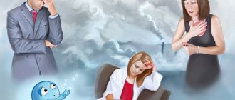
Muscles twitch all over the body (causes)
The condition, which causes muscles throughout the body to twitch, can be a medical emergency. This is how tonic convulsions begin with dehydration (hypovolemic shock), extensive blood loss and an emergency drop in blood pressure. Naturally, the reasons why muscles twitch throughout the body are not always so critical.
Such conditions must be preceded by certain events, for example, injury, or long-term diarrhea with vomiting. If there were no extraordinary events, but chaotic muscle twitching is observed throughout the body, then most likely there is a violation of innervation. They can be local or generalized.
Local problems are often observed when nerve impulse conduction is disrupted due to osteochondrosis, carpal tunnel syndrome, deformation of large joints, etc. Generalized seizures occur when the structure of the autonomic and central nervous systems is damaged. This may be the consequences of a transient disorder of cerebral circulation, spasms of cerebral blood vessels, discirculatory neuropathy.
Please note that very often the causes of twitching muscles throughout the body are hidden in the cervical spine. This is vertebral artery syndrome, in which due to deformation of the passages for cerebral blood vessels, their compression begins. Blood flow decreases, the posterior structures of the brain (namely, they are responsible for regulating the activity of the autonomic nervous system) suffer from impaired trophism. Clonic muscle twitching is the first characteristic sign of this disease.
Kinds
The nature of muscle contractions depends on the order of contractions and the frequency of nerve stimuli occurring.
Single
In the case of direct irritation of a muscle or through the nerve that innervates it, a single muscle contraction occurs.
There are 3 phases in this mechanism:
- latent period, from the beginning of the impulse to the muscle response, during which the action potential (AP) develops;
- period of muscle contraction or shortening;
- period of relaxation.
Muscle excitability in this case changes in accordance with certain phases. But in life, a person does not receive single impulses, but follow each other in series, at certain intervals. The muscle responds to these impulses with a prolonged contraction.
Tetanic
When ongoing muscle contraction is maintained continuously without relaxation, it is called tetanic contraction. Muscles can shorten, lengthen, or remain constant in length.
In this case, the movement is stimulated by multiple impulses with a fairly high frequency. An impulse coinciding with the relaxation phase contributes to the appearance of a whole series of successive single contractile movements.
If the frequency of impulses increases, then the relaxation phase of the previous cycle may combine with a new incoming impulse.
In this case, they speak of serrated tetanus, in which a prolonged contraction occurs, interrupted by periods of incomplete relaxation. Another type of tetanic contraction is smooth tetanus, when prolonged muscle contraction is not interrupted by periods of relaxation.
Milder tetanic contraction is normal and occurs as a normal physiological phenomenon during activities such as lifting heavy objects. In severe cases, during inflammatory and infectious conditions, tonic spasms of symmetrical muscle groups occur.
Transformations
Physiologically, a person is a biochemical engine, because food is a fuel that is broken down into its components: glucose (the smallest building blocks of carbohydrates), amino acids and fats. The mechanism of muscle contraction occurs through the use of glucose and energy provided by ATP.
Skeletal muscle transforms chemical energy into mechanical energy. Thus, the process of muscle contraction occurs under the influence of a 2-stage molecular transformation: electromechanical and chemomechanical.
Electrochemical
All living cells have membranes with proteins embedded in them. The membrane serves as a diffusion barrier and insulator for the movement of positively and negatively charged ions.
Neurons and muscle cells control the movement of charged ions through their membranes by selectively opening and closing ion channels. In this way, electrical currents and electrical signals are generated. Although the currents created by ions moving through channel proteins are very small, they form the basis of both neuronal signaling and muscle contraction.
Nerve impulses are electrically excitable, meaning they are capable of generating an action potential (AP), a special type of electrical signal that travels across the cell membrane as a wave that quickly and accurately transmits the signal over long distances.
This first stage of muscle contraction occurs due to the excitation of the muscle fiber membrane along which the action potential circulates. This property is called excitability when AP generates neurons and muscle cells.
Chemomechanical
Contractile muscle work occurs due to the energy released during the oxidation of carbohydrates or lipids.
In the process of a mechanochemical reaction, a gradual transformation of chemical energy into mechanical energy occurs:
- first, the process of binding troponin with Ca2+ ions occurs;
- the next step is the interaction of the myosin heavy chain head with actin;
- actin and myosin slide relative to each other,