Essence and basic principles
The Epley maneuver or exercises are a special maneuver used to treat benign paroxysmal positional vertigo (BPPV).
This procedure is often carried out in a physiotherapy office, but it can also be done at home after a specialist shows all the basics of the technique. This maneuver was developed by Dr. John Epley, after whom it was named. The first time it was talked about was back in the 80s of the last century.
The Epley maneuver is a series of exercises that help relieve symptoms of dizziness. Numerous studies have proven that this technique is the simplest, safest and most effective in the treatment of dizziness caused by the deposition of calcium salts in the inner canal of the ear.
All these exercises do not eliminate the presence of otoliths, but only change their location. Such manipulations cause them to move to other areas of the inner ear that do not cause dizziness.
The essence of the technique is to place your head at an angle at which gravity helps relieve unpleasant symptoms. Tilt of the head helps remove crystals from the semicircular ear canals. This means that they stop displacing fluid, which is what causes nausea and dizziness in a person.
As a result, it turns out that this maneuver relieves all symptoms, but you need to do the set of exercises several times. The thing is that the first time the crystals can move a small distance.
It is worth remembering that the Epley maneuver requires special concentration from a person, all actions must be precise, no sudden movements.
There are a number of rules that must be followed:
- classes should take place in a well-lit room;
- there should not be any objects with sharp corners, carpet paths, or objects that could cause a person to fall near the patient;
- It is better to perform the exercise on a soft surface; a couch, yoga mat, or thick-pile carpet are suitable for this;
- the clothes a person wears should be loose, not restricting movements, so that he can move easily and freely;
- shoes on feet should be without heels;
- while performing exercises, someone must be present next to the person; it is unknown how his body will react to the exercise; someone needs to provide backup;
- if a person feels unwell or has symptoms of other diseases, then therapy is postponed until the person feels better;
- Even after the Epley maneuver is successfully performed, when the results sought are obtained, you will need to visit the doctor several more times to prevent a relapse.
Experts who recommend the Epley maneuver to their patients for symptoms such as dizziness note that after the first use of a set of exercises, serious changes are observed. The symptoms go away, but only if you follow all the prescribed recommendations for several days after them.
Treatment
The choice of treatment for this disease depends on the type of damaged canal. In modern medicine, vestibular gymnastics and changing the position of otoliths are used to eliminate symptoms. Special therapeutic techniques will help alleviate the condition of BPPV. Physician-recommended physical therapy for benign paroxysmal positional vertigo reduces the severity of attacks. Treatment should take into account the location of the otolith crystal in the semicircular tubules of the inner ear. The positional Epley maneuver is the most studied method and controls seizures in posterior and lateral pathologies. The essence of treatment is to change the position of the otoliths. The fixed crystal dissolves, thereby contributing to the disappearance of symptoms.
Indications for use
The Epley maneuver is prescribed by a doctor only after examination, examination and an accurate diagnosis. It is also worth remembering that these exercises can only be recommended for stable, non-progressive illness.
Among these pathologies:
- period after a stroke;
- benign positional paroxysmal vertigo;
- violation of movement coordination;
- osteochondrosis;
- recovery period after injury to the spine or brain;
- ear diseases;
- infectious pathologies.
Gymnastics does not affect muscle function in any way. Its main goal is to teach a person to control attention, so regular use does not cause any unwanted manifestations.
Diagnostic measures
To make a definitive diagnosis of benign paroxysmal positional vertigo, specially developed Dix-Hallpike functional diagnostic tests are used.
To carry out this test, the doctor lays the patient down on the bed, then takes the head with both hands and rotates it in front to the sides around then, holding the head, lays it on the bed. After the exercise, the doctor should ask how the patient is feeling.
Objectively, a patient has nystagmus that is directed toward the floor, to the side or to the top; this depends on the direct localization of the pathological process in the semicircular canals of the inner ear.
In case of a negative effect, the exercise should be repeated a few minutes after rest. Sometimes it happens that after conducting a diagnostic test in a supine position, a positive result cannot be achieved, but the condition manifests itself after the patient gets up from the couch and the body acquires a sitting position.
When repeating positional tests, the severity of the results as a rule decreases somewhat; this also must be taken into account when making a diagnosis. As an addition to the positional test, you can use not only rotation towards the head, but also the entire body.
The most difficult for patients to tolerate is a change in body position from lying to standing.
The study of equilibrium functions is carried out through a comprehensive examination.
Physical examination methods:
- Detailed anamnesis. The nature and type of equilibrium disorder is determined.
- Study of spontaneous vestibular phenomena: standing, when walking, deviation of the limbs (the patient should stand straight, walk in a straight line for several steps, etc. - this allows you to control which side he is leaning towards, what feelings of dizziness he has). The examination provides important information about the external manifestations of balance disorders.
Instrumental research methods:
- If an organic cause is suspected, X-rays, CT, MRI (damage to the brain stem and cerebellum) are performed.
- Orientation study of brain nerve function to compare the functions of the right and left equilibrium apparatus.
- Stimulation testing of equilibrium functions (electronystagmography) usually requires special devices.
- Audiometric examination.
The test allows you to determine damage to the posterior and anterior semicircular canals with BPPV, while to determine damage to the horizontal canal (no more than 10% of all cases of the disease), the Maclure-Pagnini test is used. In this case, a positive test should be assessed in the presence of a characteristic clinical picture - recurrent systemic dizziness when changing body position in space, which lasts no more than 2 minutes, because dizziness with BPPV cannot be excluded as the only cause of this symptom, there is a possibility of the presence of two diseases simultaneously (BPPV and Meniere's disease, for example).
Contraindications for use
The Epley maneuver for dizziness is a safe therapeutic procedure, but it does have a number of contraindications.
It is prohibited to carry out manipulations if the patient:
- with active manifestations of symptoms: confusion, disorientation;
- respiratory function is impaired if there are problems with the functioning of the heart, blood vessels, and respiratory organs;
- worsening after exercise.
In other cases, the maneuver can be carried out, but it is better to do this under the supervision of a specialist.
Diagnostics
The diagnosis of BPPV can be made by a doctor based on the patient’s medical history. Additional neurological diagnostic methods are the Dix-Hallpike test and the rotation test.
Benign vertigo is diagnosed through a comprehensive evaluation of the patient suffering from imbalance, including evaluation of other medical disciplines (especially neurology). Diagnostic goals:
- The main result of the auditory-vestibular examination of a patient with a balance disorder should be a clear conclusion whether the dizziness is of otogenic origin or not.
- The examination serves to understand the equilibrium reactivity of both labyrinths, especially their mutual cooperation. The balance or asymmetry of the bilateral function is determined (hyperreflexivity, hyporeflexivity).
- Determination of the peripheral or central origin of the predominant part of the symptoms.
- If possible, determine the most likely cause of the condition.
It is carried out with other diseases, the main symptom of which is dizziness:
- Central positional vertigo - the clinic is supplemented by focal neurological disorders (weakness in the arm or leg, numbness in the limbs, asymmetry of the facial muscles).
- Vestibular neuronitis - neuropathy of the vestibular nerve - occurs as episodic, acute dizziness lasting up to a day. The clinic develops after an infectious disease, especially otitis media, labyrinthitis. Dizziness is intense, with pronounced vegetative symptoms. There are no focal neurological signs, hearing does not change.
- Meniere's disease - the basis of the pathological process is an increase in the volume of fluid in the semicircular tubules, causing stretching of the walls of the labyrinthine canals. Women aged 30–40 years are more likely to get sick. The basis of the symptoms are repeated attacks of intense systemic dizziness, roaring noise in the ear on the affected side. The duration of the attack is up to 24 hours. The clinic is complemented by balance disorders and autonomic disorders. After an attack, hearing loss and unsteadiness of gait may persist for some time.
- Vertebro-basilar insufficiency is caused by deterioration of blood supply to the brain stem and cerebellum (structures responsible for balance). Symptoms increase slowly over several years. Along with dizziness, patients experience headaches in the occipital region, impaired balance, decreased hearing in both ears, decreased memory, attention, and emotional instability.
Useful tips for patients
There are some recommendations that will help you complete the procedure successfully with maximum results and without any inconvenience:
- Before starting the exercises, the specialist recommends that the patient take a vestibulolytic drug. It will help reduce the severity of symptoms. Take dimenhydrinate in a dosage of 100 mg half an hour before the start of the procedure.
Take 2 Dramamine tablets 30 minutes before the Epley maneuver for dizziness. - All exercises should be performed quickly, but without sudden movements.
- When performing the maneuver, the neck should be extended as much as possible, thereby protecting against re-entry of calcium salts into the semicircular canal.
- The patient may need to perform the maneuver several times in 1 session.
Main complex
Back in the early 80s of the last century, a set of exercises was developed, which today is called the Epley maneuver.
Its use allows calcium carbonate crystals to move out of the semicircular tubules under the influence of gravity. During this maneuver, the patient often experiences an increase in autonomic symptoms, which can be explained by the fact that otolith fragments are forced to move.
It is better to perform the maneuver for the first time only in the presence of a specialist. Its main feature is a clear trajectory, slow movement from one position to another, without any sudden movements.
The Epley maneuver for dizziness requires precise adherence to all exercise techniques:
- The patient sits straight on the couch, turns his head towards the ear (45˚), in which an accumulation of otoliths is detected.
- Without turning your head, you need to lie down on the couch with your head thrown back slightly. You need to stay in this position for at least 1 minute.
- Turn your head 90˚ towards your healthy ear and hold for 30 seconds.
- The body and head in a fixed position are turned in the same direction by another 90˚. The patient's face at this moment should be directed downward. Stops again for 30 seconds.
- Again the person returns to the starting position, sitting on the couch.
While performing the maneuver, the person should feel slightly dizzy. If such a symptom appears, it means that he is doing everything correctly. All the exercises described above should be performed several times for 10 minutes. Only in this case can the full effect be achieved.
The Epley maneuver for dizziness does not accept any sudden movements; all manipulations must be smooth. After exercise, you should definitely rest. Regular exercise will allow you to gradually dissolve all the salts and forget about dizziness.
If after the first session of the Epley maneuver there are no results or the patient experiences frequent relapses, then in this case a modified procedure can be used at home. If you follow all the rules for doing the exercises, the symptoms disappear after a few days.
The Epley maneuver can also be used as a diagnostic tool in people who cannot be definitively diagnosed with BPPV but are strongly suspected to have it. But it is carried out only if the Hallpike tests are negative.
Step by step guide
The physician performing the Epley maneuver manually moves the person into a series of positions. This can also be done at home by a person who is experiencing symptoms of CDB. The steps for both versions are detailed below.
Steps of the Epley maneuver performed by a physician.
When a doctor performs the Epley maneuver, he or she does the following:
- Have the person sit upright on the examination table with their legs fully extended in front of them.
- Turn the patient's head at a 45-degree angle to the side of the dizziness where they experience the worst dizziness.
- Quickly move the person back so that their shoulders are touching the table. The person's head is pointed in the direction most susceptible to dizziness, but now at an angle of 30 degrees, so that it is raised slightly from the table. The doctor holds the patient in this position for 30 seconds to 2 minutes until the dizziness stops.
- Rotate the person's head 90 degrees in the opposite direction, stopping when the opposite ear is 30 degrees away from the table. Again, the doctor holds the patient in this position for 30 seconds to 2 minutes until the dizziness stops.
- They then turn the person in the same direction they are facing, onto their side. The side they experience the worst dizziness on will be facing upward. The doctor holds the patient in this position for 30 seconds to 2 minutes until the dizziness stops.
- Finally, the doctor returns the patient to a sitting position.
- The entire process is repeated up to three times until the patient's symptoms disappear.
Steps of the Epley maneuver performed at home.
It is best to have a doctor perform the Epley maneuver if the person experiencing BPSD has not previously used this technique.
After the doctor performs the Epley maneuver, the patient may want to repeat the procedure at home if they experience other symptoms.
A person experiencing HPV symptoms can follow these steps to get relief at home:
- Sit in bed with their legs extended in front of them and turn their head 45 degrees to the side where they feel most dizzy.
- Lie down with your head turned to the side and raised at a 30-degree angle from the bed. They should remain in this position for 30 seconds to 2 minutes until the dizziness stops.
- They should then turn their head 90 degrees to the other side and stop when it is 30 degrees away from the bed on the other side. Again, the person should hold this position for 30 seconds to 2 minutes until the dizziness stops.
- They should now roll onto their side towards the head, maintaining this position until the dizziness stops.
Consolidate the result
After exercise, you need to sit for 10 minutes. This is necessary to ensure that all the contents of the inner ear remain in place and do not move. This is the only way to protect yourself from repeated attacks of dizziness. You should wear a soft pillow around your neck for the rest of the day. With its help, you can limit head movements and record the results of the Epley maneuver for a long time.
In the days following the exercises, you need to sleep with your shoulders and head straightened. It is better to sleep in a position where your head is turned 45˚. During the day, after the Epley maneuver, the head should be kept upright. You should not visit a hairdresser or dentist at this time, who will ask you to tilt your head back.
You should not perform exercises that require sudden movements and throwing your head back.
After the Epley maneuver, you should wait a week and not provoke attacks of dizziness during this period. Check to see if symptoms appear if you take a position that previously caused them. If there is no recurrence of symptoms, then the procedure was successful.
Forecast
The prognosis for recovery is usually favorable; this condition does not pose a particular danger to the patient’s life. Depending on what disease or injury could have triggered the development of this condition, further recovery and the effect of the treatment will depend.
The prognosis for full recovery also depends on how promptly the patient sought qualified medical help.
The danger of this disease lies in the fact that it is quite difficult to carry out diagnostic measures, and if the disease is provoked by an infectious disease of the inner ear, when the infectious process is advanced, the infection can spread into the cranial cavity and lead to death for the patient.
When to expect an effect
After the first session, within 3 days, and after a month, the patient is recommended to visit a specialist. If symptoms do not go away, you may need to repeat the maneuver or need to look for other causes and remedies to get rid of dizziness.
Relapses occur in 20 patients out of 100.
The Epley maneuver may not produce the desired results if:
- neck extension is insufficient, due to which calcium salts return to their place;
- the patient has blockage of the posterior semicircular canal with a conglomerate;
- the diagnosis was incorrectly made and the patient does not have canalolithiasis of the posterior semicircular canal, but cupulolithiasis, as a result of which, after performing the maneuver, the pressure goes in the opposite direction;
- transformation of BPPV of the posterior canal into BPPV of the anterior canal.
The table below shows other types of vestibular maneuvers that can be used if the Epley maneuver does not give the desired result. But they are used only on the recommendation of a doctor.
| Maneuver name | Peculiarities |
| Semonta | It is carried out only under the supervision of medical personnel. Often during it, patients experience a pronounced autonomic reaction, manifested in the form of nausea, vomiting and heart rhythm disturbances. It is carried out like this: sitting on the couch, legs hanging, the patient turns his head towards the healthy ear. The head in this position must be fixed with the help of hands. The patient in this position moves to a lying position on the side of the sore ear. Stay for a couple of minutes, relax. Afterwards, the patient quickly sits down again, the head remains fixed, and turns over to the other side. Head down, lie down again for 2 minutes, and then return to the starting position. |
| Lempert | This maneuver is very similar in technique to the Epley maneuver. The difference is that after turning the patient’s torso on its side, with the healthy ear down, the rotation of the torso continues. The patient lies on his stomach face down, and then on the sore eye with his ear down. At the end of the complex, he returns to a sitting position. Its actions strongly resemble rotation around its axis. After performing this maneuver, you should avoid bending your body for several days and sleep only on a raised head of the bed or on a hard pillow. |
In rare cases, relapses may occur or the complex needs to be repeated, but after talking with the patient it turns out that he did not adhere to the recommendations. The maneuver is truly an excellent opportunity to remove otoliths from the inner ear, because they are the ones that cause an unpleasant symptom, but a favorable result is guaranteed only if all the rules are followed.
The Epley maneuver is an effective complex that helps a person get rid of dizziness without any harm to health.
Thanks to this technique, it is possible to normalize the vestibular apparatus if the problems are associated with the deposition of calcium salts in the inner ear. But it should only be carried out on the recommendation of a doctor, there is no self-medication and it is better to do everything the first time in the presence of a specialist.
Article design: Vladimir the Great
The structure of the vestibular apparatus and the nature of BPPV
The term "dizziness" is not precisely defined, but it is used to describe feelings such as:
- leaning to the side;
- lack of confidence when walking;
- feeling of impending fall;
- balance disorder (swaying, weakness, darkness before the eyes).
Dizziness is sometimes accompanied by a number of symptoms, including:
- nausea;
- sweating;
- nystagmus;
- walking disorder (ataxia);
- heartbeat;
- other vegetative symptoms.
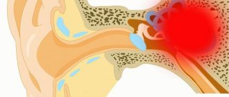
The equilibrium device (vestibular apparatus) consists of several subsystems that work effectively only in harmony. Receptors that respond to a specific stimulus include:
- direct equilibrium device = labyrinth located in the inner ear;
- visual apparatus;
- deep perception receptors;
- through the corresponding nerve, information from these receptors is transferred to the centers that process signals (the centers mainly belong to the area of the vestibular nuclei in the brain stem and cerebellum).
The ability to maintain balance depends on the correct response of the central nervous system to the information received. In practice, the division into peripheral and central systems is important. The peripheral balance device is located in the pyramid of the temporal bone. It is an integral part of the structure of the inner ear, with which it is organically connected.
