What is cerebral venous angioma
The disease is not aggressive, but its presence should not be ignored. The interweaving of blood vessels are fused balls that expand over time, provoking the development of inflammatory processes in the brain.
The more the brain is affected, the greater the health risk caused by the presence of the formation. This pathology is dangerous for the body due to the rapid growth of angioma, which, affecting the tissues of nearby organs, leads to disruptions in the functioning of the entire body.
It is the brain that is responsible for the quality functioning of all systems. Pathological changes occur and, if not treated in a timely manner, destruction occurs. A patient with a tumor is at high risk of cerebral hemorrhage, which can be fatal.
At the first symptoms of the disease, it is necessary to begin treatment immediately. Therapy in the early stages of tumor development has a favorable outcome. At an advanced stage of the disease, drug and surgical interventions will not give positive results.
Causes
One of the probable reasons for the development of angioma is genetic disorders.
So far, the reasons for the development of such tumors have not been fully studied. According to statistics, children are most prone to the appearance of vascular neoplasms in the brain, and this fact is explained by the immaturity of their internal organs and systems.
In 95% of cases, brain angiomas are congenital and develop due to some genetic abnormalities. The remaining 5% are caused by infectious lesions of the cerebral vessels or are the consequences of trauma. Especially often, angiomas form after severe traumatic brain injury.
In addition, scientists suggest that the development of such vascular neoplasms can be provoked by various serious diseases (for example, cirrhosis of the liver) or highly oncogenic tumors developing in other organs.
All of the above reasons can cause either the appearance of one angioma or lead to the development of angiomatosis (the formation of multiple neoplasms).
Mechanism of angioma development
Normally, an arterial vessel first divides into smaller arterioles, which subsequently branch into even smaller vessels - capillaries. They disperse in a network and then form venules and veins.
With angioma, such separation of vessels does not occur, and the artery immediately turns into a vein. Such abnormal formation of the bloodstream leads to circulatory disorders, because the pathological vessel “robs” the normal vascular network and the area of the brain does not receive sufficient nutrition. As a result, certain neurological symptoms appear, the manifestations of which depend on the location of the angioma in a particular area of the brain. In addition, when the tumor reaches a large size, it compresses the tissues of this vital organ and disrupts their functioning.
Kinds
Venous angioma is an enlarged venous column of vessels collected in a tangle. Depending on the location of the formations, the disease is divided into several types.
Frontal lobe angioma
She is responsible for initiative, correct decision-making, responsibility and showing interest. Therefore, when education is localized in this area, the character of a person changes greatly.
He becomes apathetic, a change in self-esteem occurs, speech is impaired, and inadequacy appears in his behavior and actions.
It is difficult for a person to think logically; he often commits unconscious actions. The patient cannot take responsibility and is unable to independently solve various problems.
Performance decreases, the emotional state is unstable, and over time the person suffers from depression. The patient does not feel cold or heat when touched.
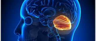
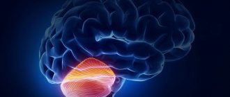
Cerebellar angioma
If there is a tumor in this area of the brain, functional disturbances occur in the body. The patient's muscle movements are not coordinated, he cannot perform smooth movements, so all actions are abrupt. A person’s motor ability is greatly slowed down, all actions are performed in the form of jerks.
There are disturbances in speech, there are serious problems when writing words and sentences. Breathing is impaired, resulting in problems with the respiratory system and skeletal muscles.
It is impossible to cure the disease using medications. With this form of angioma, surgical intervention is indicated.
Angioma of the left hemisphere
There is inconsistency in the work of the muscles of the arms and legs. The patient has speech impairment and changes in gait.
On this topic
- central nervous system
How does a headache with a brain tumor hurt?
- Natalya Gennadievna Butsyk
- December 3, 2021
Taste preferences change completely. Vision decreases, nystagmus of the extraocular muscles is observed.
Due to impaired blood flow, a person is susceptible to epileptic seizures and convulsions. At an advanced stage of the disease, partial paralysis may occur.
Angioma of the right hemisphere
With this type of disease, the patient experiences a movement disorder. It becomes slow and sharp. Speech becomes slow, writing style changes. A person is bothered by tremors of the arms and legs.
Angioma of the parietal lobe
The patient's coordination of movements is impaired, tactile sensations and pain threshold change, and speech abilities may deteriorate.
Angioma
First of all, a distinction is made between blood vessel angiomas (hemangiomas) and lymphatic vessel angiomas (lymphangiomas).
From a histological point of view, monomorphic and polymorphic angiomas are distinguished. Monomorphic angiomas are true vascular formations emanating from one or another element of the blood vessel (hemangioendotheliomas, hemangiopericytomas, leiomyomas). A sign of polymorphic angioma is a combination of various elements of the vascular wall; a transition from one type of tumor to another is possible.
Types of hemangiomas
Based on the type of structure, simple, cavernous, branched, combined and mixed angiomas are distinguished.
Simple (capillary, hypertrophic) hemangioma is a proliferation of newly formed capillaries, small arterial and venous vessels. Capillary hemangiomas are localized on the skin or mucous membranes in the form of a spot of bright red (arterial angiomas) or bluish-purple (venous angiomas) color. The sizes of capillary hemangiomas vary - from limited to giant. When you press on a vascular tumor, its color fades. Capillary hemangioma extremely rarely transforms into malignant hemangioendothelioma.
Cavernous (cavernous) hemangiomas are formed by wide spongy cavities filled with blood. Externally, such an angioma is a node of purple-bluish color, with a lumpy surface and a soft-elastic consistency. By palpation or x-ray, angiolites or phleboliths—dense, spherical, decalcified blood clots—can be detected in the thickness of the angioma. Cavernous hemangiomas usually have a subcutaneous location. A symptom of temperature asymmetry is typical for them - the vascular tumor feels hotter than the surrounding tissues to the touch. When pressing on the tumor, due to the outflow of blood, the hemangioma collapses and turns pale, and when straining, it becomes tense and enlarges (the so-called erectile symptom caused by the influx of blood).
Branched (racellous) hemangioma is represented by a plexus of dilated, tortuous vascular trunks. A characteristic feature of this type of angioma is the pulsation, trembling and noise detected above it, as above an aneurysm. It is rare, mainly localized on the extremities, sometimes on the face. The slightest trauma to the angioma can lead to threatening bleeding.
Combined hemangiomas combine superficial and subcutaneous location (simple and cavernous angioma). Clinical manifestations depend on the predominance of one or another component of the angioma. Hemangiomas of mixed structure originate from vessels and other tissues (hemymphangiomas, angiofibromas, angioneuromas, etc.).
The following types of angiomas are distinguished by shape: stellate, flat, nodular, serpiginous. Separately among the vascular tumors stand senile angiomas, which are multiple small round formations of pink-red color. Senile angiomas appear after 40 years.
Types of lymphangiomas
Among lymphangiomas, simple, cavernous and cystic vascular formations are distinguished. Simple lymphangiomas include enlarged tissue gaps lined with endothelium and filled with lymph. This type of angiomas develops mainly in the muscles of the tongue and lips and externally appears as a soft, colorless tumor.
Cavernous lymphangiomas are multi-chamber cavities formed by lymphatic vessels, with thick walls of muscle and fibrous tissue. Cystic lymphangiomas grow like chylous cysts and can reach significant sizes. They are found in the neck, groin, intestinal mesenteries, and retroperitoneal tissue. The addition of a secondary infection can cause the formation of fistulas and prolonged, debilitating lymphorrhea.
Symptoms
In addition to the differences between different types of angiomas, there are common symptoms that are characteristic of all types of cerebral venous angioma:
- frequent fainting;
- dizziness;
- paralysis;
- noise in the head;
- tremor of hands and feet;
- disorders ;
- headache ;
- imbalance and coordination of movements ;
- disturbances in the functioning of the cardiovascular system;
- epilepsy attacks
- problems ;
- convulsions;
- disorders ;
- blurred vision;
- decreased mental activity;
- noise in the head;
- change in taste preferences;
- impairment ;
- increased fatigue;
- nausea and vomiting;
- loss of ability to work.
Treatment methods
Treatment for venous angioma is chosen depending on the location of the tumor in the brain tissue, its size, and the nature of the course of the disease. In the fight against pathology, various methods are used, including minimally invasive operations.
An effective method of therapy is electrosurgery (electrocoagulation), which involves the use of an electric needle. With its help, an electric current is supplied to pathologically altered structures, and they are destroyed. Other treatments:
- X-ray irradiation.
- Radiosurgery using Gamma Knife or Cyber Knife installations.
- Surgical laser treatment.
- Conservative therapy.
Traditional medicine methods can alleviate the patient’s condition and prevent complications by reducing risk factors for hemorrhage. For this purpose, infusions and decoctions of medicinal plants are used, which normalize blood pressure and strengthen the vascular wall. Medicinal mixtures based on the herb St. John's wort, coltsfoot, tansy, celandine, yarrow, plantain, and calendula flowers are shown.
Conservative therapy is carried out when neurological symptoms are clearly expressed and surgical intervention is not possible for any reason. Prescribed drugs:
- Steroid hormones.
- Sedatives (calming).
- Angioprotectors.
- Painkillers.
- Anticonvulsants.
If the tumor does not grow, the symptoms are irregular and mild, monitoring the course of the disease is indicated. To monitor the growth of the tumor, an instrumental examination is performed every 6-12 months.
Surgical intervention is performed in different ways, which include a conventional operation to excise the tumor or a single exposure to pathologically altered tissue with targeted high-power radioactive radiation (Gamma Knife, CyberKnife radiosurgery). Other methods:
- Sclerotherapy. Introduction into the cavity of pathologically altered structures of special compounds that make the vessels impassable, excluding the tumor from the general system of cerebral blood flow.
- Embolization. Introduction into the vascular cavity of a special spiral or drugs that cause blockage of the lumen.
- Angioplasty. Implantation of stents and balloons into the vascular cavity to restore normal blood flow.
When choosing a method, the neurosurgeon gives preference to minimally invasive forms of intervention that provide the least trauma to surrounding tissues. The success of surgical treatment largely depends on the location and size of the tumor.
Diagnostics
To perform an accurate analysis, a specialist carries out diagnostic measures.
First of all, laboratory tests are prescribed. The patient must undergo a general and biochemical blood test, as well as a general and biochemical urine test.
If the norm deviates, the patient is then prescribed an ultrasound examination to confirm the diagnosis. Magnetic resonance imaging will determine the presence of formations, identify the location and size of the angioma.
It is also possible to perform a computed tomography, angiography or x-ray. Only after all diagnostic procedures have been completed can treatment be prescribed.
Treatment
Treatment of education includes taking medications, using traditional medicine, and in advanced cases, surgery.
Drug treatment
It is impossible to cure venous angioma by taking medications. This type of therapy is used only to alleviate the patient’s condition.
Drug therapy is prescribed if there is no threat of hemorrhage. To improve blood circulation, vascular drugs are indicated: Cerebrolysin, Cavinton, Bravinton, Vinpocetine, Telektol.
To improve the functions of cerebral vessels, the following drugs are effective: Actovegin, Mexidol, Emoxipin. Aspirin and Heparin are used to thin the blood. To increase blood supply to the brain, Tanakan and Bilobil are indicated.
On this topic
- central nervous system
What are the dangers of a porencephalic cyst of the brain?
- Olga Vladimirovna Khazova
- May 27, 2021
Sedatives occupy an important place. The most effective are: Persil, Sedavit, Fenozepam, valerian tincture, Persen, Tenoten, Deprim.
To eliminate headaches, drugs such as Nurofen, Tramadol, Ketanov, Diclofenac are indicated.
All drugs will only alleviate the patient’s condition, but will not contribute to recovery. A benign formation can only be removed surgically.
Surgical intervention
There are several methods of surgical treatment of the disease.
In severe cases of the disease, surgical removal of the accumulation of blood vessels is performed. A plastic spiral is inserted into the patient's lumen.
If the sclerotherapy method is chosen, a special substance is injected using a special catheter, which clogs the lumens of the vascular bundle and promotes scarring of the tumor. The operation is painful and lengthy.
It is possible to apply a superficial radiation beam to the tumor using a gamma knife. With its help, blood vessels are blocked. Education stops growing and developing, no longer presenting a danger to human life and health.
Liquid embolism can be injected into the collection of vessels, which easily penetrates into the smallest cavities and prevents the growth of angioma. Over time, the area where the tumor grew is replaced by connective tissue.
Diet
Diet plays a big role in treating the disease. The patient should exclude fatty and fried foods from his diet. Minimize the consumption of salt and sugar, coffee, chocolate and baked goods.
Avoid eating butter, pork, full-fat milk, liver and kidneys. It is necessary to eat vegetables and fruits, fish and seafood, cereals, dried fruits, and herbs.
Brain angioma is a serious disease. Therefore, it is strictly forbidden to self-medicate.
Treatment and diagnosis
It is impossible to select a therapeutic regimen without diagnostic results, since it is difficult to predict the further development of cerebral angioma. Treatment with medications is preventative and helps relieve some symptoms. Treatment options include
- Analgesics. Help relieve headaches: “Analgin”, “Pentalgin”, “Sumatriptan”.
- Vascular agents. Normalize blood pressure, thereby preventing the development of stroke: Diazepam, Nifedipine.
- Sedatives. Relieves cramps, calms the patient, improves sleep: valerian, peony tincture, motherwort.
- Hormone therapy. Prescribed by a doctor to reduce the size of a tumor before removal.
If the tumor is small and does not bother the patient, he has every chance of living to a ripe old age. But as the pathology progresses, medication alone is not enough. In such cases, doctors recommend surgery to remove the tumor.
Three types of operations are performed:
- Embolization of the “tangle”. A sclerotizing substance is injected into the tumor through a vascular catheter, with the help of which the capillaries are sealed and scarred. The operation is lengthy and requires anesthesia with subsequent rehabilitation of the patient.
- Radiosurgery. It is performed without opening the skull by irradiating the tumor with a gamma knife. The radiation clogs the vessels, blood circulation in the bundle stops and, accordingly, its growth stops.
- Mechanical removal. It is performed using craniotomy, but is only suitable for those angiomas that are located close to the surface of the meninges.
Neurosurgeons prefer minimally invasive operations that do not injure surrounding tissues. This method does not require long-term rehabilitation, and the risk of complications is minimal.
Can it develop into cancer?
Venous angioma of the brain is a benign formation. Therefore, it does not degenerate into a cancerous tumor.
On this topic
- central nervous system
Everything you need to know about pituitary adenoma
- Olga Vladimirovna Khazova
- February 28, 2021
Over time, it can lead to negative consequences and threaten human life. Therefore, it is important to consult a doctor in a timely manner for diagnosis and adequate treatment.
If the tumor does not manifest itself in any way, the person does not have behavioral disturbances or deterioration in health, you can live with this type of disease until old age. But in most cases, when diagnosing a tumor, surgical intervention is indicated.
Causes of angioma
The etiology of angiomas is constantly being studied as the disease becomes more common. There are several possible ways for this type of formation to appear in the body:
- congenital defect (hereditary factor), in which the epithelial tissue of the vascular walls grows uncontrollably;
- mechanical injury;
- diseases in which demyelination of the membranes of the brain occurs;
- previous infectious brain disease;
- constant contact with chemicals;
- other types of cancer that have migrated to the brain.
Uncontrolled division of vascular cells (mutation) can stop or accelerate under the influence of external factors (hormonal changes, chemical exposure).
Consequently, symptoms can appear at any age, but it is worth paying special attention to neurological complaints during transitional periods (puberty, menopausal disorders, pregnancy).
Degeneration into a malignant tumor occurs if there has been prolonged contact with a negative environment, a hormonal imbalance has occurred, or malignant neoplasms have appeared in another organ.
D18 is a code according to ICD 10, which means any type of angioma by location and origin.
For successful treatment, it makes sense to distinguish between venous and cavernous angiomas. The first type occurs in the veins, as an expansion of one or more venous vessels. The pressure in the veins is low and the walls are quite strong, so the risk of rupture is low.
The cavernous type includes in its structure not only blood vessels, but also small cavities of the brain. The tumor completely fills the caverns, and its boundaries are thin membranes. With this type of disease, there remains a high risk of hemorrhagic stroke with profuse hemorrhage.
For treatment, the location of the tumor, the type of benign or malignant, and the activity of changes in size (constantly growing or has clear, unchanged boundaries) are important.
Reliable causes of angiomas have not been found to date. However, according to existing data, they are congenital in nature and are associated with dysregulation of the process of angiogenesis in the fetus at an early stage of pregnancy (5–8 weeks) and are caused by the loss of signals that inhibit angiogenesis or the predominance of signals that inhibit apoptosis or promote angiogenesis.
It is assumed that factors such as severe constant stress, extensive and frequent injuries, alcohol consumption during pregnancy, and taking certain medications can affect the growth of angioma.
Forecast
An intact angioma may not be detected throughout life and may not cause serious trouble to the patient.
But if a hemorrhagic stroke or vasospasm occurs, the person may become disabled for life or die. Often, rupture of the vascular bundle leads to irreversible processes in brain activity.
Depending on the patient’s age, his general health, and the presence of vascular diseases, a prognosis for the course of the disease is made. A properly prescribed treatment regimen and timely surgical intervention will allow the patient to lead a healthy and fulfilling life for a long time.
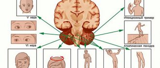
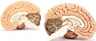
Localization and symptoms
Most often, venous angiomas are located in the white matter of the cerebellum and cerebral hemispheres, near the walls of the ventricles. The temporal lobes are very rarely affected.
In the human body, angiomas can be single or multiple (from 2 to 9% of all cases), then they speak of the angiomatous type of structure of the venous system. The larger the tumor, the more pronounced the symptoms of the disease.
Neurological symptoms that appear over a long period of time and cannot be relieved “help” in making a diagnosis.
A patient with an angioma periodically experiences headaches, non-food nausea, sensory disturbances, and fainting. Depending on which part of the brain the neoplasm is localized and what its nature is (veins, capillaries, or caverns are involved), specific symptoms will appear:
- Venous angioma of the occipital lobe:
- spasmodic sensations in the muscles of the back of the head;
- visual hallucinations (the appearance of long-term images and short-term sparkling).
- Venous angioma of the right temporal lobe:
- Damage to the left temporal lobe, just like the right, disrupts speech and the ability to perceive it. Strong emotional surges are added for no apparent external reasons.
- Venous angioma of the right parietal lobe:
- lack of “right-left” distinction when exposed to stimuli;
- unconscious paralysis.
- Damage to the left parietal lobe leads to loss of orientation not relative to one’s body, but in space (inability to determine distances or read a map).
- Venous angioma of the right frontal lobe:
- emotional instability;
- inability to control your actions;
- euphoric mood;
- loss of the ability to competently construct sentences of any length.
- With a tumor of the left frontal lobe, the patient cannot move on to the verbal formulation of thoughts (he thinks, but cannot speak), and has little control over his own behavior.
In general, capillary angiomas can occur in any part of the brain, and venous angiomas mainly in the cerebellum, the white matter of the brain.
The prognosis depends on the size of the tumor, its benign or malignant quality, and location. Most formations are treatable.