CT scan of the brain is an informative and common research method. With its help, various pathological conditions are diagnosed. Accessibility, painlessness, accuracy of the study and quick results are the undoubted advantages of computed tomography. Using CT, a three-dimensional image of the brain is obtained with the ability to study the organ layer by layer. Contrast enhancement increases the diagnostic information content of the method.
Yusupov Hospital conducts all types of computed tomography of the brain. The clinic is equipped with modern equipment that allows diagnostics to be carried out in the shortest possible time.
The essence of the method
Computed tomography of the brain is a research method based on the use of x-rays. Using a series of images, a three-dimensional image of the brain is created. Various pathological conditions are diagnosed in this way. The advantage of CT is the ability to study the organ layer by layer. Tomography is used in various fields of medicine. The method has a small number of contraindications, which allows it to be used for various conditions. To determine damage to the blood vessels of the brain, a computed tomography scan is performed using a contrast agent. This type of study allows you to most accurately determine the affected area.
Advantages
Computed tomography has some advantages over other methods of studying the brain. The main ones include:
- Painless. Some discomfort may occur only after the administration of a contrast agent. This must be reported to your doctor.
- Accuracy. A layer-by-layer study of the brain allows us to detect initial changes in its structure. Contrast enhancement increases the information content of the method.
- Speed of research. The procedure takes on average one hour. Within an hour after diagnosis, the results are delivered to your hands.
- Possible for people suffering from claustrophobia.
What does a CT scan show?
Computed tomography of the brain is prescribed for various conditions. Depending on the reason for the study, diagnostics are performed with or without a contrast agent. This diagnostic method is prescribed to identify the following conditions:
- bleeding;
- strokes and hemorrhages;
- neoplasms in the head and neck area;
- fractures of the base of the skull;
- hematomas and injuries;
- inflammatory process;
- pathologies of tissue development;
- the presence of foreign bodies in the area being examined.
Use of contrast agent
This type of procedure is needed in situations where doctors suspect the appearance of cancer cells in the head. The patient is injected with a liquid containing iodine, which collects more in those places where the tumor appears. Iodine in the blood creates increased contrast, the image becomes clear and accurate. The extent of the disease is being assessed.
The technique makes it possible to detect tumors at the primary stages of development. Treatment is much simpler. Because the process is labor-intensive and time-consuming, the research requires more money.
Unlike simple tomography, diagnostics using a contrast agent have a preparatory stage, during which the patient is injected with iodine-containing liquid, after which the patient may feel a little dizzy. Contrast material should not be administered to patients with an allergy to iodine.
Indications
A CT scan of the brain is possible with or without a contrast agent. Computed tomography without contrast is indicated for the following conditions:
- Head injuries. This group of indications includes bruises, concussions, and damage to the bones of the skull.
- Dental examination. The dentist may prescribe a computed tomography scan to determine developmental disorders of the maxillofacial area. In addition, research is necessary before implantation.
- Acute cerebrovascular accident. If acute cerebrovascular accident is suspected, a CT scan of the brain is performed to confirm the diagnosis.
- Diseases accompanied by loss of consciousness. Syncope, accompanied by convulsive syndrome, requires a computed tomography scan of the brain.
A CT scan of the brain using a contrast agent is called angiography. It allows you to most accurately examine pathological formations in the brain area. This research method has the following indications:
- Determination of the area of stenosis. Angiography allows you to determine the area of vascular narrowing, as well as the degree of stenosis.
- Diagnosis of cerebral aneurysms.
- Suspicion of ischemic stroke.
- Checking for vascular patency. Using tomography, obstruction is diagnosed due to thrombus formation or blockage of the lumen of the vessel by an embolus.
CT scan of the brain
Main diagnostic features
Features of computed tomography
CT scan of the brain is an examination that is more accessible than magnetic resonance imaging, allowing the visualization of many brain pathologies, while the price of a CT scan of the brain is usually lower than that of an MRI. With computed tomography, bone structures are better visible for study; with the use of contrast, vessels are better visible; with magnetic resonance imaging, soft tissues of the brain and blood vessels are better visible. A tumor, hydrocephalus and many other pathologies can be detected using tomography; it is prescribed if the patient develops neurological symptoms that were not there before. New headaches or meningeal symptoms (symptoms that occur when intracranial pressure increases), as well as changes in the patient's behavior.
Computed tomography allows you to view the structures of the body in a non-invasive way; the duration of the procedure is extremely short, so a child will undergo a CT scan of the brain without fear or concern. Brain CT scans in Moscow are performed in our clinic by appointment online or by phone.
Preparation for CT diagnostics
Brain scanning using computed tomography is painless and does not require special preparation.
It is important to conduct the study on an empty stomach; this will help the doctor decide on the need for unscheduled use of a contrast agent, and prevent the possible onset of nausea and other unpleasant sensations.
Patients with diabetes should consult with the doctor conducting the examination, because in their case it is possible to take food and drink with them.
How to behave during a CT scan of the brain?
- Before the examination, you must remove all metal jewelry. You should also put away your mobile phone.
- During a CT scan of the head, you do not need to take off your clothes; it is enough to get rid of all the metal elements of clothing that are in the way.
- Before the examination, you should inform your doctor about any allergies, if any, this is important since a contrast agent is often injected during a CT scan of the head.
Computed tomography with contrast is much more effective because it gives a more accurate picture of the disease. Examinations using contrast take longer. Only the doctor decides whether to administer contrast or not, and indications for this type of examination are changes in the brain, laryngological diseases (tinnitus) and suspicion of neoplasms.
During the examination, the patient has constant opportunity to contact the radiologist, who should be immediately informed if:
- Any sudden discomfort (eg, feeling claustrophobic); any symptoms after administration of intravenous contrast agent (shortness of breath, dizziness, nausea).
- After completing the test, to facilitate the removal of the contrast agent from the body, it is recommended to drink plenty of fluids (about 2.5 liters of still water or unsweetened drinks over the next 24 hours).
The price for a CT scan of the brain vessels with contrast will be higher than for a similar procedure without contrast.
Computed tomography technique
MSCT of the brain
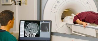
The procedure for a CT scan of the brain may seem stressful at first glance, but it is definitely not something to be afraid of. The procedure itself lasts several minutes. The subject is placed inside the apparatus on a special moving table (couch). During diagnosis, it is very important for the patient to lie still and not breathe while the scanning beams are working, which the radiologist conducting the study will warn about in advance. By adhering to these rules, a CT scan of the head will be carried out correctly and accurately.
The following diagnostic procedures are performed as part of tomography of head structures:
- Orbital tomography (search and assessment of small intraocular changes when ultrasound diagnostics is ineffective, primary neoplasms of the eyeball, metastases to the eyeball);
- Craniofacial tomography (preoperative assessment of the craniofacial bone, assessment of post-traumatic changes in the craniofacial bones, assessment of neoplastic tumors, assessment of inflammatory changes);
- Tomography of the pyramids of the temporal bone (post-traumatic changes, congenital anomalies, inflammatory processes, proliferative processes, structures of the middle and inner ear).
Indications for CT examination
CT scans are prescribed for people who have suffered traumatic brain injuries. Most often victims of road accidents. First of all, there is a suspicion of bleeding inside the skull. The cause may be trauma to the head and spinal canal. Other indications for CT scanning of the brain include the following pathologies:
- brain abscess;
- cerebral hematoma;
- diseases of the skull bones;
- brain tumors;
- pathological changes in the vessels of the brain, encephalitis, meningitis, aneurysm, thrombosis, stroke, neoplasms of various types.
Computed tomography of the brain is also prescribed for a child and an adult if it is necessary to study congenital anomalies, for example, in order to track the progress of changes in development and, on this basis, develop an individual treatment and rehabilitation plan. Computed tomography with contrast successfully diagnoses any changes and the smallest pathologies of the blood vessels of the brain. The price for CT angiography of vessels in our clinic is one of the best in the city.
Contraindications
Frequent repetition of the study is harmful to the body, despite the fact that the radiation dose when used on modern tomographs is extremely small, CT scans are not recommended to be performed more than 3 times a year. For the same reasons, CT scanning is not recommended for pregnant women. If a woman is in the 2nd half of her cycle and suspects that she could become pregnant, without vital indications she should not undergo a CT scan, as this can cause complications for the health of the fetus, it is better to wait until the 2-3rd trimester.
Advantages and disadvantages of tomography
Computed tomography has the following advantages:
CT angiography of vessels
Visualization. CT examination is widely used in almost all areas of medicine and allows obtaining informative images of scanned organs. The undeniable advantage of the method is the short diagnostic time compared to other hardware methods. Scanning the area under study takes only a few tens of seconds, which expands the category of patients for whom this method is suitable and increases the comfort of the procedure. CT scans are often used in emergency departments where accident victims are admitted. In emergency situations, it allows you to immediately visualize hemorrhages and internal injuries. An undoubted advantage of the method is also its non-invasiveness; no mechanical impact on the patient is required, which reduces the risk of complications.
Computed tomography simultaneously creates images of internal organs, bones and blood vessels, with high diagnostic capabilities and spatial resolution, which distinguishes the method from other radiation studies. Accurately visualizes the anatomy and pathology of the brain, allows you to assess its contours, dimensions and internal state. Another significant advantage of computed tomography is the ability to perform it on patients with implanted devices: pacemaker, neurostimulator, and others. Examinations using a tomograph allow images to be obtained on a monitor in real time, which makes it an invaluable tool in monitoring biopsies of various organs.
Research results
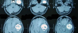
The result of computed tomography of the brain is highly informative images:
- bone tissue of the head;
- nervous tissue of the brain (divided into white and gray matter);
- fluid-filled spaces of the brain;
- ventricular system;
- arachnoid reservoir systems.
Thanks to this, the doctor can diagnose diseases such as intracranial hemorrhage (acute and chronic), ischemic and hemorrhagic strokes, epilepsy, hydrocephalus, benign and malignant brain tumors. The cost of computed tomography of the brain is more than offset by the accuracy and non-invasiveness of the examination. In addition to the images, the patient is given a CD with the examination results.
Sign up for Computer Diagnostics
Diagnostics in our medical center are carried out using powerful, modern equipment, by highly qualified specialists with many years of experience in performing computed tomography. This allows us to guarantee high service and quality of services provided.
You can sign up for a CT scan at the Open Clinic by calling the phone number provided on the website, using the feedback form, or directly at the clinic administrator. During the phone call, if necessary, you will be further advised and told about the procedure. We have one of the best prices in Moscow for computed tomography of the brain.
Indications for CT scan of the head and neck
Computed tomography of the head carries a certain radiation load on the body, so scanning is recommended no more than 3 times a year for emergency indications.
The interval between studies should not be less than 4 weeks. For preventive purposes, experts do not recommend undergoing examination, as the X-ray load on the body increases. The main indications for CT of the head and neck are:
- dizziness and tinnitus, the causes of which have not been established;
- anomalies in the development of bone structures of the head, blood vessels;
- suspicion of the development of a hernia in the cervical spine;
- neck and head injuries;
- suspected tumors of the head and neck;
- headache;
- identifying dilation of blood vessels in the neck and head;
- signs of cerebrovascular insufficiency.
When contacting specialists at the Yusupov Hospital with these signs, patients receive advice on the scope of diagnostic measures to identify the causes of the pathology, after which they are sent for diagnosis.
Make an appointment
Indications for use
CT scans are prescribed to detect inflammation and tumors of the head, hemorrhages, and post-traumatic disorders. Computed tomography allows you to monitor the effectiveness of the therapy or rehabilitation used for other pathologies. The images clearly show the cranial bones, structures and membranes of the brain, blood vessels and paranasal sinuses.
A neurologist prescribes a CT scan in the following situations:
- Suspicion of swelling, inflammation, hematoma after injury.
- Problems with blood circulation.
- Abscess and cyst.
- Infections.
- Oncopathology.
- Problems with brain development.
- Vascular diseases.
- Hydrocephalus.
- Convulsions, vision problems, dizziness, deterioration of sensitivity.
- Unreasonable pain in the head without a clear reason for 2-3 months.
- Preoperative studies.
- Contraindications for MRI.
Contraindications
Despite the well-tolerated procedure and its painlessness, computed tomography of the brain has some contraindications. These include:
- Pregnancy. Since the research method is based on the use of X-rays, there is a possibility of negative effects on the fetus. Carrying out a CT scan of the brain in a pregnant woman is justified only in cases of threat to her life.
- Excessively heavy weight. Obesity, in which weight reaches above 200 kg, is a limitation for performing computed tomography of the brain.
- Mental illnesses. During the CT scan, you must remain motionless. Mental illnesses accompanied by inadequate reactions make diagnosis difficult.
- Individual intolerance to the components of the contrast agent.
- The presence of severe renal or liver failure. This contraindication is due to the need to remove the contrast agent. In cases of insufficient liver or kidney function, excretion becomes difficult.
Where can I get tested?
A CT scan of the brain can be performed in most medical institutions. To display the most accurate results, you need to take into account the following key factors:
- The quality of the technology used.
- Qualifications of specialists.
For many patients, the determining factors are the speed of results and the cost of diagnosis. It is advisable to clarify such information in advance.
Many private diagnostic centers meet quality standards and provide medical services. Some use modern European technology, making it possible to obtain the most accurate and fastest results. Radiation exposure in this situation is underestimated. Staff can take care of patient comfort at different stages. Before the procedure, the radiologist conducts a consultation.
When the analysis is completed, the doctor informs the patient in detail about his state of health and advises him to undergo additional examinations if necessary.
Preparation
No special preparation is required to perform a CT scan of the brain. If it is necessary to use a contrast agent, the last meal should be 5 hours before the examination. Before a CT scan of the brain, it is recommended to consult with your doctor about the need to stop taking medications during the study.
Progress of the study
The procedure for performing a CT scan of the brain consists of the following steps:
- The patient is placed on a conveyor.
- The head is fixed using special devices. This is necessary to ensure maximum immobility during the examination.
- After starting the tomograph, a series of images are taken, with the help of which a three-dimensional image of the brain is formed.
- Computed tomography is a painless diagnostic method. During the examination, the patient hears only clicks. This factor is eliminated with the help of earplugs.
- If contrast enhancement is necessary, a special substance is injected into the body. It stains the blood vessels, which allows for a more accurate diagnosis of the brain. The contrast agent may cause a metallic taste in the mouth, nausea, or headache. This must be reported to the doctor conducting the study.
On average, a CT scan of the brain takes from 30 minutes to an hour. The duration of the study depends on the type of diagnosis and the general condition of the patient. The results are deciphered immediately after the procedure. It takes about 1-1.5 hours depending on the complexity of the tomography.
How does a CT scan differ from an MRI of the brain?
| COMPUTER TOMOGRAPHER | MAGNETIC RESONANCE TOMOGRAPHER |
The principle of operation of a magnetic resonance imaging scanner
Diagnosis of brain diseases using MRI is one of the most harmless and harmless examination methods.
Magnetic resonance imaging is based on the ability of high-frequency radio waves to cause vibrations in the hydrogen atoms present in the body's cells when they are exposed to a powerful magnetic field. The MRI machine records the received impulses, which are then processed by a computer. Due to the fact that different tissues of the body are characterized by signals of varying intensities, high-frequency images of the substance, meninges and vascular bed are formed with high spatial resolution. The increased power and ultra-sensitivity of modern tomograph models have made it possible for doctors to diagnose millimeter changes at an early stage of their occurrence. An MRI machine, depending on the type of model (open or closed), resembles a large installation with a narrow pipe in the center or a magnetic canopy. During the examination, the patient lies, as still as possible, on a mobile table that is placed inside the tomograph. An MRI scan lasts from 20 to 40 minutes, depending on the purpose and examination protocol. It is quite comfortable for the patient. Only loud noises from the machine can cause slight concern. This noise is safe for the patient. It occurs due to vibration of the metal coils in the device, which is caused by rapid pulses of electricity. The induction power of a tomograph (fullness) is one of the most important parameters of a tomographic installation. It is measured in Tesla. There are three types of devices that differ in magnetic field strength: low-field (0.3-0.5 Tesla), high-field (1-1.5 Tesla) and ultra-high-field (3 Tesla or more). The higher the induction level of the tomograph, the better the quality of the images it produces.
Initial appointment with a NEUROLOGIST
ONLY 1800 rubles!
(more about prices below)
The principle of operation of a computed tomograph
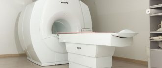
CT scans of the brain are performed using a device called a spiral CT scanner.
During the examination, the patient's body is exposed to several beams of X-rays released by the emitter sensor at different angles. Unlike conventional radiography, which offers only a flat image from several positions, a CT scan of the brain produces a set of images in the form of small sections in different projections. To do this, the device's sensors move in a spiral around the patient's body, recording information received from different angles. On a computer, this information is combined into three-dimensional models (tomograms) of a specific area or lobe of the brain. The power of a CT scanner depends on the number of slices the scanner makes per revolution of the gantry ring. The higher the shearing ability of the unit, the more accurate the scanning results will be. In St. Petersburg clinics today you can undergo examination using 4-340 slice computed tomographs. A high-quality CT scan of the brain will require a device of 32 slices or higher.
To obtain CT images of the vessels of the head, the patient is injected intravenously with an iodine-containing contrast agent, which stains the vascular bed well. Without it, high-quality visualization of the vascular system of the head and neck is impossible. The CT scan procedure is absolutely silent and fast. The average scan duration is about 5-7 minutes.
Decoding the results
After the tomography, the doctor evaluates the resulting layer-by-layer images. The presence or absence of neoplasms, areas of hemorrhage or ischemia are taken into account, and brain structures are assessed. A computed tomography scan is considered normal if there are no signs of pathological neoplasms, brain structures are age-appropriate, there are no areas of fluid accumulation, and there are no violations of the integrity of bone tissue.
Any deviations from the norm are recorded in the conclusion and require further consultation with a specialist. Doctors at the Yusupov Hospital are deciphering all types of computed tomography of the brain. The latest equipment makes it possible to accurately determine the initial changes in the organ.
Price
The cost of a head and neck CT scan varies depending on the qualifications of the specialist, the equipment used and other factors, which include the administration of a contrast agent. At the Yusupov Hospital, patients have access to a wide range of diagnostic services, and also have the opportunity to undergo scanning with contrast.
Specialists at the Yusupov Hospital guarantee obtaining clear, high-quality images that will allow you to fully evaluate the area under study. If you need a head CT scan, there are many diagnostic centers in Moscow. However, services that meet European standards are provided at the Yusupov multidisciplinary hospital located on Nagornaya Street. If you need a specialist consultation for a head CT scan, please make an appointment by phone.
Make an appointment