Hypothalamic-pituitary system: general information
Within this structure, a close humoral and nervous connection is established between the main elements. It is customary to distinguish three parts in the hypothalamus: posterior, middle and anterior sections. The latter is involved in regulating the parasympathetic nervous system. The middle one provides control over trophic and endocrine functions. The tasks of the posterior section include regulating the nervous sympathetic system. The nuclei of the hypothalamus produce some steroids, which are then concentrated in the pituitary gland. In this regard, damage to one department usually leads to damage to another. The hypothalamic-pituitary system thus acts as a structure whose elements exist in close interaction.
Connecting to the Brain
A feature of the vascularization of the hypothalamus is the intensity of the capillary blood supply. It significantly exceeds the speed in other parts of the brain. Due to vascularization, vascular permeability increases. This, in turn, ensures the passage into the brain from the blood of various humoral compounds that signal the state of the body. The hypothalamus is closely connected with the cerebral cortex, reticular formation and subcortical formations. The hypothalamus is involved in the regulation of humoral and endocrine processes. They, in turn, ensure the body’s adaptation to constantly changing conditions of the internal and external environment. The role of the hypothalamic-pituitary system in the body is vital. This structure is an important link, a key element of the limbic-reticular organization of the cerebral integrative mechanism. It ensures the integrity of the formation of activities.
Magnetic therapy as an optimal treatment method
Biomag low-frequency pulsed magnetic therapy is an optimal method that combines several effects:
- effect of pharmacotherapy – improvement of microcirculation of oxygenated blood and nutrients in the central nervous system;
- the effect of physiotherapeutic treatment is correction of muscle tone, acceleration of recovery.
The good effects of magnetic therapy have been documented even in severe conditions and impaired mobility in children. It is recommended to apply it to the entire area of the spine and head (lower spine → occipital area → head area). In case of movement disorders, it is advisable to add exposure to the affected area. Magnetic therapy is a suitable daily and long-term treatment as part of comprehensive care.
In adults who have suffered a stroke (after treatment of the acute stage aimed at maintaining vital functions, decongestant, thrombolytic therapy, normalization of blood pressure), magnetic therapy is indicated in the initial phases of rehabilitation.
By using Biomag low-frequency pulsed magnetic therapy, direct stimulation of neurons, vasodilation of precapillaries and capillaries in damaged tissues of the central nervous system is achieved. This leads to a significant improvement in microcirculation, supply of oxygen and nutrients to the brain, and an anti-edematous effect. In general, metabolic processes are improved, the normal function of nerve cells is stimulated, and further targeted physical rehabilitation is improved.
Disruption of activity
Diencephalic syndrome is a consequence of the action of pathogenic factors. One of them is increased vascular permeability. It facilitates the penetration of viruses and toxins present and circulating in the blood into the brain. Closed TBI is also important. When the fluid column moves, the walls of the third ventricle are injured, the ependyma of which covers the nuclei. Diencephalic syndrome is also observed when affected by a tumor. This may be pinealoma, subcortical glioma, basal meningioma, craniopharyngioma. Diencephalic syndrome can result from prolonged pathologies of internal organs and endocrine disorders. Mental trauma, along with other provoking factors, also has a certain significance. All this suggests that diencephalic syndrome is based not only on structural and anatomical damage, but also on functional disorders.
Manifestations in adolescence and adulthood
Symptoms, as in childhood, are varied. These include:
- inattention;
- anxiety;
- ease of distraction;
- impulsiveness;
- moodiness;
- explosive character;
- inability to express an opinion;
- disobedience;
- aggression;
- learning difficulties.
Damage to the mediobasal structures is manifested by impaired speech, analytical abilities, and concentration.
When mesencephalic structures are affected, parkinsonism, cerebellar disorders, and hyperkinesis occur. Dysfunction of the midbrain structures can cause epileptic seizures along with impaired speech development.
Some (more severe) brain dysfunctions are a reason to delay military service or be completely discharged from military service. Manifestations of damage to mesodiencephalic structures concern behavior, thinking, and emotional life. During puberty, some symptoms may even intensify.
Children diagnosed with brain dysfunction may be unable to cope with both work and social needs later in life. Men are more prone to conflicts with the law due to impulsive behavior; women are more likely to experience mood changes and sleep disturbances. Other symptoms may include:
- inability to rest;
- low self-esteem;
- inability to establish and maintain long-term relationships;
- increased affectivity, hot temper, etc.
Only hyperactivity decreases with age.
Clinical picture
Diencephalic syndrome, the symptoms of which are extremely polymorphic, can manifest itself immediately or after a long period after pathogenic exposure. Most naturally, when affected, there is a disorder in the activity of the vascular structure and internal organs, thermoregulation, and metabolic processes (protein, mineral, water, fat). Dysfunction of the intrasecretory glands and disruption of wakefulness and sleep are observed. A varied combination of these disorders determines one or another nature of the clinical picture. Typical symptoms include thirst, headache, changes in appetite (anorexia or bulimia), difficulty breathing, insomnia or drowsiness, and palpitations.
Minimal brain dysfunction in children
The child may also experience brain dysfunction. Most often it manifests as minimal brain dysfunction. This is a very common pathology, and every 5 children can be diagnosed with it. The reasons why brain dysfunction begins to develop in children are as follows:
- difficult pregnancy.
- difficult and long labor process.
- exposure of the child to harmful and toxic substances.
- infectious diseases.
Symptoms of dysfunction of the midline structures of the brain in children are quite striking and manifest themselves as follows:
- severe headaches that are systematic.
- there is excessive activity, as well as hyperexcitability.
- There is constant nervousness and irritability.
- motor and speech functions are noticeably impaired and slowed down.
- retardation in development.
- disturbance of attention and memory.
- rapid fatigue and fatigue.
When this disease begins to develop, then, accordingly, the symptoms become more pronounced and appear much more intense. Such violations can provoke other, more serious consequences. For example, epilepsy or dangerous nervous disorders.
Foreign doctors practice such treatment as constant monitoring of the child by an osteopath. He must constantly monitor the baby’s condition and monitor whether there are any changes or deterioration in his condition. If dysfunctions of the midline brain structures are detected in the early stages, the situation can be easily corrected and the disease can be cured without subsequent harmful and negative consequences.
Classification
Pathology can be primary or secondary. This or that type is determined in accordance with the causes of the syndrome. Neuroinfections and injuries act as provoking factors for the primary disease. The secondary type of pathology is caused by a disorder of metabolic processes. This is evidenced by obesity. There is also a classification according to severity: severe, moderate, mild. In accordance with the predominant clinical symptom during the pathology, diencephalic syndrome is distinguished with:
- obesity;
- neuroendocrine disorders;
- signs of hypercortisolism;
- neurocirculatory disorders.
How is the diagnosis made?
The main method of instrumental diagnostics is electroencephalography of the brain. During such an examination, zones of excitation of the brain and brain stem in particular are detected.
During the EEG, basic and additional tests are carried out, which allow you to objectively assess the activity and type of waves, their average amplitude and dominant frequency. The correspondence of clinical symptoms and the characteristics of EEG waves is a guarantee of a correct diagnosis by a pediatric neurologist.
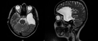
However, in addition to the EEG, the doctor may need to obtain a layer-by-layer analysis of the structure of the soft tissues of the brain , which are visible on computed tomography or MRI images. This is often necessary in cases where a person with the described symptoms has no history of injury, bruise or other provoking factor.
In this case, the doctor, visually noting signs of brain disorders, including dysfunction of stem structures, must find out the mechanism of their development. MRI and CT are methods by which various types of accumulations of tissues and cells are identified, not excluding atypical elements.
In addition, such studies make it possible to identify concomitant pathologies, for example, parallel venous dysfunction - a condition in which the outflow of blood from the brain is impaired due to vascular disorders. Clinically, headaches, fainting, cyanosis of the facial part of the head, darkening in the eyes, swelling of the soft tissues of the face are added to the symptoms of damage of the stem and diencephalic nature.
Further clarification of such irritations is carried out using additional studies, for example, angiography or ultrasound of cerebral vessels.
The doctor receives a certain amount of information from laboratory blood tests for the presence of glial neurotrophic substance. This is a type of enzyme immunoassay. The concentration required to confirm the diagnosis is amounts greater than 17.98 pg/l.
Neuroendocrine type
This category is considered the most common form of pathology. It is usually based on pluriglandular dysfunction, which is combined with autonomic disorders. This group includes a number of outlined clinical forms, in particular:
- adipose-genital dystrophy,
- Itsenko-Cushing syndrome,
- diabetes insipidus,
- dysfunction of the gonads (impotence, early menopause).
Vegetative-vascular disorders
The clinical picture in this case includes symptoms such as:
- high vascular excitability (unstable blood pressure, tendency to palpitations),
- increased sweating,
- spasms in peripheral, cerebral and cardiac vessels.
There is also instability in the functioning of the digestive system. This type of diencephalic syndrome is also characterized by periodic vegetative-vascular paroxysms. Crises may occur. In some patients they are rare (once every few months), in others they are frequent (up to several times a day). Typically, this form is characterized by severe emotional disturbances.
What does this diagnosis mean?
The brain stem is a continuation of the spinal cord, and there is no clear boundary between them. It is located in the area of the opening of the occipital lobe of the skull and normally has a size of no more than 7 cm. This small section contains the midbrain, medulla oblongata and pons. According to some sources, the diencephalon and cerebellum are also included in the trunk .
Pathological changes in the structure and functioning of the trunk can occur both in general and in sections in particular. Depending on the location of the problem, the patient may be diagnosed with:
- Dysfunction of diencephalic structures . Typical complaints: sleep disturbances, poor appetite, fluctuations in body temperature, disturbances in self-regulation and metabolism processes. This is the most commonly diagnosed form of neurological disorder. Its typical example is vegetative vascular dystonia, familiar to many, affecting 30% of the female population.
- Dysfunction of brain stem structures . Patients note uneven breathing and disruptions in muscle tone. This group of pathologies also includes weakening of the vocal cords and problems in the functioning of the speech apparatus (dysphonia), difficulty swallowing and frequent choking (dysphagia), and poor speech perception (dysarthria).
- Dysfunction of midline structures . Causes emotional disorders, unbalanced behavior, sudden mood swings, vegetative forms of somatic disorders.
Normally, the processes of regulation of human life activity from the side of the brain stem are clearly established and do not require correction. However, under the influence of certain risk factors, diseases arise, which, depending on the degree of complexity, can be expressed in vivid or subtle clinical manifestations.
Neurodystrophic form
It is relatively rare. The clinical picture includes:
- Trophic skin and muscle disorders (bedsores, neurodermatitis, dryness and itching).
- Damage to internal organs (bleeding and ulcers along the gastrointestinal tract).
- Bone damage (sclerosation, osteomalacia).
There are disturbances in salt metabolism. As a result, in some cases, ossification of the muscles and interstitial swelling occur. In some cases, there are sleep and wakefulness disorders, constant low-grade fever, accompanied by hyperthermic attacks. Phenomena of an astheno-neurotic nature are also detected. They accompany trophic, endocrine and autonomic disorders. The neurological clinical picture is presented as mild scattered signs.
About the clinical manifestations of midline dysfunction
Depending on the location of the dysfunctional cells and tissues, the consequences of dysfunction of the midline structures may have the following clinical manifestations:
- Loss of skin sensitivity in all areas;
- there is excessive pain sensitivity with an increase in its individual threshold;
- tremor of the limbs becomes noticeable (even at rest);
- signs of puberty appear early;
- Unreasonable changes in mood appear in behavior: crying is replaced by laughter, even hysterics, and vice versa;
- the functioning of the endocrine system causes serious disruptions. Depending on the location of the lesion, symptoms of hyperthermia, as well as an increase or decrease in blood pressure, may be observed.
Such disorders can be characterized as thalamic, and the resulting syndromes are called neuroendocrine.
Diencephalic syndrome: diagnosis
Against the background of pathology, changes in a number of blood parameters are noted. Detection of the disease is carried out by determining the main hormones in the serum. The study of circadian rhythms in the process of synthesis of LH, prolactin and cortisol is a mandatory analysis when examining for diencephalic syndrome. Treatment of pathology is prescribed in accordance with the degree of metabolic disorders. The list of mandatory studies also includes determination of serum glucose concentration, glucose tolerance test and food load analysis. The level of metabolites for sex hormones in the daily urine of a patient in adolescence is of great importance when making a diagnosis.
Therapeutic measures
The main goal of treatment is to stabilize metabolic processes, restore the mechanisms involved in regulating the activity of the reproductive system, and the formation of the ovarian-menstrual cycle in girls.
The most significant stages of non-drug effects are considered to be the normalization of sleep and wakefulness, the rehabilitation of all infectious chronic foci, and the normalization of body weight. In case of pathology, physiotherapy, balneotherapy and reflexology are indicated. To eliminate the causes of the disease, surgery to remove tumors is used. Rational infectious therapy is also prescribed, the consequences of injuries are eliminated, and the effects on the primarily affected visceral and endocrine organs are carried out. As a pathogenetic treatment, vegetotropic drugs are used that reduce or increase the tone in the parasympathetic or sympathetic part of the nervous autonomic system. Ascorbic acid, vitamin B1, calcium preparations, antispasmodics, ganglion blockers (medicines Pentamin, Benzohexonium, Pachykarpin) are prescribed. To regulate the tone of the parasympathetic system, anticholinergic drugs (for example, Atropine) are recommended. Vitamin B12 and the drug “Acefen” are also prescribed. If sympathetic-adrenal pathology predominates, the drug “Pirroxan” is indicated.
What are the risk factors?
The health of brain stem areas can be negatively affected by: traumatic factors, toxic effects on brain tissue of chemicals and biological toxins, radiation exposure, environmental disasters, and infectious diseases.
If the diagnosis in question is made to a newborn child, then the neonatologist suspects the quality of childbirth and the primary postpartum period.
A hereditary predisposition to this type of pathology cannot be ruled out, as well as disturbances in the blood supply leading to cell hypoxia and atrophic phenomena in tissues in general. In adult patients with such problems, the causes may be traumatic brain injuries, bruises, poisoning, various kinds of hormonal imbalances, cancer and their consequences at various levels.
