Cerebral ischemia is a condition that often develops against the background of intrapartum (during childbirth) asphyxia. The pathology is also known as hypoxic-ischemic encephalopathy. This name fully reflects the pathological processes involved in pathogenesis - hypoxia (oxygen deficiency), ischemia (changes that occur in the nervous tissue due to oxygen deficiency), encephalopathy (brain dysfunction).
Characteristics and degrees
Cerebral ischemia is a pathology that reflects the consequences of oxygen deficiency of brain tissue, which in newborns manifests itself as neurological symptoms. Prematurity correlates with a worse clinical picture and prognosis. Irreversible damage to nerve cells leads to the development of permanent disorders in brain function. The prevalence of the pathology is about 3 cases per 1 thousand infants.
Cerebral ischemia, which occurs in a mild form (grade 1), in a newborn is manifested by a number of symptoms - excessive excitation of neurons or depression of the central nervous system with a total duration of no more than 7 days. With moderate cerebral ischemia (grade 2) in newborns, processes of excitation or depression of central nervous system functions are observed for more than 7 days. Associated symptoms include:
- Intracranial hypertension.
- Convulsive syndrome.
- Autonomic-visceral disorders (a wide range of somatoneurological and neuroendocrine disorders that arise as a result of disturbances in neurohumoral regulation, manifested by thermoregulation disorders, vascular and gastrointestinal pathological reactions).
Severe cerebral ischemia (grade 3) in newborns is characterized by a progressive decrease in brain activity lasting more than 10 days. The depression syndrome transforms into a coma, in other cases it alternates with periods of psychomotor agitation, which are characterized by seizures. Convulsive or status epilepticus can lead to coma.
Ischemia in a newborn, which occurs in a severe form, is accompanied by dysfunction of the brain stem areas, which manifests itself in disruption of vital functions - respiratory and cardiovascular activity. Other symptoms: autonomic-visceral disorders, progressive intracranial hypertension.
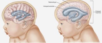
In an infant with third-degree pathology, during neuroimaging, processes of decortication (exhaustion, atrophy of the cortical layer of the brain) and decerebration (exhaustion, atrophy of the anterior brain regions) are observed. Periventricular ischemia in newborn infants is a pathology that is characterized by damage to the medulla in the area of the ventricular system, which is associated with the development of intracranial hypertension. Cerebral ischemia is listed in the list of ICD-10 diseases under the code “P91”.
massage for cerebral ischemia in newborns
Hello, I gave birth at 36 weeks via cesarean section due to prenatal rupture of amniotic fluid. There was no effect from enzoprostan stimulation. And on October 10, 2008, a child was born (girl - 2440 and height 45 cm) with the defect cloacal form of anal atresia. An operation was required, which was performed at the Rusakovskaya hospital. Many thanks to EVGENIYA VLADIMIROVNA KUZNETSOVA - Sonya’s first mother)))) It’s even a shame that I called her Picasso when I found out about her stoma - out of ignorance I took it as a cruel joke. Here are the results. 1. As a result, SONYA has a bicornuate uterus (both the uterus and bladder WERE enlarged - now they are normal). During the operation, the vagina was cut to remove the urinary fistula. It’s too early to think about it, we’ll figure it out in about 7 years. 2. Atresia of the rectum, a high degree of cloaca, that is, not only the butt, but also the other three exits are not completed as necessary. HOORAY! We had the operation and now we are the happy owners of butts!!! Many thanks to OLEG EVGENIEVICH GLIZNUTSIN - our first dad))) And also Yuri Yurich Sokolov and Vsevolod Yurievich contributed. 3. Vesicoureteral reflux (when urine from the bladder goes back to the kidneys). They were cured, although during the operation the bladder was moved and retention is still an issue. 4. Hypospadias - the urethra is underdeveloped and its length is reduced. Corrected by moving the bladder. I don’t know what or how it will work, it’s still a diaper—not much time has passed. However, I am glad that the plastic surgery to create the urethra did not involve the formation of the vaginal wall, and plastic surgery of the bladder sphincter. 5. Pyeloectasia on the left. Neurogenic bladder. Anemia. Cerebral ischemia, depression syndrome - passed in passing. Massages, drops, tablets, homeopathy, physiotherapy, oxygen. It’s probably not clear what was done where... I’m writing 1. 10.10.08-19.11.08 - an operation was performed in the neonatal period to remove a loop sigmostoma. Usually it is on the other side, but there was severe inflammation. 2. 11.21-12.02.08 - we were transferred to the infectious diseases department with rotavirus. Well, nurses don’t always wash their hands, what can you do? But the way the head of department seven behaves can be described in one word - BITCH! But if we describe a separate topic. 3. 12/23-12/24/08 – examination in Izmailovskaya regarding gynecology. 4. 01/22-03/04/09 - examination for constipation and urinary tract infection. . As a result, right-sided lower lobar pneumonia was diagnosed. And they were treated for a long time. I wish I could focus on the heart then - damn, but I don’t have a medical education. Now I know that frequent wheezing and pulmonary diseases can be a symptom of a patent ductus arteriosus. A relaparotomy, biopsy, and formation of a separate sigmostoma was performed. (They made a stoma on the other side) 5. 08/22-09/24/09 - fecal impaction, delayed motor development, rickets. 6. 30.11.-.03.12.09. They went to the plastic surgery, but a girl with chickenpox was admitted to the department and discharged. 7. 01/14/10-01/18/10 went to plastic surgery, but got sick. 8. 01.03.10-30.03.10 Plastic surgery to create the urethra, butt, vagina
Now I am preparing for the closure of the Patent Ductus Arteriosus (botalli) in Bakulevo - the most characteristic of the defect are frequent respiratory diseases and pneumonia in the first and second year of life, and retardation in physical development. When examining the patient, a systole-diastolic murmur is detected above the heart with the epicenter above the pulmonary artery. Tomorrow I’ll run to the department to get my quota (2 Shchimilovsky lane 4a, Novoslobodskaya metro station)
I will test for dysbacteriosis (m. Teply Stan), I will go to Rusakovka for a consultation (m. Sokolniki),
and to the clinic to see a neurologist (m. Vernadsky Pr. - and I’ll die in the evening)))
Causes
Ischemia is a condition that develops in newborns as a result of oxygen deficiency in the brain, which is accompanied by damage and death of neurons. Hypoxic-ischemic encephalopathy of the first degree occurs as a result of intrauterine hypoxia. Another probable cause is mild asphyxia during birth.
Moderate (second) degree cerebral ischemia in a newborn occurs as a result of perinatal hypoxic brain damage. Ischemic-hypoxic processes in the stage of intrauterine development occur against the background of infectious-inflammatory and other diseases suffered by the mother. Other influencing factors include drinking alcoholic beverages and smoking during gestation.
Intrapartum asphyxia, birth injuries to the spine, and other pathological influences and conditions can provoke the occurrence of moderate ischemia. In many newborns, pronounced ischemic changes in the nervous tissue that forms the brain arise as a result of a combination of intrauterine hypoxia and severe intrapartum asphyxia.
The following factors are involved in the pathogenesis: congenital heart disease, including a defect, respiratory distress syndrome (respiratory dysfunction due to underdevelopment of the lungs and surfactant deficiency), hypovolemic shock (rapid reduction in circulating blood volume).
Prevention
The likelihood of cerebral ischemia in a child can be reduced.
To do this, a woman must adhere to preventive measures:
- spend more time in the fresh air;
- ventilate the room in which the woman sleeps;
- eliminate bad habits;
- reduce nervous feelings;
- observe the work and rest schedule;
- do not overload the body, but also do not forget about physical activity;
- Healthy food;
- monitor body indicators (blood pressure, temperature, pulse);
- monitor body weight;
- treat diseases in a timely manner;
- avoid abortion;
- It is advisable that the birth be attended by a specialist with extensive experience;
- undergo a preventive examination by a therapist once every 6 months, and during pregnancy, once every 3 months;
- undergo a complete examination of the body;
- During pregnancy, follow the recommendations of your doctor.
Symptoms
Cerebral ischemia is a condition that manifests itself in a newborn with characteristic signs, which allows one to immediately suspect the diagnosis. Disturbances in mild ischemia correlate with the current status of the child. In full-term children, psychomotor agitation is more often detected. Ischemia occurring in premature infants is more often manifested by central nervous system depression.
Clinical manifestations are observed for no more than 7 days. In moderate forms, the disturbances last more than 7 days. The clinical picture in the moderate form is supplemented by tonic or atypical seizures, which are observed mainly in premature newborns. Atypical seizures are most often presented in the form of:
- Apnea (cessation of respiratory movements) of convulsive origin.
- Spontaneously occurring patterns of oral automatism.
- Tremor of the eyelids.
- Pathological movements of the upper limbs, reminiscent of rowing.
- Pathological movements performed by the lower extremities, reminiscent of pedaling.
- Myoclonus (short-term rapid movements) of the eyeballs.
In full-term infants, seizures are more often multifocal clonic in nature. Convulsive attacks are short-term, less often single, more often repeated. Severe ischemia (grade 3) is manifested by progressive cerebral dysfunction that lasts longer than 10 days. Typically, in the initial 12 hours of life, the baby is in a comatose state, then there is a short-term increase in the level of wakefulness. As the day passes, the level of oppression increases. Other symptoms:
- Repeated convulsive attacks, status epilepticus is possible.
- Impaired respiratory function (changes in rhythm, depth, frequency of breathing).
- Oculomotor disorders, pathological reactions of the pupils.
- Signs of damage to the hemispheres or brain stem: decortication posture (the upper limbs are bent against the background of straightened lower limbs) or decerebration (hypertonicity of the arms and legs of the extensor type). At the same time, reflexes become stronger and pathological foot signs are observed.
Autonomic-visceral disorders and intracranial hypertension are clearly expressed at this stage of the course. Depending on the degree of ischemic-hypoxic changes in the brain tissue, the following variants of the clinical picture are distinguished:
- Encephalopathy grade 1 (mild). Neurological disorders are short-term, transient, and reversible. The pathological condition suffered does not affect the further development of the child. In the initial hours of life, depression of the central nervous system functions is noted, which is soon replaced by excitation. There is a revival of reflexes in the tendon area, clonus (fast, rhythmic movements caused by involuntary contraction of a muscle group) of the feet.
- Encephalopathy grade 2 (moderate). Ischemic damage to the brain is reversible, but the duration of manifestation of neurological symptoms can reach 3 weeks. In the first hours of life, children often experience lethargy, drowsiness, and decreased muscle tone, which correlates with low spontaneous motor activity. Some children show minimal signs of brain dysfunction in the long term.
- Encephalopathy grade 3 (severe). It manifests itself as a severe lack of blood supply to the brain, the development of perivascular edema, and the appearance of multiple foci of necrosis in the nervous tissue. Progressive cerebral edema provokes seizures. The deterioration of the child’s condition can be provoked by factors: deficiency of antidiuretic hormone, damage to liver tissue of hypoxic origin. An electroencephalography study shows an increase in epileptiform activity.
Severe hypoxic-ischemic brain damage in infants is manifested by the development of coma. Associated symptoms: intermittent, irregular breathing, the need for forced mechanical ventilation, apnea, repeated convulsions. Characteristic signs indicating the severity of the condition: adynamia (loss of strength and muscle weakness, lack of spontaneous motor activity), areflexia (lack of reflexes), atony (lack of normal skeletal muscle tone).
A CT scan shows the presence of multiple foci of cerebral infarction. Dysfunction of the brain stem leads to the absence of the pupillary reflex (constriction in response to a light stimulus) and respiratory arrest. In most cases, newborns with similar symptoms die within the 1st day of life due to cerebral edema. Improvement of the condition during therapy is manifested by the following signs:
- The emergence of consciousness.
- Physical activity.
- Normalization of sleep.
- Normal Moro reflex (a protective reflex that manifests itself by spontaneously spreading the arms to the sides with fingers apart or clenching them into a fist when there is a sudden external irritant, for example, after a sharp clap of the hands next to a child or after hitting a pillow lying under the child’s head) .
- Reduced frequency or complete disappearance of seizures.
Restoration of cerebral blood flow is accompanied by correction of permeability indicators at the vascular-tissue and cellular level. As the condition improves, normalization of the tone of the vascular wall, as well as factors characterizing hemostasis (blood clotting indicators), is simultaneously observed.
Diagnostics
The diagnosis of mild cerebral ischemia is made based on the results of a diagnostic examination. Laboratory tests indicate metabolic disorders - hypercarbia (increased partial pressure of carbon dioxide), moderate hypoxemia (low oxygen concentration in the blood), acidosis (changes in the acid-base balance with an increase in acidity levels).
During instrumental studies in the format of CT, MRI, and neurosonography, changes in the morphological structure of brain tissue are not detected. A study of the cerebral blood flow system shows a compensatory increase in the volume and speed of blood flow through the main cerebral arteries.
Examination of the brain of newborns with moderate cerebral ischemia shows a pronounced, persistent disturbance of metabolic processes, as well as changes in the morphological structure of the nervous tissue. Changes caused by hypoxic-ischemic processes of moderate severity, revealed in an infant during an instrumental examination of the brain:
- Hyperechoic foci of local localization (according to neurosonography). In full-term infants, they are often located subcortically (under the cortex), in premature infants - more often in the periventricular (next to the ventricular system) zone.
- Focal damage to the parenchyma - the substance of the brain (according to MRI).
- Local focal decrease in the density of the medulla (according to CT data). In full-term infants, lesions are often located subcortically or cortically (in the cortical layer), in premature infants - more often in the periventricular zone.
A study of cerebral blood flow in newborns with moderate cerebral ischemia shows a decrease in perfusion (leakage, passage of blood through the brain tissue). In full-term infants, pathological manifestations are observed in the territory of the middle artery, in premature infants - in the territory of the anterior artery. With ischemic damage to the brain substance of the 3rd degree, the following disorders are detected:
- Increased echogenicity of the parenchyma of diffuse localization (in full-term infants according to the results of neurosonography).
- Increased echogenicity of brain structures of periventricular localization (in premature infants according to the results of neurosonography).
- Reduction in the volume of the lateral ventricles, formation of cystic cavities of the periventricular localization (according to the results of neurosonography). Premature infants show signs of atrophic changes in the hemispheres, expansion of the cerebrospinal fluid spaces without an increase in the influx of cerebrospinal fluid.
- Decreased parenchyma density (according to CT results). Multifocal damage to the medulla - areas of low density located cortically and subcortically. In full-term infants, a decrease in the density of thalamic tissue and basal ganglia structures is detected.
A study of cerebral blood flow during grade 3 ischemia shows paralysis (lack of vascular wall tone) of the main arteries with the subsequent development of a state of stable decrease in perfusion. A steady increase in the resistance index (the difference between the phases of the cardiac cycle) is revealed. A high index correlates with an increased risk of death.
Treatment
Therapy is carried out taking into account the severity of damage to brain structures, leading symptoms, the child’s condition and the results of a diagnostic examination. Main therapeutic measures:
- Control of vital functions.
- Correction and maintenance of adequate body temperature.
- Ensuring sufficient oxygenation of the lungs.
- Creating conditions for sufficient blood circulation and homeostasis.
- Prevention of hemorrhages and infections.
According to indications, the doctor prescribes drugs to correct convulsive syndrome, acid-base status, hypoglycemia (decreased glucose concentration), hypocalcemia (decreased calcium concentration), hypomagnesemia (decreased magnesium concentration). In parallel, treatment is carried out aimed at normalizing cerebrospinal fluid dynamics and eliminating cerebral edema.
The use of pharmaceuticals with vasoactive effects and nootropic drugs is almost always unjustified. Anticonvulsants (Phenobarbital, Diazepam) are indicated if there are at least 3 episodes of seizures or if the duration of a single episode exceeds 3 minutes.
Non-drug treatment methods: laser therapy, dry immersion (simulation of weightlessness conditions to simulate a child being in water), aromatherapy and music therapy, psychosensory stimulation, massage, therapeutic exercises, exercises in water. Massage and exercise therapy as part of complex treatment for cerebral ischemia are prescribed after the end of the acute period to regulate skeletal muscle tone and improve motor function.
Methods of detection and treatment
To diagnose neonatal encephalopathy, a number of studies are carried out:
- blood test for glucose and electrolyte levels;
- examination of the cerebrospinal fluid for infection;
- neurosonography through the large fontanel.
A comprehensive examination includes MRI or CT to clarify ischemia and hypoxia, encephalography for seizures, Doppler ultrasound to study the condition of blood vessels. The child requires an examination by an ophthalmologist, and with age, consultation with a psychologist and speech therapist.
Drug treatment depends on the syndromes that develop in the child:
- To eliminate movement disorders, Dibazol and Galantamine are prescribed. To reduce muscle tone - Baclofen and Mydocalm. Treatment is supplemented with electrophoresis, paraffin therapy, massages and gymnastics.
- For convulsive seizures, anticonvulsants are prescribed - Diazepam and Phenobarbital. Children with epilepsy are prohibited from swimming, gymnastics and massages.
- For increased intracranial pressure, Diacarb is prescribed, a decongestant with diuretic functions. More often, children are prescribed herbal medicine. Dehydration treatment includes Mannitol, sometimes a lumbar puncture and the use of corticosteroids are required.
- To correct restless sleep, spontaneous movements, and emotional lability, Actovegin and Partogram are used - drugs aimed at increasing brain blood flow and improving nerve conduction of cells.
Complex therapy is aimed at restoring and maintaining neuronal growth. Solutions of glucose, electrolytes, magnesium, and a complex of vitamins B and C are administered intravenously.
Drugs used to increase the metabolism of brain tissue are Vinpocetine and Cortexin. In severe cases, surgery is performed. Children with impaired sucking reflex require parenteral nutrition.
To relieve the vegetative symptoms of colic and regurgitation, it is recommended to feed more frequently, reduce portions, and position the child in an upright position after meals.
In case of hyperexcitability, the neurologist prescribes psychostimulants and refers them to behavioral therapy sessions. A visit to an osteopath can solve a child’s problems with sleep, fatigue, and headaches, since the specialist normalizes the blood supply to the brain using manual techniques.
During the recovery period, newborns with perinatal encephalopathy are usually treated on an outpatient basis or in a day hospital. The neurologist prescribes a repeated course of nootropic drugs and angioprotectors, physical therapy, swimming, massage, amplipulse and electrophoresis; for muscular dystonia - homeopathic and phytotherapeutic correction, visiting an osteopath. For mental retardation (MDD) with alalia and dysarthria syndromes, speech therapy classes are indicated.
Consequences and prognosis
In newborns, the consequences of severe ischemia of the substance that forms the brain are manifested by severe psychoneurological disorders that are associated with childhood disability. The period of restoration of brain function in moderate forms of ischemia can last up to 1 year in full-term infants and up to 24 months in premature infants. Disorders after hypoxia include:
- Hyperexcitability syndrome.
- Seizures in the form of convulsions, epileptic seizures.
- Impaired motor activity (paresis, central and peripheral muscle weakness, extrapyramidal disorders, cerebellar dysfunction).
- Hydrocephalic syndrome.
- Violation of the development of higher cortical functions - delayed development, weakness of pre-speech and cognitive functions.
Persistent, repeated convulsions and long-lasting stupor are associated with an unfavorable prognosis of the disease. Mortality is 20% of cases, about 25% of children become disabled after suffering from cerebral ischemia. Complications are associated with impaired motor (cerebral palsy) and mental development (mental retardation of varying degrees of severity).
Cerebral ischemia occurs in chronic or acute form and reflects the process of oxygen deficiency affecting brain tissue. In infants, it is often the cause of delayed motor and mental development. In severe cases it leads to disability and death.
Why is this pathology dangerous?
Ischemic disease is associated with processes of circulatory disorders in the vessels of the brain. The bloodstream delivers oxygen and nutrients to brain cells.
Failure in the contractions of the heart muscle, problems with blood vessels, and an insufficient amount of transported oxygen leads to the death of brain tissue and the appearance of lesions.
Neuropsychiatric disorders are actively beginning to form. Such violations can lead to disability of the newborn.
The consequences of ischemia are quite dangerous for the child’s life, since oxygen stops flowing to the brain.
Periventricular ischemia is the result of many unfavorable factors that cause the disease. This is insufficient oxygen supply to the fetus during pregnancy or prolonged labor. The disease can manifest itself as hydrocephalus, increased intracranial pressure, and impaired muscle tone.