Content
- MRI of head vessels
- Angiography, vasography, vascular MRI - what's the difference?
- What pathologies will an MRI of the head vessels reveal?
- Indications for MRI of head vessels
- What does MRI of head vessels show?
- Contraindications to MRI of head and neck vessels
- Description of diseases diagnosed using arteriography
- Aneurysm
- Arteriovenous malformation
- Stenosis and blockage of cerebral vessels
- MRI signs of pathologies MRI signs of arteriovenous malformation
- MRI - signs of narrowing and blockage of blood vessels
- Signs of an aneurysm
- Signs of vasculitis
Preparation for the procedure
Before performing selective angiography of cerebral vessels, a number of preparatory measures are necessary:
- No later than 5 days before diagnosis, you need to take blood tests: - general, biochemistry, coagulogram and to determine the Rh factor.
- Get a fluorography (if it was last done more than a year ago).
- Do an electrocardiogram.
- 14 days before the procedure, completely eliminate alcoholic beverages from your diet.
- 2 days before angiography, a test is performed to determine the patient’s tolerance to the components of the contrast agent (in particular iodine). If the test shows the presence of an allergic reaction, the procedure must be canceled.
- For a week, you need to exclude from the medications you are taking (if any) all drugs that thin the blood (for example, aspirin).
- The day before the procedure, in the evening, you need to take an antihistamine and a sedative. The name of the drugs will be announced by the attending physician.
- 8 hours before the procedure, do not eat or drink anything.
- The place on the body where the puncture will be made to insert the catheter must be free of hair.
Angiography, vasography, vascular MRI - what's the difference?
There is practically no difference. The essence of all manipulations is a contrast study of vessels, arteries and veins, which allows medical workers to make a conclusion about the state of the anatomical structures and tissues in the area under study.
Angiography is the collective name for manipulations (radiography, ultrasound diagnostics, fluoroscopy, MRI) used for contrast examination of blood vessels.
Vasography is a similar collective name for contrast X-ray examination of veins, which can be carried out using various technological means.
Where is the best place to do angiography of the cerebral arteries?
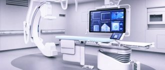
Such examinations are carried out mainly by large clinical and diagnostic centers that have the appropriate equipment, in particular:
- a fluorography unit operating at high speed;
- angiograph - a specialized system for X-ray studies of the vascular bed;
- camera for X-ray photo and video recording of results.
The most modern approach in this area is CT angiography. Devices for its implementation provide higher quality and more detailed images, and therefore more reliable results.
Find out the price
Find out the price
Error! Please fill in all required fields
Thank you! We will contact you shortly
✕
What pathologies will an MRI of the head vessels reveal?
- Cerebrovascular accidents caused by a variety of etiological factors.
- Complete or partial obstruction of blood through the vessels and veins in the area under study.
- Blockage of arteries with blood clots.
MRI examination also allows:
- Determine the stability of the spinal column if its incorrect position interferes with blood circulation.
- Assess the permeability of blood through the tissue structures of the brain.
- Detect impaired metabolism.
- Obtain a layer-by-layer detailed image of vessels, arteries, veins and capillaries in the area under study.
- Assess the condition of the cerebral cortex.
Indications for MRI of head vessels
- TBI (traumatic brain injury) or other physical impact of a traumatic nature.
- Atherosclerosis (accumulation of cholesterol in the walls of blood vessels, which causes their narrowing and blockage).
- Dilation of the walls of blood vessels and pathological forms of inflammation occurring in them.
- Compression of the great vessels.
- Aortic dissection, dystrophic changes in its tissues.
- Congenital and acquired heart defects.
- Painful reaction at the junction of the neck and skull, intensifying when turning/tilting the head.
- Headaches (usually pulsating) of unknown etiology.
- Pathological narrowing of arteries and veins.
- Strokes and heart attacks.
- Tumor processes (malignant and benign), cysts.
- Suspicion of metastasis.
- Weakness, fatigue, dark spots before the eyes, dizziness.
- Deviations from the norm in blood pressure readings.
MRI image of a child's brain vessels
Decoding the results
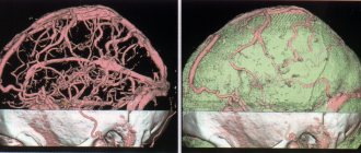
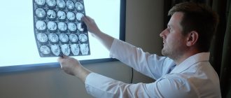
After processing the X-rays, the doctor places them on a backlit surface and examines them. Blood vessels and cerebrospinal fluid are black. They stand out clearly against the background of white bone tissue and gray brain matter.
Smooth lines of blood vessels are considered a sign of health. They smoothly narrow and expand, and also branch, giving the blood network a resemblance to a tree. The contrast agent should fill the vessels evenly.
To identify pathology, the doctor compares the X-ray image of the examined person with that of a healthy person. If the image is unclear or the doctor has doubts, he may order a repeat angiogram.
Deviations from the norm
A sharp narrowing or dilation of a vessel is considered a deviation from the norm. Aortic aneurysm and other types of this pathology cause protrusion of the wall of the blood vessel. If intracranial hemorrhage has occurred, the doctor will find dark spots surrounded by ring-shaped stripes on the image.
Over time, the thickness of the strips decreases. Therefore, the presence of thick stripes indicates that the hemorrhage occurred recently.
The tumor can be seen on the x-ray. Neoplasms displace blood vessels to the side, disrupting the uniformity of vessel branching. Tumors can compress blood vessels, impairing blood circulation in certain areas of the brain. Areas where tissues are starved of oxygen appear lighter in color. On them, the blood vessels are narrowed or not visualized.
What does MRI of head vessels show?
An MRI study of cerebral vessels will detect a violation of the blood supply in a certain area of the area being studied. To study in more detail the smallest changes in blood microcirculation, the study is carried out using a contrast agent (often gadolinium), which is injected into the patient intravenously.
MRI diagnostics allows you to timely detect the consequences of strokes and degenerative processes occurring in the tissues and arteries of the brain. This research method also makes it possible to diagnose Alzheimer's disease, multiple sclerosis, and demyelination (selective damage to the myelin sheath).
When describing the images obtained, the terms “filling defect” or “wall protrusion” are often used.
When an intraluminal thrombus was discovered during diagnosis, the contours of the aneurysm on tomograms appear as an irregularity with sharp, jagged edges. The defect itself appears to be either partial or poorly functioning. The signal from the thrombosed area of the aneurysm disappears on tomograms or has a chaotic periodicity.
Methodology
Before conducting the examination, the doctor must obtain written consent from the patient. After placing a catheter in a peripheral vein, necessary for immediate administration of drugs, the patient is premedicated. He is administered painkillers and a tranquilizer to achieve maximum patient comfort and relieve pain. The patient is connected to special devices to monitor his vital functions (oxygen concentration in the blood, pressure, heart rate).
Next, the skin is treated with an antiseptic to prevent infection, and contrast is injected into the carotid or vertebral arteries during direct angiography, and into the femoral artery during indirect angiography. If indirect angiography is performed, a catheter is also inserted into the femoral artery, which is pushed through the vessels into the desired artery in the brain. This procedure is completely painless, since the inner vascular wall has no receptors. The movement of the catheter is monitored using fluoroscopy. Most often, indirect angiography is performed.
When the catheter has reached the required location, a contrast volume of 9-10 ml is injected into it, having previously been warmed to body temperature. Sometimes, a few minutes after the administration of contrast, the patient is bothered by a feeling of heat and the appearance of an unpleasant metallic taste in the mouth. But these feelings quickly pass.
After the contrast is administered, two x-rays of the brain are taken - in the lateral and direct projections. The images are assessed by a radiologist. If there are still uncertainties, it is possible to reintroduce contrast and take two more photographs.
At the end, the catheter is removed, a sterile bandage is applied to the insertion site, and the patient is monitored for 24 hours.
6. Contraindications for MRI of head and neck vessels
This diagnostic procedure is strictly contraindicated when:
- The presence in the body of foreign bodies of any origin and nature, containing metal or elements from it. These may include surgical staples, pins, pacemakers, unrecovered shot fragments, hearing aids, insulin stimulators, dentures, and even braces.
- Chronic and acute renal failure, pregnancy, during the lactation period, with identified allergies - since the study of blood vessels, arteries and veins is often carried out using a contrast agent. It is toxic in certain doses and can negatively affect the condition of a weakened body.
- For epilepsy and claustrophobia, MRI diagnostics are not advisable.
Types of cerebral angiography
According to the established classification, this diagnostic technique is divided into 2 types:
- Selective - targeted, local. When performing selective cerebral angiography, an iodine-containing contrast agent is injected into an arterial vessel that supplies one of the brain regions.
- Survey is an extended research method. A contrast agent is injected into the area of the main artery, which is responsible for the general cerebral blood supply and nutrition. Using this technique, a specialist can carefully examine all cerebral vessels.
The optimal type and method of implementation is determined by a specialist individually, taking into account the characteristics and severity of the clinical case.
Description of diseases diagnosed using arteriography
Arteriography can only be prescribed by a specialist. Patient complaints of dizziness, migraine and tinnitus are not indications for a contrast study. With such symptoms, the patient should consult a doctor to determine the need for a procedure.
With the help of a contrast MRI study you can:
- diagnose the development of a cerebral aneurysm;
- determine blockage or degree of narrowing of blood vessels (stenosis);
- check the location of vascular clips;
- determine the presence of arteriovenous malformation;
- diagnose vasculitis.
Each of the above diseases should be considered separately.
Angiography in Turkey
The Turkish Medical Center has all the modern equipment necessary to conduct this study. Our specialists are highly qualified doctors, each of whom has extensive experience in successfully performing angiographies of the brain and other organs. Therefore, examination in our clinic will be completely safe, fast and as accurate as possible. In addition, the cost of angiography in Anadolu is noticeably lower than in diagnostic centers in other countries.
The material was prepared in agreement with medical doctors.
Aneurysm
An aneurysm is a specific section of a blood vessel that is intensively supplied with blood and grows in size. The convex wall of the vessel often compresses neighboring tissues and nerve endings. Damage to the aneurysm, which results in hemorrhage, poses a particular threat.
An aneurysm is diagnosed in any part of the brain, but is more often localized in the area where branches originate from arteries. Aneurysms develop asymptomatically and only when they reach large sizes do they manifest clear symptoms. Main signs of pathology:
- deterioration in the quality of vision;
- severe headaches;
- nausea;
- sudden loss of consciousness.
Recommendations for patients
During the procedure, the patient may experience discomfort. Patients complain of a metallic taste in the mouth, warmth or burning sensation spreading throughout the body. Their facial skin may become red.
Such symptoms are not dangerous. They disappear after a few minutes. If the discomfort does not disappear, but intensifies, you need to tell your doctor about it.
After angiography, the patient must follow all the doctor’s recommendations. Stress and emotional overload should be avoided. If the catheterization angiography method was used, the limb into which the catheter was inserted into the blood vessel must be kept in an extended state.
It is necessary to avoid physical activity until complete recovery. If you experience any unpleasant sensations, you should immediately consult a doctor.
Arteriovenous malformation
The condition is manifested by an abnormal plexus of veins and arteries. The external appearance of the formation resembles a tumor. Malformation leads to disruption of the supply of oxygen and nutrients to the brain.
If the formation reaches a significant size, then it becomes the cause of defective functioning of the organ. Gradually, the veins stretch and weaken, which is ultimately complicated by hemorrhage.
The problem can only be dealt with surgically. In the absence of timely treatment, malformation leads to irreversible changes.
Signs of pathology depending on its type:
- Hemorrhagic. It is characterized by a sharp headache, paralysis of the limbs, weakness, difficulties in perceiving sounds and images.
- Torpidnaya. It is characterized by severe pain sensations that occur in a certain area of the brain. Long-term pathology leads to impairment of speech, thinking and visual functions.
The risk of complications from arteriovenous malformation increases in older people and pregnant women.
Classic angiography - how the procedure is performed
Classic angiography is an invasive procedure because it is accompanied by a violation of the integrity of the vessels. Therefore, diagnostic testing is carried out in a hospital. During the procedure, the subject remains on the table. The position of his body is recorded.
Before the procedure begins, the patient is given painkillers, tranquilizers and antihistamines to minimize the likelihood of adverse reactions and reduce discomfort. To eliminate pain during the injection, a local anesthetic is applied to the skin.
After the examination, a pressure bandage is applied to the injection site. The patient is prescribed bed rest. He is advised to drink a lot of water so that the body gets rid of iodine faster. The patient must remain in the hospital under the supervision of a doctor for at least 6-8 hours. Then he can return home.
Preparatory measures
- Before angiography, allergy tests with a contrast agent are performed. The subject is administered intravenously 2 ml of an iodine-containing drug. If he develops nausea, vomiting, nasal discharge, skin rash or cough within 10-15 minutes, the study is canceled.
- If no signs of allergy are found, the patient is prescribed clinical and biochemical blood tests, a general urine test, a coagulogram, as well as an analysis to determine the Rh factor and blood group.
- The subject must also do an ultrasound of the kidneys, an electrocardiogram and consult an anesthesiologist.
- After conducting laboratory and instrumental studies, the doctor finds out what chronic diseases the patient has and what medications he is taking. To prevent bleeding, the doctor stops medications that reduce blood thickness (anticoagulants).
- 10-14 days before angiography you need to give up alcohol.
- You cannot eat for 8-10 hours before the procedure. You are prohibited from drinking water for the last 4 hours before the test.
- The area of skin where the injection will be made must be carefully shaved.
- Before starting angiography, the examinee must remove all objects containing metal parts. They can distort the results of a diagnostic test.
- If the patient has previously had complications after angiography (venography, coronography, arteriography), he should inform the attending physician about this.
Contraindications to the procedure
The main contraindications to diagnostic testing are:
- allergic reaction to iodine;
- pregnancy;
- mental pathologies that do not allow the subject to lie still;
- diseases in the acute stage;
- bleeding disorders;
- renal failure;
- diabetes mellitus in the stage of decompensation;
- hyperthyroidism;
- coma.
Possible complications
Medications used for diagnostic testing sometimes cause an allergic reaction in the form of redness or rash on the skin. Some subjects experience vomiting, nausea and tachycardia. They complain of chills and loss of strength.
Hemorrhage may occur at the site where the blood vessel is punctured. It is extremely rare for people to develop more severe complications that cause kidney disease and pathologies of the cardiovascular system (stroke, myocardial infarction).
10. Stenosis and blockage of cerebral vessels
Stenosis is a narrowing of cerebral vessels that occurs due to the influence of unfavorable physiological factors, for example, deposits of calcium and cholesterol on the walls of blood vessels.
The narrowing of the intracranial arteries leads to the fact that certain areas of the brain cease to receive enough oxygen. The risk of complications increases when blood vessels narrow by more than 50%. The process causes strokes.
Signs of a problem depend on which part of the brain has undergone a pathological change (anterior, middle or posterior). The main symptoms of stenosis and occlusion include:
- unilateral or bilateral damage to the facial muscles;
- violation of speech functions;
- visual defects (flickering “fly spots”, split images);
- severe headaches that do not disappear after taking medications;
- increase in pressure.
The likelihood of recovery from stenosis and blockage of blood vessels depends on the timely detection and treatment of the pathology. The longer a person does not see a doctor, the less chance he has for a full recovery. The disruption of blood flow to the brain is temporary, i.e. it regularly recovers and worsens again.
MRI signs of pathologies
Using arteriography, a specialist can assess the condition of any vessel.
In a healthy person, these structures have smooth outlines and the same degree of narrowing throughout.
The signal coming from different structures (soft tissue, brain matter, bone structures) is not the same. The bones in the photographs are shown in white, the cerebrospinal fluid and blood vessels are shown in black. The substance of the brain is visualized in gray in the images.
Today, arteriography is considered the most informative method for identifying vascular pathologies. After the procedure, only 5% of cases experience complications associated with allergies to contrast agents.
11.1 MRI signs of arteriovenous malformation
In the photographs, the areas of the malformation are berry-shaped. The picture on T1 and T2 WI depends on the stage of development of the pathology and the rate of hemoglobin decay in the areas of the malformation.
A gradient echo allows you to identify the presence of pathology and the degree of its neglect. In patients with hereditary cavernous malformation, the study makes it possible to determine the number of intertwined arteries and veins, which cannot be detected using other diagnostic methods.
Images (SWI) are no less sensitive than gradient echo. In addition, such images make it possible to clearly see the presence of calcifications on the walls of the arteries, which cannot be diagnosed on T1 and T2 weighted images.
In the presence of recent hemorrhage, perifocal edema is visualized on T1 and T2 WI. The MP signal on T1 WI usually does not change its intensity after the administration of contrast agents.
11.2 MRI - signs of narrowing and blockage of blood vessels
Images obtained after arteriography visualize compressed vessels. The penetration of the contrast agent into this area is slowed down. Pathological areas in the images are displayed in light color.
As a result of the examination, the specialist pays attention to the following criteria:
- lengthening of arteries;
- the presence of calcifications on the walls;
- wall compaction;
- dilation of arteries and vessels in certain areas.
The combination of MRI and MRA makes it possible to identify stenosis with 100% probability and create a competent surgical plan. The image shows vasoconstriction in varying degrees.
Initial stage of stenosis
Progressive form of pathology
The disease is in an advanced stage
The degree of pathology is determined by the ratio of the diameter of the healthy area of the artery to the pathological area. With complete or partial occlusion, there may be no blood flow in the artery.
Stenosis and blockage of blood vessels according to arteriography images resembles an artifact in the cavernous segment of the carotid artery. For this reason, only highly qualified specialists should decipher images.
Rare diseases characterized by blockage of the arteries include Moya-Moya pathology. Damage to cerebral vessels is observed bilaterally and is well visualized using standard MRI. Signs of pathology in the images are a narrowing of the ICA and the presence of many dilated vessels in the deep parts of the brain.
The image shows Moya-Moya pathology on T1 WI in the sagittal plane.
11.3 Signs of an aneurysm
Arteriography allows specialists to determine the size of the aneurysm. When a contrast agent is injected, a clear image of the veins and arteries appears on the images. By making multiple sections, aneurysmal dilatation of thrombosed and nonthrombosed etiology can be determined.
Using images obtained in three-dimensional projection, the shape and size of the formation are determined.
With a thrombosed aneurysm, images obtained using MR angiography reveal areas of lack of blood flow in the basilar artery. The photo is shown below.
11.4. Signs of vasculitis
Features of the clinical picture of the disease depend on the extent of the inflammatory process and the area of the brain affected. The symptoms of the disease are varied - from muscle pain to complete dysfunction of individual organs and systems.
Using MR arteriography, stenosis of intracranial arteries and disruption of blood flow in them are determined. On T1-weighted images after contrast administration, pronounced ischemic disorders of cerebral circulation are visualized.
Vasculitis is considered a rapidly progressing pathology, with the formation of numerous foci of infarction in the structures of the brain. The gray matter in the supratentorial region also undergoes pathological changes during the disease.
Possible complications
Despite the high level of safety for patients of different ages, angiography may result in the development of negative consequences for the patient. The most commonly observed conditions are:
- release of a radiopaque substance from the vascular bed into the surrounding tissues. This situation can lead to inflammatory changes of varying severity;
- allergic reactions to the contrast agent or its individual intolerance. In such cases, the patient may experience itching, urticaria, Quincke's edema and other allergy-specific symptoms;
- acute renal dysfunction, as a complication of the examination, is observed in patients with their diseases.
To prevent complications of the procedure, it is necessary to ensure a comprehensive examination of the patient before the study.
Related posts:
- Psychology and psychiatry, what is the difference? A modern person’s entire life is accompanied by a mass of disturbing phenomena, accompanied by stress...
- Hyperkinesis with developmental delay Translated from Greek, “hyperkinesis” means “supermovement”, which accurately reflects...
- Burning in the sternum, what it means and what it can lead to. Pain and burning in the chest are quite common symptoms,…
- Plaque on the tongue: a signal of a serious illness? A white coating on the tongue means that something is happening in the oral cavity...
