Certain centers of the brain are responsible for the performance of any mental, physical or sensorimotor function. Modern scientists compare it with a perfect computer, capable of efficiently processing a large flow of information and performing many actions at the same time. Each individual lobe performs important functions, including the medulla oblongata, the part of the brain that connects it to the spinal cord. It has long been known that its main function is not just connective. Physiological scientists have found that this small area plays a huge and important role for the entire organism as a whole. To understand what its role is in human life, let’s look at the anatomy and main functions of this brain area in more detail.
The main functionality of an important department
Experts consider the functions of the medulla oblongata to be vital, given that even a minor disruption or failure in its functioning can lead to complex consequences.
| Functions | Content | Implementation mechanism |
| Sensory | Analysis of taste and auditory sensations Transmission of sensitivity of the facial nerves The work of vestibular stimuli | Processing and sending impulses received from external stimuli to the subcortex |
| Conductivity | Work of ascending and descending paths | Transporting impulses to important brain areas |
| Reflex | Vital | Sucking, chewing, swallowing Static, statokinetic |
| Minor | Increased and decreased heart rate, Increased salivation |
Structure
The structure and functions of the medulla oblongata, characterized by its small size and unremarkable appearance, are in fact closely interrelated. It is this small area of the brain that has many nuclei in its structure, as well as many ascending and descending pathways that serve as conductors of signals and impulses.
The structure of the medulla oblongata is distinguished by the presence of important nerve receptors and centers in this area:
- glossopharyngeal nerve;
- accessory nerve;
- vagus nerve;
- hypoglossal nerve;
- part of the vestibulocochlear nerve.
Experts draw attention to the fact that even minor injuries and lesions that the medulla oblongata can take on can lead not only to complex consequences, but also to death.
Nuclear structure of the medulla oblongata
For a brief description and determination of the level of damage, it is necessary to know about the symptoms that develop during pathological processes in the posterior cranial fossa. The medulla oblongata has a specific structure and functions determined by the location of the nuclei of 5, 8, 9, 10, 11, 12 pairs of nerves.
Nuclear damage to the trigeminal nerve is manifested by a violation of pain and temperature types of sensitivity. The sensation of a light touch does not suffer. This is most typical for syringomyelia.
With nuclear damage to the vestibulocochlear nerve, dizziness and nystagmus appear, and the friendly rotation of the eyes in the direction opposite to the head suffers.
The glossopharyngeal and vagus nerves have common nuclei. The functional state of these cranial nerves is checked together. They innervate the larynx, pharynx, posterior third of the tongue, internal organs of the abdominal and thoracic cavities, tonsils, hearing organs, dura mater, and heart.
The medulla oblongata regulates the vital functions of the body, so bilateral damage to these nerves in combination with the sublingual may be incompatible with life, since bulbar syndrome develops.
https://youtube.com/watch?v=JX3OT2DFyeQ
The latter is characterized by impaired swallowing, voice, breathing, and disorders of cardiovascular activity. This situation develops with tumors, amyotrophic lateral sclerosis, pseudorabies, polio, and diphtheria.
With strokes, pseudobulbar palsy develops, which, in addition to the above symptoms, is manifested by violent emotional reactions in the form of laughter or crying, the appearance of pathological pyramidal symptoms, a decrease in productive mental activity, impaired coordination of movements, and central paralysis of the limbs.
Knowing the location of the nuclei in the medulla oblongata, one can clearly understand at what level the damage occurred.
The nerves suffer on the side of the pathological process, and on the opposite side sensitivity and motor functions are impaired. This phenomenon is caused by the intersection of motor and sensory pathways at the level of the pyramids. As a rule, such symptoms occur with vascular pathology in the system of the carotid, vertebral, and spinal arteries.
Sensory functional regulation
The activity of the sensory functions of the medulla oblongata is aimed at receiving signals from sensory receptors that respond to changes in the external or internal environment.
Experts identify several main areas of internal reactions:
- Reception and analysis of sensory signals sent by the respiratory system. The medulla oblongata processes the information received and analyzes not only the state of the respiratory organs, but also the quality of metabolic processes. It is on the basis of analytical results that the brain center makes a decision to change the cyclicity, duration or reflex activity of the respiratory organs.
- Recognition and analysis of signals coming from taste and digestive receptors. In the medulla oblongata, a step-by-step analysis of the complex combination of the state of chewing, gustatory and digestive functions takes place, which is then analyzed by the main brain centers.
The medulla oblongata region of the brain is also capable of analyzing and transporting sensory signals coming from external stimuli:
- Changes in ambient temperature, overheating, hypothermia.
- Damage to the skin, irritation of pain receptors.
- Auditory, tactile and visual signals of varying intensity and frequency.
Main nuclei of the medulla oblongata
The nerve centers of the medulla oblongata organize pairs of cranial nerve nuclei:
- Pair IX - glossopharyngeal nerves, are composed of three parts: motor, affective and autonomic. The motor region is responsible for the movements of the muscles of the pharyngeal canal and the oral cavity. The affective region receives signals from the gustatory sensory system at the back of the tongue. Vegetative regulates the secretion of saliva.
- The X pair is the vagus nerve, which includes three nuclei: the autonomic one is responsible for the regulation of the larynx, esophagus, cardiovascular system, gastrointestinal tract and digestive glands. The nerve contains afferent and efferent fibers. The sensitive nucleus catches signals from receptors in the lungs and other internal systems. The motor nucleus controls contractions of the oral muscles during swallowing. There is also a mutual nucleus (n. ambiguus), the axons of which are activated when a person coughs, sneezes, vomits the contents of the stomach and changes the intonation of the voice.
- The XI pair is an accessory nerve, divided into 2 parts: the first is closely interconnected with the vagus nerve, and the second is directed to the muscles of the sternum, key and trapezius muscles. With pathology of the XI pair, disturbances in head movements occur - it throws back or moves to the side.
- XII pair is the hypoglossal nerve, responsible for the motility of the tongue. Regulates muscles such as the styloglossus, mentalis, as well as the rectus and transverse muscles of the tongue. The functions of the XII pair also include, in part, the reflexes of swallowing, chewing and sucking. The composition includes mainly motor neurons. The nuclei control lingual motor skills during the process of eating and grinding food, and the movement of the mouth and tongue during conversation.
The structure also contains the cuneate and tender nuclei, along the paths of which signals pass to the somatosensory area of the cortex. The cochlear nuclei regulate the auditory system. The nuclei of the underlying olivary control the transmission of impulses to the cerebellum.
In the underlying caudal region of the myelencephalon there is a hemodynamic center that interacts with the fibers of the 5th pair of nerves. It is assumed that it is from this area that excitatory activating signals of sympathetic fibers to the cardiovascular system are born. This fact is confirmed by studies on the intersection of the caudal areas of the medulla oblongata, after which the level of blood pressure did not change.
Also located within the structure is a section of the reticular formation. The axons of the locus coeruleus secrete the hormone norepinephrine, which affects the excitability of nerve cells. This center controls reactions such as tension and anxiety.
Respiration processes are controlled thanks to the respiratory center, which is located between the superior region of the pons and the underlying region of the medulla oblongata. Violations of this center lead to cessation of breathing and death.
Conductor functional role
Receiving signals from external and internal stimuli is not the only role function of the medulla oblongata. This brain area, after processing the received information, performs transporting or conducting functions:
- Neurons of an important section of the elongated shape transmit the necessary information to the sections of the central nervous system along descending and ascending pathways.
- The medulla oblongata ensures the transportation of signals and impulses to parts of the brain, where they are analyzed and processed for the purpose of subsequent response.
Medulla oblongata
The medulla oblongata (lat. Myelencephalon, Medulla oblongata) is one of the most important links that make up the structure of the brain. This section is represented by a continuation of the spinal cord in the form of its thickening, and also connects the brain to the spinal cord.
The oblong section looks very much like an onion. Below the medulla oblongata is the spinal cord, and above it is the pons. It turns out that this section connects the cerebellar part and the brain bridge with the help of special processes (legs).
In children in the first month of their life, this section is larger in size compared to other sections. Around the age of seven and a half, nerve fibers begin to become covered with a myelin sheath. This gives them additional protection.
Structure and structure of the oblongata section
In adults, the length of the oblong section is approximately 2.5-3.1 centimeters , which is where it got its name.
Its structure is very similar to the spinal cord and consists of gray and white brain matter:
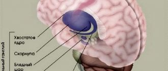
- The gray part is located in the center of the brain and forms nuclei (clumps).
- The white part is located on top and envelops the gray matter. It consists of fibers (long and short).
The nuclei of the medulla oblongata are different, but they perform one function and connect it with other parts.
Types of kernels:
- olive-like kernels;
- Burdach and Gaulle kernels;
- nuclei of nerve endings and cells.
These kernels include:
- sublingual;
- accessory vagus;
- glossopharyngeal and descending nuclei of the ternary nerves.
Pathways (descending and ascending) connect the main brain with the spinal cord, as well as with some parts. For example, with the reticular pharmacy, striopalidal system, cerebral cortex, limbic system and upper parts of the brain.
The medulla oblongata acts as a conductor for some reflex functions of the body.
These include:
- vascular;
- cardiac;
- digestive;
- vestibular;
- skeletal;
- protective.
It also houses some regulatory centers .
These include:
- management of respiratory functions;
- regulation of saliva secretion;
- regulation of vasomotor functions.
Availability of main centers
The functions of the medulla oblongata directly depend on the structure and presence of reflex centers and their work.
The center responsible for digestion is capable of taking on important functions:
- regulation and regulation of salivation;
- sucking and chewing functions;
- production and transportation of gastric juice;
- swallowing.
The protective center, which performs a number of important functions for the body, is responsible for the following processes:
- lacrimation to moisturize and wash the organs of vision;
- cough spasm;
- sneezing reflex;
- blinking to protect the eye from drying out;
- gag reflex for timely cleansing of the digestive system from toxic sources.
The centers of the medulla oblongata, aimed at regulating the tone of skeletal muscles, are located in this important section and perform the following functions:
- control and regulation of human behavior;
- formation of stability in space;
- coordination of movements;
- facial functions.
The anatomy of the vegetative centers allows them to perform the following functions:
- the function of breathing regulation is aimed at supporting the normal activity of the respiratory muscles;
- Cardiovascular function ensures the functioning of the heart organ, normalization of arterial parameters, and optimization of the condition of arterial vessels.
The external and internal structure of the vital part of the brain called the medulla oblongata is characterized by complex completeness. Thanks to this, important transport and metabolic functions are ensured in this area and it communicates with other areas and parts of the brain and components of the central nervous system.
What are the functions of the medulla oblongata
The medulla oblongata of the human brain is responsible for maintaining vital functions - breathing and blood circulation. Damage to the structure results in instant death.
The oblongata has age-related characteristics - at the time of birth, cells are developed only to regulate respiratory function, maintain blood circulation and control digestive processes. The cells mature as the child grows; the structure is fully functional from about 7 years of age.
The bulb comes from the rhomboid part of the brain and is the main conductor for incoming information from the analyzers.
The main functions of the oblong structure:
- Control over the circulatory system (work of the heart muscle, stabilization of blood pressure);
- Management of digestive processes (enzyme production);
- Control of muscle tone;
- Control of unconditioned reflexes;
- Controlling the respiratory center (gas exchange).
The functional set of the medulla oblongata is determined by the peculiarities of the internal structure (nucleus) and various pathways (long, short) that connect the central nervous system into a single management mechanism.
Based on this, the functionality of the bulb is divided into 4 groups:
- Sensory;
- Conductor;
- Reflex;
- Integrative.
All unconditioned reflexes are generated from the medulla oblongata, which in turn transmits impulses to the posterior brain structure.
Table of the functional orientation of centers in the medulla oblongata:
| Name | Purpose |
| Glossopharyngeal nerve | Controls secretion in the parotid gland and regulates the stylopharyngeal muscle, which elevates the larynx. Responsible for triggering protective reflexes in the oral cavity and larynx. Regulates taste sensitivity on the tongue. |
| Nervus vagus | Innervates transversely striated and smooth muscles. The muscles in the oral cavity are responsible for the motor capabilities. Participates in the production of secretion in the digestive organs. |
| Accessory nerve | Controls complex motor capabilities of muscles (turning the head in the opposite direction, bringing the shoulder blades together, raising one shoulder higher than the other). |
| Hypoglossal nerve | Regulates all muscles of the tongue and is partially responsible for innate reflexes (swallowing, sucking, chewing). |
| Olive Kernel | The intermediate vestibular center is responsible for the unconditioned muscle reflex when losing balance. |
| Gaulle and Burdach bundles | Responsible for sensitivity in the limb. |
| blue spot | Responsible for the response to anxiety or other irritant. |
There are connections in brain structures - these are short and long pathways. Therefore, the functional affiliations of the centers of the medulla oblongata and other structures may overlap with each other. Centers regulate the work of only a specific organ or system.
The centers in the oblong structure are formed by a cluster of nuclei and surrounding cells that interact with each other and control reflexes.
The functions of the medulla oblongata are divided into:
- Primary – maintaining vital unconditioned reflexes (breathing, heart function);
- Secondary – reception and transmission of information from sensory analyzers, control of autonomic reflexes.
After analyzing the information, an impulse is transmitted to the organ or system in order to increase activity or, conversely, suppress it (inhibition).
Sensory
The bulb processes the flow of information that comes from all sensory analyzers.
Function regulation:
- Reading information from skin receptors (on the face);
- Recognition of taste sensations;
- Monitoring the functioning of the vestibular apparatus and regulating changes;
- Perception of sound waves from auditory receptors.
After receiving information in the bulb, a primary analysis takes place, which takes into account the strength of the incoming signal. After processing, impulses with information are transmitted to subcortical structures, where sensations are read and distributed according to biological significance.
Conductor
The spinal cord tracts pass through the medulla oblongata - ascending and descending, as well as the origin of the tracts. Therefore, the structure takes part in the control of muscle contractions and tonic reactions.
The medulla oblongata structure has bilateral nerve connections with other parts of the brain and the cortex (corticoreticular connection). This connection allows the bulb to take part in the control of skeletal muscles and manage autonomic reactions.
The white matter of the bulb contains motor pathways that regulate the functioning of the facial muscles and tongue. Near the pyramids, the nerve fibers form a cross - this allows the threads to move to the opposite side and descend to the spinal cord canal.
Integrative
The medulla oblongata controls complex reflexes through nerve connections and interaction with other centers.
Communication with the vestibular centers and the oculomotor system helps control the eyes when the head is deviated. Some neurons in the brainstem region perform automatic functions - they provide tone and support the active work of many centers in the medulla.
Reflexes
Important reflexes of the medulla oblongata are provided in conjunction with other structures of the brain stem. Few people realize that habitual reflex motor actions are easily supported by the anatomy of the medulla oblongata.
- Reflexes, called postural reflexes by physiologists, are regulated by the centers of the medulla oblongata in conjunction with the control of higher levels of the central nervous system. It is with the help of this complex that a person easily changes postures and body position, including during sleep.
- Rectifying reflexes include the work of the vestibular apparatus and neck muscles. It is these reflexes that ensure normal upright posture of the body and regulation of balance.
- Labyrinthine reflexes regulate the position of the head relative to the body, control the distribution of tone in all muscle parts, maintain body balance, and control instant changes in muscle position.
- Cervical indicators, in turn, control the constant position of the head, its turns and tilts. When the medulla oblongata is damaged, disorders also occur in the cervical muscles.
Physiology of the medulla oblongata and pons
The medulla oblongata and the pons perform two functions - reflex and conductive. Through the sensitive fibers of the roots of the cranial nerves, it receives impulses - information from the receptors of the scalp, mucous membranes of the eyes, nose, mouth (including taste buds), from the organ of hearing, the vestibular apparatus (organ of balance), from the receptors of the larynx, trachea, lungs, as well as from interoreceptors of the cardiovascular system and digestive apparatus.
Through the medulla oblongata, many simple and complex reflexes are carried out, covering not individual metameres of the body, but organ systems, for example, the digestive, respiratory, and circulatory systems. The reflex activity of the medulla oblongata can be observed in a bulbar cat, that is, a cat in which the brain stem above the medulla oblongata has been cut. The reflex activity of such a cat is complex and diverse.
The following reflexes occur through the medulla oblongata: 1) protective: coughing, sneezing, blinking, lacrimation, vomiting; 2) food: sucking, swallowing, secretion of juice from the digestive glands; 3) cardiovascular, regulating the activity of the heart and blood vessels; 4) in the medulla oblongata there is an automatically working respiratory center that provides ventilation to the lungs; 5) the vestibular nuclei are located in the medulla oblongata and the pons.
From the vestibular nuclei of the medulla oblongata begins the descending vestibulospinal tract, which is involved in the implementation of posture reflexes, namely in the redistribution of muscle tone. A bulbar cat can neither stand nor walk, but the medulla oblongata and cervical segments of the spinal cord provide those complex reflexes that are elements of standing and walking. All reflexes associated with the standing function are called positioning reflexes. Thanks to them, the animal holds its body, usually with the crown up.
The special importance of this part of the central nervous system is determined by the fact that the medulla oblongata contains vital centers: respiratory, cardiovascular. Therefore, not only removal, but even damage to the medulla oblongata ends in death.
In addition to the reflex function, the medulla oblongata performs a conductive function. Conducting pathways pass through it, connecting the cortex, diencephalon, midbrain, cerebellum and spinal cord with a two-way connection.
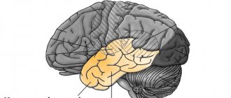
The cerebellum is located dorsal to the pons and medulla oblongata. It has two hemispheres and a middle part - the worm. The surface of the cerebellum is covered with a layer of gray matter (cerebellar cortex) and forms narrow convolutions separated by grooves. With their help, the surface of the cerebellum is divided into lobules. The central part of the cerebellum consists of white matter, which contains accumulations of gray matter - the cerebellar nuclei. The largest of them is the dentate nucleus. The cerebellum is connected to the brain stem by three pairs of peduncles: the upper ones connect it to the midbrain, the middle ones to the pons, and the lower ones to the medulla oblongata. The peduncles contain bundles of fibers connecting the cerebellum to various parts of the brain and spinal cord.
During development, the isthmus of the rhombencephalon forms the boundary between the hindbrain and midbrain. From it develop the superior cerebellar peduncles, the superior medullary velum located between them and the triangles of the loop, lying outward from the superior cerebellar peduncles.
During development, the fourth (IV) ventricle (ventriculus quartus) is a common cavity of the medulla oblongata and hindbrain. At the bottom, the IV ventricle communicates with the central canal of the spinal cord, at the top it passes into the cerebral aqueduct of the midbrain, and in the roof area it is connected by three openings to the subarachnoid space of the brain. Its anterior (ventral) wall—the bottom of the fourth ventricle—is called the rhomboid fossa. The lower part of the rhomboid fossa is formed by the medulla oblongata, and the upper part by the pons and isthmus. The posterior (dorsal) wall - the roof of the IV ventricle - is formed by the superior and inferior medullary sails and is supplemented at the back by a plate of the pia mater lined with ependyma. This area contains a large number of blood vessels that form the choroid plexus of the fourth ventricle. The convergence of the superior and inferior sails protrudes into the cerebellum and forms a tent. The rhomboid fossa is of vital importance, since most of the nuclei of the cranial nerves (V - XII pairs) are located in this area.
Physiology of the cerebellum
The cerebellum is a suprasegmental part of the central nervous system that does not have a direct connection with the receptors and effectors of the body. It is connected in numerous ways to all parts of the central nervous system. Afferent pathways are sent to it, carrying impulses from proprioceptors of muscles, tendons, vestibular nuclei of the medulla oblongata, subcortical nuclei and cerebral cortex. In turn, the cerebellum sends impulses to all parts of the central nervous system.
The functions of the cerebellum are studied by irritating it, partially or completely removing it, and studying bioelectrical phenomena. The Italian physiologist Luciani characterized the consequences of removal of the cerebellum and loss of its functions with the famous triad A: astasia, atony and asthenia. Subsequent researchers added another symptom: ataxia.
A dog without a cerebellum stands on widely spaced legs and makes continuous rocking movements (astasia). She has impaired proper distribution of flexor and extensor muscle tone (atony). Movements are poorly coordinated, sweeping, disproportionate, abrupt. When walking, the paws are thrown beyond the midline (ataxia), which is not observed in normal animals. Ataxia is explained by the fact that movement control is impaired. Analysis of signals from proprioceptors of muscles and tendons is missing. The dog cannot get its muzzle into the food bowl. Tilt of the head downwards or to the side causes a strong opposite movement.
The movements are very tiring: the animal, after walking a few steps, lies down and rests. This symptom is called asthenia.
Over time, movement disorders in a cerebellar dog smooth out. She eats on her own and her gait is almost normal. Only biased observation reveals some violations (compensation phase).
As E. A. Asratyan showed, compensation of functions occurs due to the cerebral cortex. If the bark of such a dog is removed, then all the violations are revealed again and are never compensated.
The cerebellum is involved in the regulation of movements, making them smooth, precise, proportionate. According to the figurative expression of L. A. Orbeli, the cerebellum is an assistant to the cerebral cortex in controlling skeletal muscles and the activity of autonomic organs. As studies by L.A. Orbeli have shown, cerebellar dogs have impaired autonomic functions. Blood constants, vascular tone, the functioning of the digestive tract and other autonomic functions become very unstable and easily shift under the influence of certain reasons (food intake, muscle work, temperature changes, etc.).
When half of the cerebellum is removed, motor functions on the side of the operation are impaired. This is explained by the fact that the cerebellar pathways either do not cross at all or cross twice.
Midbrain
The midbrain includes the cerebral peduncles, located ventrally, and the roof plate (lamina tecti), or quadrigemina, lying dorsally. The cavity of the midbrain is the cerebral aqueduct. The roof plate consists of two superior and two inferior colliculi, which contain the nuclei of gray matter. The superior colliculi are associated with the visual pathway, the inferior colliculi with the auditory pathway. From them originates the motor pathway that goes to the cells of the anterior horns of the spinal cord. On a cross section of the midbrain, three of its sections are clearly visible: the roof, the tegmentum and the base of the cerebral peduncle (Fig. 114). Between the tire and the base there is a black substance. The tegmentum contains two large nuclei - the red nuclei and the nuclei of the reticular formation. The cerebral aqueduct is surrounded by central gray matter, which contains the nuclei of the III and IV pairs of cranial nerves. The base of the cerebral peduncles is formed by fibers of the pyramidal tracts and tracts connecting the cerebral cortex with the nuclei of the bridge and the cerebellum. The tegmentum contains systems of ascending pathways that form a bundle called the medial (sensitive) loop. The fibers of the medial lemniscus begin in the medulla oblongata from the cells of the nuclei of the thin and cuneate fasciculi and end in the nuclei of the thalamus. The lateral (auditory) loop consists of fibers of the auditory tract running from the pons to the inferior colliculi of the pontine tegmentum (quadrigeminal) and the medial geniculate bodies of the diencephalon.
Rice. 114. Cross section of the midbrain (diagram). 1 - cerebral peduncle; 2 - black substance; 3 — roof plate; 4 - red core; 5 - nucleus of the oculomotor nerve; 6 - oculomotor nerve; 7 - cerebral aqueduct
Physiology of the midbrain
The midbrain plays an important role in regulating muscle tone and implementing the righting and righting reflexes, which make standing and walking possible.
The role of the midbrain in the regulation of muscle tone is best observed in a cat in which a transverse incision is made between the medulla oblongata and the midbrain. Such a cat has a sharp increase in muscle tone, especially extensor muscles. The head is thrown back, the paws are sharply straightened. The muscles are so strongly contracted that an attempt to bend the limb ends in failure - it immediately straightens. An animal placed on outstretched paws like sticks can stand. This condition is called decerebrate rigidity. If the incision is made above the midbrain, then decerebrate rigidity does not occur. After about 2 hours, such a cat makes an effort to get up. First she raises her head, then her body, then stands on her paws and can begin to walk. Consequently, the nervous apparatus for regulating muscle tone and the functions of standing and walking are located in the midbrain.
The phenomena of decerebrate rigidity are explained by the fact that the red nuclei and reticular formation are separated from the medulla oblongata and spinal cord by transection. The red nuclei do not have a direct connection with receptors and effectors, but they are connected with all parts of the central nervous system. They are approached by nerve fibers from the cerebellum, basal ganglia, and cerebral cortex. The descending rubrospinal tract begins from the red nuclei, through which impulses are transmitted to the motor neurons of the spinal cord. It is called the extrapyramidal tract
The sensitive nuclei of the midbrain perform a number of important reflex functions. The nuclei located in the superior colliculi are the primary visual centers. They receive impulses from the retina and participate in the orientation reflex, i.e. turning the head towards the light. At the same time, the width of the pupil and the curvature of the lens (accommodation) change, which contributes to clear vision of the object.
The nuclei of the inferior colliculi are the primary auditory centers. They participate in the orienting reflex to sound - turning the head in the direction of the sound. Sudden sound and light stimulation cause a complex alarm reaction (start reflex), mobilizing the animal for a quick response.
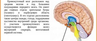
Diencephalon
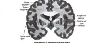
The diencephalon is located under the corpus callosum and fornix, fused on the sides with the cerebral hemispheres. It includes the thalamus (visual thalamus), epithalamus (supra-tubercular region), metathalamus (sub-tubercular region) and hypothalamus (sub-tubercular region). The cavity of the diencephalon is the third ventricle.
The thalamus is a paired, ovoid collection of gray matter covered by a layer of white matter. The anterior sections are adjacent to the interventricular foramina, the posterior sections are expanded to the quadrigeminal. The lateral surfaces of the thalamus grow together with the hemispheres and border the caudate nucleus and the internal capsule. The medial surfaces form the walls of the third ventricle, the lower ones continue into the hypothalamus. In the thalamus, there are three main groups of nuclei: anterior, lateral and medial, and there are 40 nuclei in total. In the epithalamus lies the upper appendage of the brain - the pineal gland, or pineal body, suspended on two leashes in the recess between the upper colliculi of the roof plate. The metathalamus is represented by the medial and lateral geniculate bodies, connected by bundles of fibers (handles of the colliculi) with the superior (lateral) and inferior (medial) colliculi of the roof plate. They contain nuclei that are reflex centers of vision and hearing.
The hypothalamus is located ventral to the thalamus and includes the subcutaneous region itself and a number of formations located at the base of the brain. These include: the terminal plate, the optic chiasm, the gray tubercle, the infundibulum with the lower appendage of the brain extending from it - the pituitary gland and the mastoid bodies. In the hypothalamic region there are nuclei (supravisceral, periventricular, etc.) containing large nerve cells capable of secreting a secretion (neurosecretion) that flows along their axons into the posterior lobe of the pituitary gland and then into the blood. In the posterior part of the hypothalamus lie nuclei formed by small nerve cells, which are connected to the anterior lobe of the pituitary gland by a special system of blood vessels.
The third (III) ventricle is located in the midline and is a narrow vertical slit. Its lateral walls are formed by the medial surfaces of the thalamus and the subtubercular region, the anterior - by the columns of the fornix and the anterior commissure, the lower - by the formations of the hypothalamus, and the posterior - by the cerebral peduncles and the supracuberous region. The upper wall - the lid of the third ventricle - is the thinnest and consists of the soft membrane of the brain, lined on the side of the ventricular cavity with an epithelial plate (ependyma). The soft shell has a large number of blood vessels here, forming the choroid plexus. In front, the third ventricle communicates with the lateral ventricles (I - II) through the interventricular foramina, and behind it passes into the cerebral aqueduct.
Protective reflex functions
Reflexes related to regulators of muscle tone, maintaining posture and organizing movements are considered important for maintaining and normalizing orientation in space and performing coordination functions.
No less important functions of the medulla oblongata are considered to be protective reflexes:
- The sneezing reflex is necessary to cleanse the mucous membrane from the entry of dust particles, microbes, viruses, and allergic agents into the mucous membrane of the nasopharynx.
- The gag reflex refers to a reflexive urge aimed at removing contents from the stomach. This happens against the backdrop of the need to rid the body of poor quality food and toxic products. In some situations, such cleansing may be necessary to normalize a person’s condition.
- The swallowing reflex and sucking reflex are normally activated in a child immediately after birth and accompany the person for the rest of his life. These reflexes are considered vital, as they are actively involved in food consumption and subsequent digestion. Otherwise, a person is deprived of the opportunity to consume food naturally.
The role of the medulla oblongata in ensuring and normalizing human life is difficult to overestimate. Deprived of the normal functioning of the medulla oblongata, the body loses many important functions and vital capabilities.