Learn more about diseases starting with the letter “P”: Panic attacks; Duchenne-Erb's palsy; Todd's palsy; Parasomnias; Paroxysmal hemicrania; Paroxysmal myoplegia, Paroxysmal positional vertigo; Peripheral autonomic failure; Hepatic encephalopathy; Pineoblastoma; Pineocytoma; Writer's cramp; Platybasia; Plexites; Brachial plexitis; Humeroscapular periarthrosis; Pneumococcal meningitis Pneumocephalus; Subacute sclerosing panencephalitis; Spinal cord injury.
Description of the disease
Duchenne-Erb's palsy is a degeneration of the nerve plexus in the shoulder area. As the anomaly develops, there is a violation of muscle tone, loss of sensitivity and atrophy of the muscles in this area. The most common is the secondary congenital form of the disease, which develops after injury to the child during obstetrics. Clinical manifestations include stiffness when trying to abduct, lift, or bend the upper limb at the elbow. Sensitivity and motor functionality in the fingers are preserved.
The term and definition of pathology were introduced and described by European doctors in the early 70s of the 19th century. The disease was named after them. Both specialists identified a direct connection between motor function disorders in the superior brachial plexus in infants and assisted births. In modern medical literature there is also such a definition as “proximal upper paralysis”. According to statistical data, the disorder has an incidence of 1-2 cases per 1000 children born. A number of specialists from various fields of medicine are developing methods to prevent injury to infants: neurologists, orthopedists, traumatologists, pediatricians, neonatologists, gynecologists and obstetricians.
Symptoms
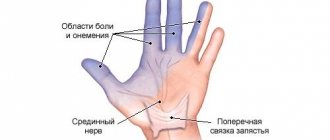
The brachial bundle carries impulses to the musculocutaneous, axillary and partially radial nerve. Therefore, when it is damaged, their functioning is disrupted, which is accompanied by loss of sensitivity, numbness in the proximal parts of the limb, a crawling sensation, and a burning sensation.
Disruption of the innervation of the biceps, brachialis, brachioradialis and deltoid muscles is expressed by disruption of their work and severe muscle weakness.
The clinical course of the disease and the severity of symptoms depend on the degree of nerve damage. If the damage is minor, the disease may be virtually asymptomatic, manifesting itself with slight weakness. For more extensive disorders or when the bundle ruptures, the following symptoms are characteristic:
- it is impossible to bend the elbow joint independently;
- due to limited mobility of the shoulder joint, the limb hangs freely along the body. The patient cannot take it to the side or lift it up on his own;
- the arm hanging along the body cannot be turned outward. Because of this, the palm is turned back from the body;
- it is difficult for the patient to straighten the hand, so the palm is constantly bent;
- straightening the fingers is difficult, sometimes impossible;
- reduced skin sensitivity on the outer side of the lower limb;
- there is no flexion-elbow reflex;
- decreased muscle tone;
- decreased skin temperature and pallor.
Erb's paresis is characterized by pain at Erb's point when pressing on it. It is localized above the collarbone at the attachment point of the sternocleidomastoid muscle.
Such symptoms are typical for children and adults. The initial examination by a neonatologist plays a huge role in determining the symptoms of a newborn. The following signs and symptoms are typical for young patients:
- when lifting a child and holding it horizontally in a “column” position, the arm hangs down freely;
- lack of active movements in the injured arm and its free hanging or lying along the body when the child lies on his stomach or back;
- the grasping reflex is not evoked;
- there is no palmar-oral reflex.
Total obstetric Duchenne-Erb palsy is characterized by complete loss of sensation and lack of active movements in the hand. A healthy limb functions normally.
Also, the severity of symptoms depends on the stage of the disease. There are three of them:
- Acute – a month from the onset of the disease. The symptoms are pronounced.
- Rehabilitation – duration 1-3 years. With a mild course and non-serious, low-grade nerve damage, the lost functions are restored and the patient’s condition is normalized. If the damage is severe and the necessary treatment is not carried out, irreversible changes may develop: muscle atrophy, limb shortening, joint contractures.
- Residual effects – after 3 years. Shoulder dislocations and subluxations may occur against the background of muscle atrophy. At the junction of the shoulder and the body, a “puppet” groove develops. Also a common complication of the disease is curvature of the spine and abnormal placement of the scapula.
Reference. It is important to determine the signs by comparing reflexes, muscle tone and strength of the affected arm with the healthy one.
Etiology of Duchenne-Erb's palsy
As stated earlier, the most common occurrence is upper obstetric paralysis, which develops as a result of birth injury to the fetus. In other age categories, the disease can be caused by damage to the brachial spinal roots located in the area of the 5th and 6th vertebrae. Nerve fibers can be damaged by a strong upper blow, a fall to the side of the arm, a bruise on a static object, by “pulling” the arm to the side, or a penetrating wound into the specified area.
Infantile paralysis is formed when the nerve trunk of the brachial plexus is stretched during turning by the leg, pulling the fetus by the arms, releasing the shoulder from the birth canal, or traction by the pelvis. The described manipulations are necessary in case of incorrect presentation of the child, insufficiently intense labor, a protracted period of delivery, narrow anatomy of the mother’s pelvis, or a large fetus. Often, along with acquired paralysis, there is a combination with torticollis.
The primary manifestations of the injury are inflammation and hematoma in the damaged area. A tear or complete rupture of nerve fibers, vascular walls, and muscles occurs. Over time, acute manifestations disappear, however, at the site of the violation of tissue integrity, scars form, which put pressure on the nerve plexus and the vessels feeding the department.
Characteristic features of the disease
Duchenne-Erb paresis is an acquired disease, the development of which is provoked by damage to the spinal cord at the level of the V-VI segments. The pathology is manifested by a significant decrease in the functional activity of the arm, the inability to fully flex and abduct it. It also occurs in adults with severe trauma, but is more often diagnosed in infants if obstetric instruments were used by medical personnel during passage through the birth canal. The development of paralysis in newborns is not necessarily the fault of medical professionals. Often such obstetric manipulations help save the lives of both the child and the mother.
Since the nerve plexuses are affected, the innervation is disrupted. There is a violation of the regulation of the muscles and ligamentous-tendon apparatus of the shoulder by the central nervous system. Impulses from it arrive untimely, in a chaotic order. This innervation disorder is manifested by the following pathological conditions:
- severe muscle weakness in areas localized close to the forearm;
- decreased strength, loss of sensitivity;
- limitation of range of motion;
- lack of reflex to grasp objects in newborns;
- impaired flexion of the arm at the elbow joint as a result of a disorder in the conduction of nerve impulses in the biceps muscle.
In adults, the situation is aggravated by ruptures of muscle fibers, ligaments and tendons of the shoulder joint. Extensive inflammatory swelling forms, pressing on sensitive nerve endings. This further upsets the innervation and causes severe discomfort.
ON A NOTE! The volume of lesions that occur affects the intensity of Erb's birth palsy. But in the vast majority of cases, lost functions of the upper limb are restored with competent, adequate treatment.
Signs of pathology
Clinical manifestations of the disease are divided into three stages, characterized by different symptoms:
1. Acute stage. It begins immediately after injury and lasts for a month after it. During this period, the arm is in one position - extended at the elbow, pressed to the body, fingers brought to the palm. The patient cannot independently move his arm forward, up, to the side, or rotate the inner surface of the limb to the front position. Any manipulations or attempts to change the position of the limb cause pain and discomfort. The biceps reflex is not recorded on the affected side. Infants lack a grasp reflex on the side of paralysis. When the child is placed in a horizontal position, a “limp” hanging of the arm is observed. Muscle tone is reduced, the skin is cold and discolored, and the sensory system is little susceptible to pain applied to the skin.
2. Recovery stage. Depending on the severity of the injury, the timeliness of therapeutic care and the adequacy of medical manipulations, the following symptoms will appear. With mild paralysis, sensitivity and activity are gradually restored to full functionality. However, in children by the age of three, a slight shortening of the affected arm and muscle weakness are noticeable.
3. Stage of residual effects. Since functionality does not return to full extent during the recovery period, subsequent disorders develop. If the paralysis is moderate or severe, the patient complains of “doll hand” syndrome. It is manifested by a clearly defined fold between the sternum and shoulder area. The structure of muscle tissue changes, the elbow joint loses its flexion ability. A pronounced rotation of the scapula is clearly visible, the fingers remain in palmar presentation. Since the muscles atrophy, subsequent chronic dislocation of the shoulder area is possible. The consequence of the altered anatomy of the upper spinal column is the curvature of the latter, scoliosis manifestations.
Causes and manifestations of the disease
Trauma during childbirth leads to motor disorders of the upper limbs in 2 babies out of 1000 newborns. The occurrence of traumatic muscle damage is caused by excessive stretching of the shoulder region with a fixed position of the head and neck. The functionality of the muscles that abduct the shoulder, rotate it outward and lift it upward, the forearm flexors and supinators is impaired. Movements in the hand are almost always preserved. The severity of obstetric Duchenne-Erb paresis is determined by the number of damaged roots of the brachial plexus.
Reasons that can lead to injury:
- large fetal weight (over 4 kg), which does not coincide with the size of the birth canal;
- protracted or too rapid labor , contributing to the unnatural development of processes;
- first birth , during which an unprepared woman experiences abnormal breathing and pushing behavior;
- breech presentation (buttocks forward), which increases the risk of Duchene-Erb's palsy when the head is released;
- the obstetrician uses special tools to extract the fetus, releasing the baby’s shoulders with his fingers.
Clinical picture
The nature of the lesion of the brachial plexus depends on the traumatic factor, the duration of its impact, the pressure applied and the location of application.
In Duchenne-Erb syndrome, birth paralysis from a minor traumatic injury only leads to stretching of the nerve plexus. Severe trauma during childbirth can cause nerve roots to be torn from the spinal cord. A timely visit to a doctor to determine the nature and extent of the lesion will allow you to develop a plan of treatment measures that will contribute to the successful correction of developmental pathology after birth trauma. Symptoms of upper Duchenne-Erb palsy include the following:
- limited or lack of mobility in the shoulder and elbow joints;
- weakening of the dorsiflexion of the hand while maintaining mobility in the fingers;
- individual reflexes are not evoked: palmo-oral, biceps, rhomboid and other muscles;
- the grasping reflex is weakened on the affected side;
- the arm is flaccid, straightened at the joints, turned inward at the shoulder, lies along the body with a bent palm, lowered shoulder and head tilted towards it, muscle tone is reduced.
How to diagnose the disease?
Preliminary collection of anamnesis, the presence of notes in the child’s medical history about difficult childbirth, and keeping the arm in a constant extended position helps specialists make a correct diagnosis. The disease in adulthood requires differentiation from other pathologies with similar symptoms.
The diagnosis is made by neurologists and orthopedists. The congenital disorder is diagnosed by a neonatologist in the first days of a baby’s life. Hardware methods for diagnosing the disease include:
- X-ray examination. Helps to detect changes occurring in the bones of the studied area. In the first months of a small patient’s life, a discrepancy in the space of the intra-articular area is visible. Signs of bone atrophy may be detected.
- Ultrasonography. Used as a confirmatory method.
- Myelography.
- Electroneurography. The intensity of the impulse passing through the trunk of the nerve canal is assessed.
- Magnetic resonance imaging. Used as a companion diagnostic to assess the condition of soft tissues.
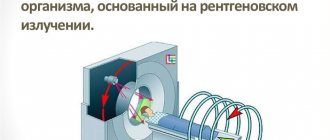
- Computer scanning. The most informative method for a planned operation. Equally effectively visualizes both bone formations and nerve pathways.
Tomographic hardware examination can only be carried out in a medical organization that has the necessary equipment and experienced functional diagnosticians. Not every district clinic is able to provide appropriate services. To quickly search for diagnostic MRI[/anchor] and CT, our portal provides a list of rating clinics located in each district of the capital. Users of the portal can study detailed contact information, compare prices for diagnostic services, get a free consultation and sign up for a scanning procedure by calling the hotline located at the top of the page.
Diagnostics
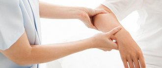
The disease is diagnosed based on the collected medical history: severe complicated childbirth, use of special equipment, fall, injury, etc., and a characteristic clinical picture. In infants, Duchenne-Erb paresis is often diagnosed after the first examination by a neonatologist. If a hand dysfunction is detected, additional consultation with a neurologist and orthopedist is required. In adults, the diagnosis is made by traumatologists together with neurologists.
Additionally, testing is carried out to determine the level of sensitivity loss and reflexes are checked. Doctors also perform x-rays of the shoulder and electroneuromyography. The degree of damage to the nerve plexus can be determined using CT myelography. It allows you to determine the degree of disruption of the nerve and the location of the damage.
Treatment of Duchenne-Erb's palsy
In the acute period of the disease, doctors resort to complete restriction of movements in the affected limb. To do this, an immobilization splint is applied. Parallel restorative manipulations are:
- visiting a massage therapist;
- exercise therapy classes;
- physiotherapeutic measures.
At this stage, pharmacological painkillers, anti-inflammatory drugs, medicinal substances that improve blood circulation and metabolic processes can be prescribed.
If therapeutic measures are poorly effective, the patient is referred for consultation to a neurosurgeon. The specialist evaluates the feasibility of surgical intervention. In childhood, instrumental correction is best performed at 6-7 months of life. Therefore, after three months of conservative treatment, you need to think about possible surgery. During neurosurgical manipulations, corrective plastic surgery of the nerve trunk is performed. Only microsurgical techniques are used.
With prolonged treatment in the stage of residual effects, it is no longer possible to restore the full functionality of the limb. Orthopedic interventions aimed at restoring the anatomical position of the hand can improve the condition. Some motor abilities that were previously lost may also be restored.
Treatment
Treatment of upper Erb-Duchenne palsy in the case of a mild lesion is not difficult and begins in the maternity hospital, before the child is discharged home. Special splints are applied to the affected limb to hold the arms in a certain position. Many splint models have the ability to change their fixing position as clinical manifestations decrease.
From the very first days, the child is prescribed neuroprotective vitamin therapy, ATP preparations, and in some cases aloe, lidase, and proserin. At the same time, therapeutic massage and gymnastics are carried out.
In case of mild lesions, treatment takes about a month; for more severe lesions, it may take longer. The prognosis for recovery is usually favorable. But even in the case of severe paralysis, regular exercise therapy, massage courses and neuroprotective drug therapy lead to positive changes.
Alexey Borisov (neurologist)
Practicing neurologist, otoneurologist (specialist in dizziness).
Graduated from Irkutsk State Medical University. Head of the dizziness office. Regularly takes advanced training courses, participates and makes presentations at educational conferences, including those with international participation. He has a large number of printed scientific publications.
More about me
Source
ICD-10 Category: P14.0
ICD-10 / P00-P96 CLASS XVI Selected conditions arising in the perinatal period / P10-P15 Birth trauma / P14 Birth trauma of the peripheral nervous system
Definition and general information[edit]
Erb-Duchenne palsy
Upper flaccid paresis of the arm associated with damage to the brachial plexus due to birth trauma.
Epidemiology
Mainly observed in full-term infants. The incidence of brachial plexus injuries is 0.5-2 per 1000 live births; among them, 90% of cases are Erb-Duchenne palsy. The most common injury to the brachial plexus is associated with shoulder dystocia.
Forecast and preventive measures
With minor damage to the shoulder and timely initiation of therapy, complete restoration of functionality is possible with conservative treatment. Statistically, 70% of positive treatment results are recorded in infancy. A fifth of those recovering received 100% regeneration of tissues and nerve fibers. If a complete rupture of the plexus occurs, therapy is powerless and surgical treatment is necessary.
As preventive measures for obstetric paralysis, close monitoring of pregnancy and childbirth and an adequate choice of method of delivery are recommended. To prevent adult paralysis, injury should be avoided.
Treatment of the disease
It is recommended to treat Duchenne palsy using special techniques, which will have a positive effect on this process. With timely treatment of the pathological process and the correct selection of a set of measures, the prognosis will be favorable. A specialist can determine Duchenne palsy by external signs during the initial examination of the newborn.
If the cause of pathology in adult patients is trauma, then this requires the same approach to treatment. The patient is only advised to increase the dosage of the prescribed drug in accordance with individual characteristics.