Arachnoiditis is an inflammation of the arachnoid membrane of the brain or spinal cord. This pathology accounts for about 5% of the total number of organic lesions of the nervous system. As a rule, arachnoiditis is diagnosed in patients under the age of 40 (it can often be detected in a child).
In the true (diffuse) type of the disease, the autoimmune component dominates. Residual conditions of arachnoiditis are the consequences of fibrosis of the membranes after traumatic injuries or infections.
The disease code according to ICD 10 is G03.9.
General information
Today in neurology, a distinction is made between true arachnoiditis, which has an autoimmune genesis, and residual conditions caused by fibrous changes in the arachnoid membrane after a traumatic brain injury or neuroinfection (neurosyphilis, brucellosis, botulism, tuberculosis, etc.).
In the first case, arachnoiditis is diffuse in nature and characterized by a progressive or intermittent course, in the second, it is often local in nature and is not accompanied by a progressive course. Arachnoiditis
Etiology
Arachnoiditis occurs as a result of acute and chronic infections, inflammatory diseases of the paranasal sinuses, chronic intoxication (alcohol, lead, arsenic), injuries (usually in the residual period). Arachnoiditis can also occur as a result of reactive inflammation with slowly growing tumors, encephalitis. In many cases, the cause of arachnoiditis remains unclear.
Morphologically, arachnoiditis is characterized by clouding and thickening of the arachnoid membrane, accompanied in more severe cases by fibrinoid deposits. In the further course of arachnoiditis, adhesions occur between the arachnoid and choroid, leading to disruption of the circulation of cerebrospinal fluid and the formation of arachnoid cysts.
Arachnoiditis can occur due to acute or more often chronic purulent otitis media (as a result of low-virulent microbes or toxins), as well as complications of purulent otitis media - labyrinthitis, petrositis, sinus thrombosis, as a consequence of cured purulent meningitis or brain abscesses and, finally, can be combined with non-purulent otogenic encephalitis.
Arachnoiditis is divided into diffuse and limited. The latter are extremely rare. Essentially, we are talking about more severe local changes against the background of a diffuse process in arachnoiditis.
Disruption of the normal circulation of cerebrospinal fluid, leading to the occurrence of hydrocephalus, is based on two mechanisms in arachnoiditis:
- impaired outflow of fluid from the ventricular system (occlusive hydrocephalus)
- impaired absorption of fluid through the dura mater during a diffuse adhesive process (aresorptive hydrocephalus)
Causes
The main cause of the pathology is various types of brain injuries or previous infectious diseases. The basis for the development of the inflammatory process may be:
- Measles. A fairly common disease, one of the consequences of which can be arachnoiditis.
- Flu. It is caused by viruses that provoke inflammation. If the disease is not treated in time, the pathological process spreads to areas of the brain.
- Meningitis, which is viral in nature. Meningitis is characterized by inflammation that can affect certain areas of the brain.
- Chicken pox. Many people do not consider it a dangerous disease, but in some cases, the chickenpox virus can trigger the development of arachnoiditis.
Diagnosis of meningitis
An infectious and inflammatory disease in the soft meninges, accompanied by an acute course, is called meningitis.
In addition, the disease can occur as a result of purulent inflammations, which are chronic in nature, and foci form in the skull. These include:
- Otitis. A pathological process that occurs in the ear cavity, if left untreated, spreads to the brain tissue.
- Sinusitis and rhinitis. Pus forms in the nasal cavity.
- Mastoiditis. Inflammation is observed in the temple area. The cause of development is various infectious diseases of the ears.
- Tonsillitis.
- Infectious diseases in the oral cavity. These also include various diseases of the gums and teeth, which are characterized by inflammation and the formation of pus.
Arachnoiditis in 30% of cases begins to develop as a result of traumatic brain injury. This occurs in cases where the injury is accompanied by significant hemorrhage.
In medical practice, there are also cases when doctors cannot determine the exact cause of the pathology. Based on the data obtained, after many years of research, specialists were able to establish a number of factors that can trigger the development of arachnoiditis.
These include:
- Frequent colds .
- Intoxication after drinking alcohol or prolonged use of drugs.
- Weakening of protective functions.
- Chronic fatigue. It often causes vegetative-vascular dystonia, which is characterized by impaired blood circulation to the brain. This may be the basis for the development of arachnoiditis.
- Difficult working conditions.
Timely treatment of colds and maintaining immunity will help to avoid the development of pathology.
Causes of arachnoiditis
In approximately 55-60% of patients, arachnoiditis is associated with a previous infectious disease.
Viral infections:
Chronic purulent foci in the skull area:
In 30% of cases, arachnoiditis is a consequence of a traumatic brain injury, most often subarachnoid hemorrhage or brain contusion, although the likelihood of arachnoiditis does not depend on the severity of the injuries received.
In 10-15% of cases, arachnoiditis does not have a clearly established etiology.
Factors predisposing to the development of arachnoiditis are chronic fatigue, various intoxications (including alcoholism), heavy physical labor in unfavorable climatic conditions, frequent acute respiratory viral infections, and repeated injuries, regardless of their location.
Etiology, pathogenesis. Past trauma, meningitis, and subarachnoid hemorrhage are important. Of the cerebral arachnoiditis, only arachnoiditis of the posterior cranial fossa is of practical importance, usually simulating a tumor of this localization. With spinal localization, arachnoiditis usually repeats the clinical picture of spinal cord compression.
Neither clinical blood tests nor examination of cerebrospinal fluid reveal specific changes. Only myelography helps to recognize arachnoiditis, which gives a characteristic picture that is different from the myepogram for a spinal cord tumor. The overwhelming majority of patients who have been treated for many years for “arachnoiditis” actually have neurosis-like (mainly asthenic, asthenodepressive) conditions, including persistent headache, and individual organic microsymptoms identified in this case become the starting point for erroneous diagnosis.
Etiological factors:
- previous infections (up to 60% of cases);
- tonsillitis;
- rhinosinusitis;
- otitis;
- contusions of the brain and spinal cord;
- frequent intoxication.
Please note: In 10% of cases, the exact cause of this disease affecting the brain cannot be identified.
Etiology of the disease, causes
It is impossible to single out one cause of the disease. Arachnoiditis of the brain can be either an independent nosology or a consequence of a previously suffered inflammatory process .
Most often it is provoked by tonsillitis, rheumatism, chronic inflammatory processes of the ENT organs, childhood infections that occur in adults, and traumatic brain injuries.
A number of cases have been described in which arachnoiditis manifested itself against the background of chronic intoxication with salts of heavy metals, oncology of the brain or spinal cord.
This disease was first classified and given its modern name in 1845, thanks to the work of A. T. Tarasenkov. It is also worth noting that the World Health Organization, when revising ICD-10, does not allocate a separate code for arachnoiditis, but classifies it as meningitis.
Symptoms of arachnoiditis
The disease develops subacutely with the transition to a chronic form. Clinical manifestations are a combination of general cerebral disorders, more often associated with intracranial hypertension, less often with liquor hypotension, and symptoms reflecting the predominant localization of the meningeal process. Depending on the predominance of general or local symptoms, the first manifestations may be different.
Among the general cerebral symptoms, headache is common, most intense in the early morning hours and sometimes accompanied by nausea and vomiting. The headache can be local, aggravated by straining, straining, or awkward movement with firm support on the heels (a symptom of jumping is a local headache when jumping with an unabsorbed descent on the heels).
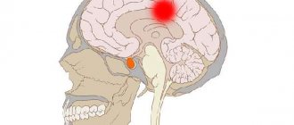
Focal symptoms depend on the location of arachnoiditis. Convexital arachnoiditis is characterized for the most part by a predominance of the phenomena of brain irritation over signs of loss of function. One of the leading symptoms is generalized and Jacksonian epileptic seizures. With basal arachnoiditis, general cerebral symptoms and dysfunction of the nerves located at the base of the skull are observed.
A decrease in acuity and changes in visual fields can be detected with opticochiasmatic arachnoiditis. Clinical manifestations and fundus images may resemble symptoms of optic neuritis. These manifestations are often accompanied by symptoms of autonomic dysfunction, severe dermographism, enhanced pilomotor reflex, profuse sweating, acrocyanosis, sometimes thirst, increased urination, hyperglycemia, adiposisogenital obesity).
In some cases, a decrease in sense of smell may be detected. Arachnoiditis in the area of the cerebral peduncles is characterized by the appearance of pyramidal symptoms, signs of damage to the oculomotor nerves, and meningeal signs. With arachnoiditis of the cerebellopontine angle, headaches in the occipital region, noise in the ear and paroxysmal dizziness, and sometimes vomiting occur.
Symptoms of damage to the auditory, trigeminal, abducens and facial nerves may be observed. Arachnoiditis of the large (occipital) cistern develops acutely, the temperature rises, vomiting, pain in the back of the head and neck appears, aggravated by turning the head, sudden movements and coughing; damage to the cranial nerves (IX, X, XII pairs), nystagmus, increased tendon reflexes, pyramidal and meningeal symptoms.
With arachnoiditis of the posterior cranial fossa, damage to the V, VI, VII, VIII pairs of cranial nerves is possible. Intracranial hypertension, cerebellar and pyramidal symptoms are often observed. Differential diagnosis with tumors of the posterior cranial fossa is mandatory. Lumbar puncture is performed only in the absence of congestion in the fundus.
The disease is characterized (without taking into account the nature, morphology and localization of the lesion):
- headache;
- sleep disorders (insomnia (insomnia at night) or daytime sleepiness);
- visual impairment;
- decreased performance.
Arachnoiditis of the brain is accompanied by a constant headache, which is further aggravated by overexertion. Intense pain syndrome under the influence of thermal factors (in hot or cool weather). Neurological disorders develop that are characteristic of the area where the inflammatory focus is located.
The convexital (damage to the frontal lobes of the brain) variety is characterized by focal (individual) or general convulsive seizures, accompanied by loss of consciousness.
With the basal variety, the arachnoid membrane of the trunk suffers. Based on localization, arachnoiditis of the posterior cranial fossa, opto-chiasmatic arachnoiditis and damage to the cerebellopontine angle are distinguished.
In the optic-chiasmal form, the optic nerve and fundus of the eye are affected. Consequence: complete loss of vision, severe metabolic disorders.
When the inflammatory focus is localized in the posterior cranial fossa, the cranial nerves are affected. The pain syndrome is localized in the back of the head. The patient has increased intracranial pressure. Congestion in the fundus cannot be ruled out. Symptoms are similar to clinical manifestations of neoplasms in the central nervous system.
Damage to the cerebellopontine angle is characterized by neuritis of the seventh pair of cranial nerves and a sharp decrease in hearing (on one side). As the disease progresses, cerebellar disorders develop. The appearance of clinical paresis (paralysis) of the limbs is possible.
With the development of cystic adhesive arachnoiditis, the main symptom is intense headache. Cystic arachnoiditis is characterized by seizures caused by irritation of the medulla in the area of formed adhesions and cavities between them.
Post-traumatic arachnoiditis that affects the spinal cord is characterized by weakness in the limbs.
Important: With complicated arachnoiditis, disability is possible!
The clinical picture of arachnoiditis unfolds after a significant period of time from the influence of the factor that caused it. This time is determined by the ongoing autoimmune processes and may differ depending on what exactly caused the arachnoiditis. So, after suffering from the flu, arachnoiditis manifests itself 3-12 months later, and after a traumatic brain injury, on average, after 1-2 years. In typical cases, arachnoiditis is characterized by a gradual, subtle onset with the appearance and increase of symptoms characteristic of asthenia or neurasthenia: increased fatigue, weakness, sleep disturbances, irritability, increased emotional lability. Against this background, the appearance of epileptic seizures is possible. Over time, cerebral and local (focal) symptoms accompanying arachnoiditis begin to appear.
General cerebral symptoms of arachnoiditis
General cerebral symptoms are caused by impaired cerebrospinal fluid dynamics and in most cases manifest themselves as cerebrospinal fluid hypertension syndrome. In 80% of cases, patients with arachnoiditis complain of a fairly intense bursting headache, most pronounced in the morning and aggravated by coughing, straining, and physical effort. Increased intracranial pressure is also associated with pain when moving the eyeballs, a feeling of pressure on the eyes, nausea, and vomiting.
Arachnoiditis is often accompanied by tinnitus, decreased hearing and non-systemic dizziness, which requires the patient to exclude ear diseases (cochlear neuritis, chronic otitis media, adhesive otitis, labyrinthitis). Excessive sensory excitability (poor tolerance of sharp sounds, noise, bright light), autonomic disorders and autonomic crises typical for vegetative-vascular dystonia may occur.
Often, arachnoiditis is accompanied by a periodically occurring sharp worsening of liquor-dynamic disorders, which clinically manifests itself in the form of a liquor-dynamic crisis - a sudden attack of intense headache with nausea, dizziness and vomiting. Such attacks can occur up to 1-2 times a month (arachnoiditis with rare crises), 3-4 times a month (arachnoiditis with moderate crises) and more than 4 times a month (arachnoiditis with frequent crises). Depending on the severity of symptoms, liquorodynamic crises are divided into mild, moderate and severe. A severe liquorodynamic crisis can last up to 2 days, accompanied by general weakness and repeated vomiting.
Focal symptoms of arachnoiditis
The focal symptoms of arachnoiditis may vary depending on its predominant localization.
Convexital arachnoiditis can manifest as mild to moderate impairment of motor activity and sensation in one or both extremities on the opposite side. In 35% of cases, arachnoiditis of this localization is accompanied by epileptic seizures. Usually there is polymorphism of epileptic seizures. Along with primary and secondary generalized ones, psychomotor simple and complex seizures are observed. After an attack, temporary neurological deficits may occur.
Basilar arachnoiditis can be widespread or localized primarily in the opticochiasmatic region, anterior or middle cranial fossa. Its clinical picture is mainly due to damage to the I, III and IV pairs of cranial nerves located at the base of the brain. Signs of pyramidal insufficiency may occur. Arachnoiditis of the anterior cranial fossa often occurs with impaired memory and attention, and decreased mental performance. Opticochiasmatic arachnoiditis is characterized by a progressive decrease in visual acuity and narrowing of the visual fields. These changes are most often bilateral in nature. Opticochiasmatic arachnoiditis can be accompanied by damage to the pituitary gland located in this area and lead to the appearance of an endocrine metabolic syndrome similar to the manifestations of a pituitary adenoma.
Arachnoiditis of the posterior cranial fossa often has a severe course, similar to brain tumors of this location. Arachnoiditis of the cerebellopontine angle, as a rule, begins to manifest as damage to the auditory nerve. However, it may begin with trigeminal neuralgia. Then symptoms of central neuritis of the facial nerve appear. With arachnoiditis of the cistern magna, a pronounced liquor-hypertensive syndrome with severe liquor-dynamic crises comes to the fore. Cerebellar disorders are characteristic: coordination disorders, nystagmus and cerebellar ataxia. Arachnoiditis in the area of the cistern magna can be complicated by the development of occlusive hydrocephalus and the formation of a syringomyelitic cyst.
Types of arachnoiditis
There are several types of arachnoiditis:
- Cerebral (ICD-10 code G00). Damage to the meninges occurs in various areas. This type is characterized by the presence of headaches of a hypertensive or meningeal nature. A person notices pain constantly; there are periods of its intensification after prolonged overheating and hypothermia. The manifestation of neurological syndromes will depend on the area of the lesion. Sometimes cerebral arachnoiditis is accompanied by attacks of convulsive focal seizures. With severe lesions, generalized convulsive seizures with loss of consciousness appear, which can lead to the development of epileptic seizures. Compression of the centers responsible for sensory and motor functions leads to disorders of sensitivity and movement such as mono- and hemiparesis. Depending on the affected area, cerebral arachnoiditis can be convexital (damage to the convex surface of the cerebral hemispheres), basal (at the base of the brain), opto-chiasmal (at the site of the optic chiasm), posterior cranial fossa and cerebellopontine angle.
- Spinal. The membranes of the spinal cord are damaged. The cause of the disease is purulent abscesses and boils. Sometimes spinal arachnoiditis is post-traumatic in nature. The inflammatory process spreads along the posterior surface of the spinal cord, which explains the presence of pain in the limbs. The disease is asymptomatic for a long time. The spinal type of arachnoiditis is divided into subtypes: cystic, adhesive and cystic-adhesive. These subspecies differ from each other in the nature of the process and symptoms:
- Cystic arachnoiditis is characterized by inflammation of the membranes of the spinal cord and is accompanied by the formation of cysts. Its manifestations sometimes resemble a tumor process. Patients complain of severe back pain and difficulty moving.
- Adhesive arachnoiditis is the spread of purulent exudate in the spinal cord, which inevitably leads to the formation of adhesions and the development of spinal compression syndrome.
- The cystic-adhesive type of arachnoiditis is characterized by the formation of zones of adhesion of the membranes to the substance of the brain. This process leads to constant irritation of the cerebral cortex, contributing to the development of seizures.
Pathogenesis of arachnoiditis
The arachnoid membrane is located between the dura mater and the pia mater. It is not fused with them, but fits tightly to the pia mater in places where the latter covers the convex surface of the convolutions of the brain. Unlike the pia mater, the arachnoid does not enter the cerebral convolutions, and under it in this area subarachnoid spaces filled with cerebrospinal fluid are formed. These spaces communicate with each other and with the cavity of the fourth ventricle. From the subarachnoid spaces, through the granulations of the arachnoid membrane, as well as through the perineural and perivascular slits, cerebrospinal fluid outflows from the cranial cavity.
Under the influence of various etiofactors, the body begins to produce antibodies to its own arachnoid membrane, causing its autoimmune inflammation - arachnoiditis. Arachnoiditis is accompanied by thickening and clouding of the arachnoid membrane, the formation of connective tissue adhesions and cystic expansions in it. Adhesions, the formation of which is characterized by arachnoiditis, lead to obliteration of the indicated outflow tracts of cerebrospinal fluid with the development of hydrocephalus and cerebrospinal fluid-hypertensive crises, causing the occurrence of cerebral symptoms. The focal symptoms accompanying arachnoiditis are associated with irritation and involvement of the underlying brain structures in the adhesive process.
Treatment of arachnoiditis
It is necessary to eliminate the source of infection (otitis media, sinusitis, etc.). Antibiotics are prescribed in therapeutic doses. Desensitizing and antihistamine drugs are indicated (diphenhydramine, diazolin, suprastin, tavegil, pipolfen, calcium chloride, histaglobulin). Pathogenetic therapy is designed for long-term course treatment with absorbable agents, normalization of intracranial pressure, improvement of cerebral circulation and metabolism.
Biogenic stimulants (aloe, vitreous, FiBS) and iodide preparations (bioquinol, potassium iodide) are used. Lidase is also used in the form of subcutaneous injections of 0.1 g of dry matter, dissolved in 1 ml of 0.5% novocaine solution every other day, for a course of 15 injections. The courses are repeated after 4-5 months. Pyrogenal has a resolving effect.
The first intramuscular injections of pyrogenal begin with a dose of 25 MTD, in subsequent days the dose is increased daily by 50 MTD and brought to 1000 MTD; for a course of treatment up to 30 injections. When intracranial pressure increases, decongestants and diuretics are used (mannitol, furosemide, diacarb, glycerin, etc.).
For convulsive syndromes, antiepileptic drugs are used. Metabolic therapy is carried out (glutamic acid, piracetam, aminalon, cerebrolysin). Symptomatic agents are used according to indications. Lack of improvement after treatment, increase in intracranial pressure and focal symptoms, opticochiasmatic arachnoiditis with a steady decrease in vision are indications for surgical intervention.
Forecast. In relation to life, usually favorable. Arachnoiditis of the posterior cranial fossa with occlusive hydrocephalus may pose a danger. The labor prognosis worsens with frequent relapses or a progressive course with frequent hypertensive crises, epileptic seizures, and the optico-chiasmatic form.
Treatment for arachnoiditis is usually carried out in a hospital. It depends on the etiology and degree of disease activity. The drug treatment regimen for patients with arachnoiditis may include:
- anti-inflammatory therapy with glucocorticosteroid drugs (methylprednisolone, prednisolone), absorbable agents (hyaluronidase, quinine iodobismuthate, pyrogenal)
- antiepileptic drugs (carbamazepine, levetiracetam, etc.)
- dehydration drugs (depending on the degree of increase in intracranial pressure - mannitol, acetazolamide, furosemide)
- neuroprotectors and metabolites (piracetam, meldonium, ginkgo biloba, pig brain hydrolysate, etc.)
- antiallergic medications (clemastine, loratadine, mebhydrolin, hifenadine)
- psychotropics (antidepressants, tranquilizers, sedatives).
An obligatory point in the treatment of arachnoiditis is the sanitation of existing foci of purulent infection (otitis media, sinusitis, etc.).
Severe optico-chaosmal arachnoiditis or arachnoiditis of the posterior cranial fossa in the case of progressive decrease in vision or occlusive hydrocephalus are an indication for surgical treatment. The operation may involve restoring the patency of the main cerebrospinal fluid pathways, removing cysts, or separating adhesions that lead to compression of nearby brain structures. In order to reduce hydrocephalus in arachnoiditis, it is possible to use shunt operations aimed at creating alternative pathways for the outflow of cerebrospinal fluid: cystoperitoneal, ventriculoperitoneal or lumboperitoneal shunting.
Treatment. Arachnoiditis of the posterior cranial fossa and spinal cord, occurring with a pseudotumorous picture, is usually treated surgically.
Diagnosis code according to ICD-10 • G03.9
Treatment of arachnoiditis is complex drug therapy carried out in a hospital setting.
Groups of medications prescribed for treatment:
- anti-inflammatory (glucocorticoids);
- anticonvulsants;
- dehydration agents (diuretics);
- absorbable;
- desensitizing agents (antihistamines);
- neuroprotective;
- general strengthening (vitamin complexes).
In case of acute infectious process, rational antibiotic therapy is carried out. Sanitation of possible foci of infection is imperative!
For some adhesive arachnoiditis, neurosurgical surgery is indicated.
Treatment
Treatment tactics are determined depending on the form and course of the pathology. In cases of severe and acute forms, therapy is carried out only in a hospital setting under the supervision of specialists.
Self-medication is dangerous with complications!
Attention
Despite the fact that our articles are based on trusted sources and have been tested by practicing doctors, the same symptoms can be signs of different diseases, and the disease may not proceed according to the textbook.
Pros of seeing a doctor:
- Only a specialist will prescribe suitable medications.
- Recovery will be easier and faster.
- The doctor will monitor the course of the disease and help avoid complications.
find a doctor
Do not try to treat yourself - consult a specialist.
For symptomatic treatment, hormonal, antibacterial and antiviral drugs are prescribed in small doses.
The drug is selected depending on the type of pathogen. To reduce swelling of brain tissue, antihistamines are prescribed, as well as medications to stimulate brain function and reduce intracranial pressure.
To relieve symptoms, the following groups of drugs are indicated:
- Analgesics to reduce pain.
- Antiepileptics to correct behavioral characteristics.
Test for meningitis
Meningitis is an inflammatory disease that occurs acutely in the membranes of the spinal cord and brain.
Drug therapy, when administered on time, allows the patient to return to an almost normal life. Surgical intervention is prescribed in cases where medications do not have the desired effect, as well as when an opto-chiasmal type is established.
A direct indication for surgery is also the cystic form, since drugs only relieve symptoms, but are not a solution to the problem.
Work ability
Patients are recognized as disabled group III if employment or transfer to light work leads to a decrease in the volume of production activities. Group II disability is established in the presence of frequent epileptic seizures, a significant decrease in visual acuity in both eyes (from 0.04 to 0.08 with correction).
Group I disabilities are patients with optico-chiasmatic arachnoiditis, accompanied by blindness. Patients with liquorodynamic disorders, epileptic seizures and vestibular crises are contraindicated from working at heights, near fire, near moving machinery, or in transport. Work in unfavorable meteorological conditions, in noisy rooms, in contact with toxic substances and in conditions of altered atmospheric pressure, as well as work associated with constant vibration and changes in head position are contraindicated.
Diagnosis of arachnoiditis
A neurologist can establish true arachnoiditis only after a comprehensive examination of the patient and comparison of anamnestic data, the results of a neurological examination and instrumental studies. When collecting anamnesis, attention is paid to the gradual development of symptoms of the disease and their progressive nature, recent infections or traumatic brain injuries. A study of the neurological status makes it possible to identify disorders of the cranial nerves, determine focal neurological deficits, psycho-emotional and mnestic disorders.
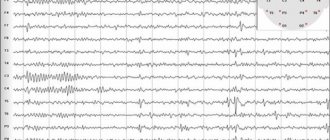
X-ray of the skull in the diagnosis of arachnoiditis is a low-informative study. It can only detect signs of long-standing intracranial hypertension: digital impressions, osteoporosis of the dorsum sella. The presence of hydrocephalus can be judged by echo-EG data. Using EEG, focal irritation and epileptic activity are detected in patients with convexital arachnoiditis.
Patients with suspected arachnoiditis must be examined by an ophthalmologist. In half of the patients with arachnoiditis of the posterior cranial fossa, ophthalmoscopy reveals congestion in the area of the optic nerve head. Opticochiasmatic arachnoiditis is characterized by concentric or bitemporal narrowing of the visual fields detected by perimetry, as well as the presence of central scotomas.
Hearing impairment and noise in the ear are a reason to consult an otolaryngologist. The type and degree of hearing loss are determined using threshold audiometry. To determine the level of damage to the auditory analyzer, electrocochleography, auditory evoked potential testing, and acoustic impedance measurements are performed.
CT and MRI of the brain make it possible to identify morphological changes that accompany arachnoiditis (adhesions, the presence of cysts, atrophic changes), determine the nature and degree of hydrocephalus, and exclude space-occupying processes (hematoma, tumor, brain abscess). Changes in the shape of the subarachnoid spaces can be detected during CT cisternography.
Lumbar puncture provides accurate information about the amount of intracranial pressure. Examination of cerebrospinal fluid in active arachnoiditis usually reveals an increase in protein to 0.6 g/l and cell number, as well as increased levels of neurotransmitters (eg, serotonin). It helps differentiate arachnoiditis from other cerebral diseases.
Important: The arachnoid mater is not affected in isolation. During infectious genesis, the pathogen is transferred from the dura or pia mater to the arachnoid. On this basis, arachnoiditis affecting the brain is considered serous meningitis (ICD 10 code for cerebral arachnoditis - G03.9). Diagnosing the disease is difficult, even with new technologies.
To make a diagnosis, anamnesis data, an objective assessment of mental and neurological status, test results and additional instrumental studies are required. Consultation with an otolaryngologist and ophthalmologist is required.
Research methods:
- lumbar puncture followed by laboratory examination of cerebrospinal fluid;
- electroencephalogram;
- Echo-EEG;
- CT (computed tomography);
- CT cisternography;
- MRI.
Please note: X-ray examination is not very informative. MRI provides useful and objective information.
Diagnostics
Diagnosis of arachnoiditis includes consultation with specialists and instrumental examination methods. If a pathology is suspected, the doctor prescribes a consultation:
- An ophthalmologist to examine the fundus of the eye.
- An otolaryngologist to identify foci of the pathological process or treat the consequences.
- A psychiatrist to assess the patient's condition.
CSF analysis for meningitis
Neurosurgeons, neurologists and infectious disease specialists often have to perform a lombal puncture, which is the collection of cerebrospinal fluid (CSF) from a patient.
After establishing the patient’s condition, an instrumental examination is prescribed, which includes:
- X-ray of the spine and skull.
- Encephalography.
- Lumbar puncture.
- Magnetic resonance imaging of the brain.
- Pneumoencephalography.
A set of diagnostic measures makes it possible to establish the focus of the inflammatory process and the degree of damage to areas of the brain. Based on the data obtained, a course of treatment is prescribed.
Notes
| Inflammatory diseases | |
| Nervous system | |
| central nervous system | Brain abscess • Encephalitis • Myelitis • Meningitis • Meningoencephalitis • Arachnoiditis • Rasmussen's encephalitis • Tick-borne encephalitis |
| Peripheral nervous system | Neuritis |
| Eye and ear | Eye: Dacryocystitis • Episcleritis • Keratitis • Retinitis • Blepharitis • Conjunctivitis • Iridocyclitis • Uveitis Ear: Otitis (external, middle) • Labyrinthitis • Mastoiditis • Eustachitis |
| The cardiovascular system | |
| Heart | Endocarditis • Myocarditis • Pericarditis |
| Arteries, Veins, Capillaries | Arteritis • Phlebitis • Capillaritis |
| Respiratory system | |
| Airways | Upper respiratory tract: Sinusitis • Rhinitis • Pharyngitis • Laryngitis • Nasopharyngitis Lower respiratory tract: Tracheitis • Bronchitis • Bronchiolitis • Alveolitis • Pneumonia • Pleurisy (pleural empyema) • Lung abscess |
| Other | Mediastinitis |
| Digestive system | |
| Gastrointestinal tract | Oral cavity: Stomatitis • Gingivitis • Glossitis • Tonsillitis (acute, chronic) • Mumps • Pulpitis • Periostitis • Inflammation of the jaw • Retropharyngeal abscess Other parts of the gastrointestinal tract: Esophagitis • Gastritis • Enteritis • Duodenitis • Colitis • Gastroenterocolitis • Appendicitis • Appendicitis • Proctitis |
| Other | Digestive glands: Hepatitis (Viral hepatitis (A, B, C, D, E, F, G, TTV), Toxic hepatitis, Radiation hepatitis, Autoimmune hepatitis, Steatohepatitis) • Pancreatitis (acute, chronic) Biliary tract: Cholecystitis (acute, chronic) • Peritoneal cholangitis: Peritonitis |
| Genitourinary system | |
| Urinary organs | Nephritis (Glomerulonephritis, Pyelonephritis, Paranephritis) • Cystitis • Urethritis |
| Female reproductive system | Adnexitis • Endometritis • Parametritis • Cervicitis • Vaginitis • Vulvitis • Vulvovaginitis • Mastitis |
| Male reproductive system | Orchitis • Epididymitis • Prostatitis • Balanitis |
| Germ tissues | Chorioamnionitis • Omphalitis |
| Other systems and organs | |
| Musculoskeletal system | Arthritis • Myositis • Bursitis • Osteochondritis (dissecans) • Tendinitis • Fasciitis • Osteomyelitis • Epicondylitis • Panniculitis |
| Leather | Dermatitis • Hidradenitis • Seizure • Acne |
| Blood | Bacteremia • Sepsis |
| Lymph nodes, Lymphatic vessels | Lymphadenitis • Lymphangitis |
| Central nervous system diseases | |
| Brain Encephalopathy | |
| Headache | Migraine • Cluster headaches • Vascular headache • Tension headache |
| Sleep disorders | Insomnia • Hypersomnia • Sleep apnea • Narcolepsy • Cataplexy • Kleine-Lewin syndrome • Sleep-wake cycle disorders |
| Movement and extrapyramidal disorders | Dyskinesia: Dystonia • Chorea • Myoclonus • Tremor (Essential tremor, Intention tremor) • Restless legs syndrome • Muscle stiffness syndrome Basal ganglia diseases: Parkinson's disease • Neuroleptic syndrome • Pantothenate kinase-associated neurodegeneration • Progressive supranuclear palsy • Striatonigral degeneration • Hemiballismus |
| Epileptic seizures Epilepsy | Localized epilepsy • Generalized epilepsy • Status epilepticus • Myoclonic epilepsy • Tuberous sclerosis |
| Dementia | Alzheimer's disease • Frontotemporal dementia/Frontotemporal lobar degeneration • Multi-infarct dementia |
| Cerebrovascular diseases | Transient disorders of cerebral circulation (Hypertensive cerebral crisis, Transient ischemic attack) • Discirculatory encephalopathy (Cerebral atherosclerosis, Subcortical atherosclerotic encephalopathy, Chronic hypertensive encephalopathy) • Stroke (Ischemic stroke, Intracerebral hemorrhage, Subarachnoid hemorrhage) • Thrombosis sinuses of the dura mater (Thrombosis of the cavernous sinus ) |
| Inflammatory diseases | Brain abscess • Meningitis • Arachnoiditis • Encephalitis • Meningoencephalitis • Rasmussen's encephalitis • Tick-borne encephalitis |
| Demyelinating diseases | Autoimmune diseases (Multiple sclerosis, Neuromyelitis optica, Schilder's disease) • Hereditary diseases (Adrenoleukodystrophy, Krabbe disease) • Central pontine myelinolysis • Marchiafava-Bignami syndrome • Alpers syndrome |
| System atrophy | Huntington's disease • Spinal ataxia Spinal muscular atrophy: Kennedy syndrome • Spinal muscular atrophy in children • Motor neuron disease • Fazio-Londe syndrome • Amyotrophic lateral sclerosis |
| Mitochondrial diseases | Leigh syndrome |
| Tumors | Brain tumor • Tuberous sclerosis |
| Cerebrospinal fluid | Intracranial hypertension • Cerebral edema • Intracranial hypotension |
| Injuries | Traumatic brain injury (Concussion, Brain contusion, Diffuse axonal brain injury) |
| Other diseases | Spina bifida • Reye's syndrome • Hepatic coma • Toxic encephalopathy • Hematomyelia |
| Spinal Cord Myelopathy | |
| Inflammatory diseases | Meningitis • Arachnoiditis • Meningoencephalitis • Myelitis • Poliomyelitis • Demyelinating diseases • Tropical spastic paraparesis |
| Other diseases | Syringomyelia • Syringobulbia • Morvan's syndrome • Vascular myelopathy • Spinal stroke • Spinal cord compression • Encephalomyelitis |