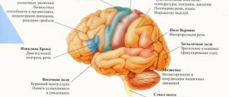
The central nervous system has delimited sections that interact with each other by transmitting and receiving impulses through thin conductors. Nerve fibers unite all structures into a single mechanism that controls the internal functioning of the body. Medulla oblongata - the functions of the department are associated with maintaining the vital functions of the body.
Division of the medulla oblongata
Structure of the medulla oblongata
The rhomboid section includes the bulb, which is an oblong structure. The bulb originates from a thick-walled canal in the spine and enters the brain stem. Anatomically, the structure is located between the exit of the first spinal nerve and the pons (superior border).
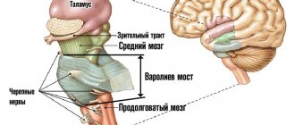
The medulla oblongata in humans is an important part of the central nervous system. The department is located at the junction of the spinal cord and brain. The spinal cord thickens at the entrance to the foramen magnum and visually becomes like an onion. Therefore, the department received a second name - onion.
The medulla oblongata combines the structure of two structures - the spinal cord and the brain. The bulb is the connecting link for the small brain and the pons.
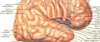
Anatomical structure and boundaries
On the surface of the medulla oblongata there is a median depression, a continuation of the spinal cord groove. Near the recess there are bundles of nerve fibers (pyramids), which pass into the spinal cords.
On the posterior surface of the oblong structure lies the dorsal part of the groove - it comes from the spinal cord. Near the recess (on the sides) lie the posterior cords - the ascending spinal tract. At the top, the cords branch in different directions and go to the cerebellum.
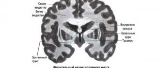
The structure of the bulb itself is heterogeneous, since it combines gray (neuron bodies) and white (processes) matter. The cell bodies of neurons are surrounded by numerous nuclei. They are covered on top by bundles of axons and dendrites. Long nerve fibers cross and pass into the spinal cord. Short processes extend from the long fibers through the oblongata (forming a medial loop) to the thalamus.
Short processes connect the nuclei of gray matter, which in turn continue to communicate with other nuclei. This connection connects all structures with each other and allows the brain to function as a single organ.
The anatomy of the bulb is divided into:
- External structure;
- Internal.
The special structure and functions of the medulla oblongata are inextricably linked with the control of all autonomic reactions in the body, and are also aimed at maintaining life support systems.
External structure
The outer part of the bulb is formed from pyramidal tracts - paired lobes in the form of cones, with an extension at the top and a gap. To the side of the pyramids there is an oval-shaped extension - an olive. It contains nuclei and is delimited by the anterolateral part of the sulcus (continuation of the spinal cord). The connection between the spinal cord and the brain has arcuate fibers.
The external structure of the back of the bulb has a dividing groove, which makes it visually similar to parts of the cylinder. Bundles of fibers coming from the outside have connections with the dorsal region.
On the back side of the oblong section there are ligaments that end in tubercles of the nuclei (thin and wedge-shaped). The back side is located near the rhomboid fossa (lower part), with a vascular tangle located nearby.
Scientists noted that the pyramids are structural features of the back of the brain that appeared during evolution, during the formation of the new cortex. This area has received the greatest development in humans, since it has connections with the cerebral cortex and the cranial nerves (cranial nerves). The pyramidal system allows a person to perform complex coordination movements.
Internal structure
The internal structure of the bulb is a cluster of nuclei that regulate deep systems in the body.
Anatomy of the gray matter nucleus:
- Plate of gray matter in the center;
- Reticular formation with nerve cells (reticular formation);
- ChMN (from 9 to 12 pairs);
- Vital centers (respiratory, blood circulation).
If we consider the oblong structure in a cross-section (transverse), then we can see the grooves, between which the pyramidal tracts are located. Minor elevations are olive trees.
The olive has a nucleus that is formed from gray matter and is connected to the cerebellum. This interaction regulates the vestibular apparatus and helps a person take an upright position.
The pyramidal tract and the olive are delimited by a depression. Communication with other parts of the brain occurs along an ascending path - the nerve fiber receives and transmits impulses (information).
Internal structure of the department
The ventral part of the structure includes nerve cells with complex connections - nerve fibers have weaves, between which the cells are located. The motor part contains vital centers.
Main nuclei of the medulla oblongata
The entire functional significance of the structure is formed from the nerve centers in the medulla oblongata. Control centers are paired nuclei of the cranial nerves and their own nuclei of the medulla oblongata.
CMN cores:
- Glossopharyngeal nerve (4 pairs) - Mixed nerve fibers, including motor, parasympathetic and sensory processes. The roots lie behind the olive, the processes are directed forward and extend to the opening of the skull. It has several thickenings due to nodes. From the skull, the nerve rushes down and passes between the carotid artery and the jugular vein. The nerve forms an arc and returns upward, innervating the root of the tongue. The nerve ganglia (superior and inferior) have multiple branches:
- From the lower node - the tympanic nerve (afferent and parasympathetic). The fiber runs in the tympanic cavity and forms one ganglion, from which multiple small branches extend to the mucous membrane. Thus, the nerve forms a plexus from which the petrosal nerve arises. It emerges from the skull through a rocky cleft;
- From the trunk there are 4 pairs - 5 branches depart, which in turn also have branches. Several branches arise from the pharyngeal nerve and form a plexus. Thin carotid branches enter the thickness of the glomus. The stylopharyngeal branches innervate the muscular layer. The branches running near the tonsils enter the mucous layer and then into the lymphoid tissue. The lingual branches, after entering the root of the tongue, break up into many small threads.
- The vagus nerve (10th pair) is the longest nerve cord, which, descending from the brain structures, passes through almost all organs (head, neck, abdominal and thoracic organs). The length of the nerve increased during evolution - the more distant the innervated organs were, the longer the fiber became. The nerve originates from the core of the medulla oblongata, descending, in the area of the jugular foramen it has two nodes, one of which is larger (thickens the fiber). Between the nodes lies a branch of the accessory nerve. The vagus cord is part of the vascular-nervous cervical bundle. In the thoracic cavity, parts of the nerve (left and right) occupy the surface of the aortic arch and subclavian vessel. Bending around the bronchi, large branches enter the esophagus and, after the formation of a plexus, break up into multiple branches. In the abdominal region, the fiber has many thin branches that innervate the organs;
- Accessory nerve (11th pair) - motor fibers, responsible for the ability to control muscles. The cord has two cores:
- The nucleus ambiguus (or cerebral) is the nucleus common to the 4th and 10th pairs of cranial nerves. Threads extend from the nucleus, which are the anatomical part of the motor nerve;
- The spinal nucleus is located in the posterolateral part of the anterior horn and extends to the 6th upper segment. Multiple roots are combined into one, which is directed along an ascending path to the brain structures. In the cranial cavity, thin branches are connected into a powerful trunk, which, when emerging from the foramen (jugular), is divided into an internal and external cord. The inner part is woven into the 10th pair and is its constituent unit. The outer part tends down to the trapezius muscle.
- Hypoglossal nerve (12th pair) - the nucleus is located in the medulla oblongata structure of the brain (motor), roots extend from it, which, passing through the grooves (between the olive and the pyramidal tract), are connected to the trunk. The trunk enters the canal and gives off branches to the dura mater, after which it leaves the cranial cavity along a descending path. Passing between the 10th pair of nerve fibers and the jugular vessel, it bends around the carotid artery and passes between them. The nerve forms an arc and extends to the submandibular region, into which the muscular layer of the tongue enters and splits into multiple terminal branches. Along the way to the tongue, the nerve cord has processes that connect it with branches of other nerves:
- Connection in the area of the cervical node;
- Connection in the region of the node 10 pair of cranial nerves;
- Included in the lingual branch (from the trigeminal cord);
- Weaved into the neck loop.
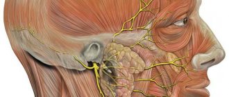
Cranial nerves of the brain
Also in the medulla oblongata there is part of the reticular nuclei (coeruleus), the processes going upward reach the upper layers of the cerebral cortex. The processes directed downwards rush into the spinal cord. High molecular weight granules (melanin) give the blue pigment to the nucleus.
The central olivary nucleus in the medulla oblongata has an additional plate of gray mass; together they form an intermediate coordination control center.
The nuclei of the Gaulle (thin) and Burdach (wedge-shaped) bundles originate from the spinal ganglia and have a system of ascending fibers. The fibers form two bundles, which are separated in the middle by a layer of nervous tissue. Closer to the middle (thin bundle) there are long conductive strands - they come from the lower extremities.
The lateral bands (wedge-shaped fasciculus) are shorter and begin in the upper limbs. Both bundles end their path in the nuclei of the bulb (the first and second path), but their axons extend to the parietal lobe of the cortex, where they end their direction (the third).
From the cochlear nuclei, processes are directed to the nuclei of the trapezoid body. The axons form a bundle of fibers called the auditory lemniscus. These cords rush to the subcortical hearing centers.
What are the functions of the medulla oblongata
The medulla oblongata of the human brain is responsible for maintaining vital functions - breathing and blood circulation. Damage to the structure results in instant death.
The oblongata has age-related characteristics - at the time of birth, cells are developed only to regulate respiratory function, maintain blood circulation and control digestive processes. The cells mature as the child grows; the structure is fully functional from about 7 years of age.
The bulb comes from the rhomboid part of the brain and is the main conductor for incoming information from the analyzers.
The main functions of the oblong structure:
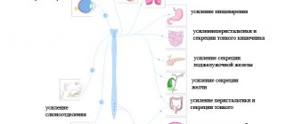
- Control over the circulatory system (work of the heart muscle, stabilization of blood pressure);
- Management of digestive processes (enzyme production);
- Control of muscle tone;
- Control of unconditioned reflexes;
- Controlling the respiratory center (gas exchange).
The functional set of the medulla oblongata is determined by the peculiarities of the internal structure (nucleus) and various pathways (long, short) that connect the central nervous system into a single management mechanism.
Based on this, the functionality of the bulb is divided into 4 groups:
- Sensory;
- Conductor;
- Reflex;
- Integrative.
All unconditioned reflexes are generated from the medulla oblongata, which in turn transmits impulses to the posterior brain structure.
Table of the functional orientation of centers in the medulla oblongata:
| Name | Purpose |
| Glossopharyngeal nerve | Controls secretion in the parotid gland and regulates the stylopharyngeal muscle, which elevates the larynx. Responsible for triggering protective reflexes in the oral cavity and larynx. Regulates taste sensitivity on the tongue. |
| Nervus vagus | Innervates transversely striated and smooth muscles. The muscles in the oral cavity are responsible for the motor capabilities. Participates in the production of secretion in the digestive organs. |
| Accessory nerve | Controls complex motor capabilities of muscles (turning the head in the opposite direction, bringing the shoulder blades together, raising one shoulder higher than the other). |
| Hypoglossal nerve | Regulates all muscles of the tongue and is partially responsible for innate reflexes (swallowing, sucking, chewing). |
| Olive Kernel | The intermediate vestibular center is responsible for the unconditioned muscle reflex when losing balance. |
| Gaulle and Burdach bundles | Responsible for sensitivity in the limb. |
| blue spot | Responsible for the response to anxiety or other irritant. |
There are connections in brain structures - these are short and long pathways. Therefore, the functional affiliations of the centers of the medulla oblongata and other structures may overlap with each other. Centers regulate the work of only a specific organ or system.
The centers in the oblong structure are formed by a cluster of nuclei and surrounding cells that interact with each other and control reflexes.
The functions of the medulla oblongata are divided into:
- Primary – maintaining vital unconditioned reflexes (breathing, heart function);
- Secondary – reception and transmission of information from sensory analyzers, control of autonomic reflexes.
After analyzing the information, an impulse is transmitted to the organ or system in order to increase activity or, conversely, suppress it (inhibition).
Sensory
The bulb processes the flow of information that comes from all sensory analyzers.
Function regulation:
- Reading information from skin receptors (on the face);
- Recognition of taste sensations;
- Monitoring the functioning of the vestibular apparatus and regulating changes;
- Perception of sound waves from auditory receptors.
After receiving information in the bulb, a primary analysis takes place, which takes into account the strength of the incoming signal. After processing, impulses with information are transmitted to subcortical structures, where sensations are read and distributed according to biological significance.
Conductor
The spinal cord tracts pass through the medulla oblongata - ascending and descending, as well as the origin of the tracts. Therefore, the structure takes part in the control of muscle contractions and tonic reactions.
The medulla oblongata structure has bilateral nerve connections with other parts of the brain and the cortex (corticoreticular connection). This connection allows the bulb to take part in the control of skeletal muscles and manage autonomic reactions.
The white matter of the bulb contains motor pathways that regulate the functioning of the facial muscles and tongue. Near the pyramids, the nerve fibers form a cross - this allows the threads to move to the opposite side and descend to the spinal cord canal.
Integrative
The medulla oblongata controls complex reflexes through nerve connections and interaction with other centers.
Communication with the vestibular centers and the oculomotor system helps control the eyes when the head is deviated. Some neurons in the brainstem region perform automatic functions - they provide tone and support the active work of many centers in the medulla.
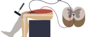
Decerebrate and waxy rigidity (contractile and plastic tone)
In an animal in which only the spinal cord is preserved, prolonged tonic reflexes can be obtained. A constant influx of impulses from proprioceptors into the nervous system maintains reflex muscle tone, thanks to efferent impulses emanating from the spinal cord and various parts of the brain (medulla oblongata, cerebellum, middle and intermediate). Transection of the afferent nerves of a limb entails the disappearance of its muscle tone. After the motor innervation of the limb is turned off, the tone of its muscles also disappears. Consequently, to obtain tone, the preservation of the reflex ring is necessary; TIC, as a tone, is caused reflexively.
After transection of the brain at the level of the anterior tubercles of the quadrigeminal, decerebrate rigidity, or contractile tone, is detected. Contractile tone is expressed in the fact that all muscles come into a state of extreme tension, especially the extensor muscles. Due to the strong tension of the neck muscles, the head is thrown back, the limbs are strongly extended. If an incision is made along the middle third of the medulla oblongata, the contractile tone disappears. Consequently, this tone is carried out by the anterior part of the medulla oblongata and the midbrain. The reflex nature of contractile tone is proven by the fact that it cannot be obtained by a limb devoid of afferent nerves, since to obtain it an influx of impulses from proprioceptors is necessary. The significance of contractile tone is that, by capturing the muscles with it, it creates a standing posture and a starting position for locomotion and working movements. However, not all animals with contractile tone have a pronounced standing posture. Depending on the type of animal, it can fix its limbs in other positions. It is regulated by the red nuclei and the reticular formation.
Irritation of skin receptors causes cessation of contractile tone in animals and rapid coordinated movements, i.e., normal reflexes. Contractile tone can last for more than a day.
After transection of the brain, another form of tone appears along the anterior border of the diencephalon - waxy rigidity, or plastic tone. Consequently, plastic tone is carried out by the diencephalon. Plastic tone is characterized by the fact that the muscles maintain their given position and the animal freezes in natural and unnatural poses. This state is similar to catalepsy, numbness, freezing of the body and its individual parts in case of poisoning with certain poisons, in diseases of the nervous system, or - in a healthy person - in a state of artificial sleep - hypnosis. With catalepsy, people lose the ability to make voluntary movements. Under the influence of suggestion, catalepsy lasts 5-10 minutes or more, and in case of disorders of the nervous system - for several hours.
Plastic tone is also a reflex that occurs when impulses arrive from proprioceptors. With the anatomical integrity of the nervous system, plastic tone suppresses contractile tone, and contractile tone, in turn, suppresses the lingering aftereffect of spinal cord reflexes. The cerebral hemispheres regulate the relationships of all types of tone, which certainly, to one degree or another, participate in locomotion.