The human eye is capable of perceiving parts of the color spectrum (different colors) in different parts of the retina. Therefore, exposure to light signals of different colors, which occurs during photostimulation, is a kind of training for the eyes. When signals of white, green, yellow, red and blue colors are applied to the retina, areas of the eye that differ in structure and role are activated. A given rhythm of signals and their specific sequence create the necessary stimuli for the organ of vision to operate in an enhanced but safe manner. At the same time, thanks to the direct connection between the eyes and the part of the brain responsible for vision, the latter better processes images coming from the eyes. Photostimulation also helps develop the ability to correctly perceive the distance between objects and evaluate their volume.
In short, the role of photostimulation comes down to the implementation of the following functions:
- New neural connections are established in the patient’s brain, and the functioning of the visual part of the brain improves.
- Blood circulation in the eye muscles improves.
- The blood supply to the retina and optic nerve of the eye improves.
As a result, by the end of the course of photostimulation, the “safety margin” of the eyes increases and they can work without getting tired in an enhanced mode for a longer time without loss of visual acuity.
Electroencephalogram of the brain
The research in question is a literal recording of the activity (namely, electrical) of certain brain structures. The results of the electroencephalogram are recorded on paper specially designed for this purpose using electrodes. The latter are applied to the patient's head in a certain order. Their task is to record the activity of individual parts of the brain. Thus, the electroencephalogram of the brain is a record of its functional activity. The study can be carried out for any patient, regardless of his age. What does the EEG show? It helps determine the level of brain activity and identify various disorders of the central nervous system, including meningitis, polio, encephalitis and others. It also becomes possible to find the source of damage and assess its extent.
When performing an electroencephalogram, the following tests are usually necessary:
- Blinking of varying speeds and intensity.
- Exposure of the patient's completely closed eyes to periodic bright flashes of light (so-called photostimulation).
- Deep breathing (rarely inhaling and exhaling) for a period of three to five minutes (hyperventilation).
The tests listed above are carried out for both children and adults. Neither diagnosis nor age affects the testing composition.
Additional studies that the doctor conducts, depending on certain factors, are the following:
- sleep deprivation for a certain period of time;
- passing a series of psychological tests;
- clenching the palm into a fist;
- monitoring the patient throughout the entire period of night sleep;
- taking certain medications;
- the patient is in the dark for about forty minutes.
Equipment
To record EEG, devices called Electroencephalographs are used. They consist of an electrode part, an amplifier system, and a recording device. Electrodes come in different types: cup and bridge. They are made from electrically conductive carbon or metal with a silver chloride coating. Such a coating is necessary so that a constant potential does not accumulate on the electrode, which causes polarization of the electrode. This results in interference. Non-metallic electrodes are least polarized.
To ensure accurate registration, parallel common-mode amplifiers with a notch filter are used. This allows you to combat network interference. In terms of their quality, amplifiers now allow recording without an electrically insulated chamber and without grounding.
Recording device. Initially, writing instruments with a paper tape feed were used as a recorder. They differed in ink devices and devices with a thermal pen. But consumables were quite expensive. Nowadays computer technology is used as a recording device. With the advent of computer technology, it became possible not only to record EEG on a non-paper medium, but also to carry out additional mathematical processing of EEG. This increased the resolution of the method.
The application of electrodes is also carried out in various ways. The international system adopted as the standard is the 10 - 20 system. Electrodes are applied as follows. Measure the distance along the sagittal line from Inion to Nasion and take it as 100%. At 10% of this distance from Inion and Nasion, the lower frontal and occipital electrodes are installed, respectively. The rest are placed at an equal distance of 20% of the distance inion - nasion. The second main line runs between the ear canals through the crown.
The lower temporal electrodes are located at 10% of this distance above the auditory canals, and the remaining electrodes of this line are located at a distance of 20% of the length of the biauricular line. The letter symbols indicate, respectively, areas of the brain and landmarks on the head: O - occipitalis, F - frontalis, A - auricularis, P - parietalis, C - centralis, T - temporalis. Odd numbers correspond to the electrodes of the left hemisphere, even numbers - to the right.
According to Young's system, frontal electrodes (Fd, Fs) are located in the upper part of the forehead at a distance of 3 - 4 cm from the midline, occipital electrodes (Od, Os) - 3 cm above the inion and 3 - 4 cm from the midline. The segments of the lines Od - Fd and Os - Fs are divided into three equal parts and central (Cd, Cs) and parietal (Pd, Ps) electrodes are installed at the division points. At the horizontal level of the upper edge of the auricle, the anterior temporal (Tad, Tas) are installed along the frontal line Cd - Cs, and the posterior temporal (Tpd, Tps) are installed along the frontal line Ps - Pd.
The advantage of the 10 - 20 system is a large number of electrodes (from 16 to 19 - 24), but this system requires more sensitive equipment, because the interelectrode distance is small and the potential is weak. Young's system provides sufficient distance and all electrodes are evenly distributed over the surface of the head, but the degree of localization during abduction is insufficient.
The method of removing potential can also be different. The generally accepted system is monopolar recording. In this case, the electrodes on the head are active and record changes in potential relative to an indifferent electrode (most often located on the earlobes). Bipolar recording detects the change in potential between two electrodes located at different points on the surface of the scalp.
All the indicated methods and techniques have their advantages and disadvantages. Therefore, in international practice, mandatory recording according to the 10-20 system is established, both in monopolar and bipolar modes. During computer recording, registration using the 10-20 system is allowed with further digital conversion of the EEG according to the selected bipolar scheme.
What does an electroencephalogram show?
What is this examination? To find out the answer, it is important to understand in detail what the EEG shows. It demonstrates the current functional state of certain structures that make up the brain. It is carried out in a variety of patient conditions, such as wakefulness, active physical work, sleep, active mental work, and so on. An electroencephalogram is an extremely safe research method, painless, simple, and one that does not require serious intervention in the functioning of the body. It allows you to accurately determine the location of cysts, tumors, mechanical damage to brain tissue, and diagnose vascular diseases, epilepsy, inflammatory diseases of the brain and its degenerative lesions.
The essence of the photostimulation technique
Photostimulation of the eyes is a long-practised, but still relevant, method of treating the organ of vision, which boils down to applying light signals to each eye in turn. In this case, the patient uses special dark glasses. The rhythm of the supplied light signals, their intensity and frequency, are selected in such a way as to have a positive effect on the visual system, through stimulation of the visual analyzer, as well as the brain that processes the information received. This method is aimed at training the internal capabilities of the body, without external interference in its work. This helps to achieve success literally in 7 or 10 sessions, which usually constitute a course of treatment.
Where to do it?
Such examinations are usually carried out in psychiatric dispensaries, neurological clinics, and sometimes in district and city hospitals. Clinics usually do not provide such services. However, it is better to find out directly on the spot. Experts recommend contacting neurology departments or psychiatric hospitals. Local doctors are sufficiently qualified and will be able to carry out the procedure in the correct way and correctly interpret the results. If we are talking about a small child, then you should contact children’s hospitals specially designed for such examinations. A similar service is also provided in private medical centers. There are no age restrictions here.
Before going for an examination, you need to get a good night's sleep and spend some time before this day in peace without stress and excessive psychomotor agitation. During the two days before the EEG, you should not drink alcohol, caffeine, sleeping pills, tranquilizers, anticonvulsants, or sedatives.
Electroencephalogram for children
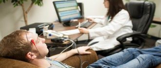
This study should be examined in more detail. After all, as a rule, parents have many questions in this regard. The baby will be forced to spend about twenty minutes in a light- and sound-proofed room, where he lies on a special couch with a cap on his head, under which the doctor places electrodes. The scalp is additionally moisturized with gel or water. Two electrodes are placed on the ears, which are not active. The current strength is so low that it cannot cause the slightest harm even to infants.
The baby's head should be level. If the baby is over three years old, he can remain awake during the procedure. You can take something with you that will distract the child and allow him to calmly wait until the end of the examination. If the patient is younger, the procedure is performed during sleep. At home, the baby needs to wash his hair and not feed. Feeding is carried out in the clinic immediately before the procedure so that he quickly falls asleep.
The frequency of the brain's alpha rhythms and other rhythms is recorded in the form of a background curve. Additional tests (eg, photostimulation, hyperventilation, rhythmic closing and opening of the eyes) are also often performed. They are suitable for everyone: both children and adults. Thus, deep inhalations and exhalations can reveal hidden epilepsy. Auxiliary studies help determine the presence or absence of developmental delays in the baby (speech, mental, mental or physical development).
Methods of rhythmic photostimulation: why, how and why?
Type 3 - peak-wave activity in the parieto-occipital region spreading to the frontal region
In 2001, a modified classification of EEG responses to RFS was proposed (Kasteleijn-Nolst Trenite et al., 2001), but it is quite complex for use in routine clinical practice and is therefore rarely used.
In different laboratories and countries, RFS is carried out differently, however, for an accurate diagnosis it is necessary to follow a single protocol developed on the basis of scientific and clinical data.
In 2012, the “European algorithm for conducting photostimulation in the EEG laboratory” was published, according to which conducting RFS implies solving two problems:
1) The main task is direct diagnosis of the phenomenon of photosensitivity in a given patient. In this case, the sensitivity of XRF as a method is high, but the specificity is low.
2) Additional task - examination according to a detailed protocol in order to identify individual characteristics of photosensitivity
In addition to direct diagnosis, RFS allows you to monitor the effectiveness of antiepileptic therapy in a particular patient.
Algorithm for conducting RFS:
Before conducting RFS it is necessary:
1) find out whether the patient took antiepileptic drugs (AEDs)
how long did you sleep at night
presence of alcohol intake
have you previously had seizures caused by flashes of light, or have you had similar seizures in your relatives?
2) no special preparation is required before conducting an EEG
Of course, for diagnostic purposes, an EEG study is most optimal when discontinuing AEDs, but this entails a greater risk of tonic-clonic or generalized seizures
3) It is necessary to obtain the patient’s informed consent for the study, since RFS is a procedure that provokes seizures, and the patient must be informed about this.
When performing RFS on young children, for safety reasons, the child can sit on the parents’ lap or in close proximity to them, but in this case the relative is also exposed to RFS.
In the event of an attack, the EEG laboratory should have emergency medications - midazolam or diazepam.
When conducting RFS:
RFS is performed upon awakening from a normal night's sleep
Sleep deprivation significantly increases the likelihood of developing seizures.
In JME, recording myoclonic seizures shortly after awakening is diagnostically important, and partial sleep deprivation can be used to increase the likelihood of this occurring.
RFS should be performed at least three minutes after or before hyperventilation.
It is considered optimal to perform RFS at the end of the EEG recording, and hyperventilation at the beginning, since hyperventilation allows the patient to relax and fall asleep during recording, and RFS, usually associated with a stressful effect on the patient’s psyche, prevents this.
The patient should be in a darkened room, in an upright position. It is important to conduct video recording.
The patient's position, on the one hand, must be as safe as possible, while, on the other hand, the doctor must be able to observe him.
Dim light is used to evaluate eye movements and avoid artifactual results. Only in patients with the fixation-off phenomenon and scotosensitivity is it necessary to conduct additional recordings in complete darkness.
1. The recording must be kept for at least 2.5 minutes with eyes open and 2.5 minutes with eyes closed before the RFS. This recording makes it possible to distinguish between spontaneous and RFS-induced discharges and to identify the fixation-off phenomenon
2. Use a lamp with a round reflector that can produce flashes of light with an intensity of at least 0.70 J.
The strobe lamp should be at a distance of 30 cm.
It is necessary to instruct the patient to look strictly at the center of the lamp and carefully follow the commands to open and close the eyes. Binocular stimulation is more effective than monocular stimulation.
Stimulation of the entire retinal area is most effective, while closing the eyes on command is a strong provoking factor, the identification of which is diagnostically important not only for the doctor, but also for the patient, since in ordinary life a similar provoking factor can be closing the eyes while watching TV or while observing behind the sun.
When conducting RFS, young children need to attract attention with a toy located above the lamp, while one of the parents or an EEG technician can close their eyes on command.
1. It is necessary to record photosensitivity in three states: with eyes closed, with eyes open, and directly when closing eyes. If there is not enough time, it is better to conduct a test with opening and closing the eyes
2. Use the following frequencies in ascending order: 1 – 2 – 8 – 10 – 15 – 18 – 20 – 25 – 40 – 50 – 60 Hz. If generalized epileptiform discharges occur at any frequency, “increasing” stimulation should be stopped and stimulation continued from the maximum frequency (60 Hz) down until a photoparoxysmal response appears
The most provocative frequencies are 15-20 Hz (see Fig. 1), but despite this, a consistent set of frequencies is extremely important.
RFS frequency, Hz
Fig.1
With standard stimulation at a single frequency of 18 Hz, the phenomenon of photosensitivity cannot be detected in 15% of patients. The use of non-standard stimulation schemes at different frequencies has never been studied in large groups of patients, and therefore the value of the results is also highly controversial.
A photoparoxysmal reaction at low stimulation frequencies is characteristic of patients with progressive forms of epilepsy. For patients sensitive to high frequencies, the stimulus that provokes attacks may be watching television.
1. If it is necessary to reproduce the photoparoxysmal response, photostimulation can be repeated, but only some time after the first attempt.
2. You should always monitor the patient and ask him about the sensations that arise during stimulation.
In RFS, myoclonus and absence seizures are most common. Almost all patients with RFS report discomfort in the eyes. There may be occipital convulsions with transition to tonic-clonic. RFS can provoke generalized seizures, but, as a rule, if the RFS technique is carried out correctly, they can be prevented.
In addition to the necessary clinical information, the patient's awareness of his own sensations and symptoms can help him identify stimuli that provoke attacks in everyday life.
Additional research methods to obtain more detailed information:
1. Installation of EMG electrodes
2. For attacks that occur while watching TV and playing computer games, it makes sense to perform RFS with color flashes.
Red color is the most epileptogenic; in addition, red and blue flashes when alternating, or red flashes in combination with geometric patterns (“Pokemon pattern”) have a good provoking ability.
3. In addition, black and white patterns, TV or computer games can be used as provoking stimuli.
This helps the patient and relatives understand how real the provoking stimuli are in the reality around them and try to avoid them.
4. Identifying the upper and lower threshold frequencies that provoke a photoparoxysmal response, studying photosensitivity using individual methods using patterns helps to develop an individual treatment program for each patient.
For example, use glasses with blue lenses, exclude provoking stimuli from everyday life, or evaluate the effectiveness of AEDs.
"Open questions:
Further work is currently required
- improvement of photostimulation techniques
- studying the mechanisms of photosensitivity
- identification of new provoking stimuli in people with photosensitivity forms of epilepsy.
The material was prepared based on the articles:
Electroencephalogram rhythms
The examination in question allows us to evaluate the following types of brain rhythms:
Each of them has certain characteristics and help assess different types of brain activity.
- The normal frequency of the alpha rhythm is in the range from 8 to 14 Hz. This should be taken into account when determining pathologies. The EEG alpha rhythm in question is recorded when the patient is awake, but his eyes are closed. As a rule, this indicator is regular. It is most quickly registered in the area of the crown and back of the head. In the presence of any motor stimuli it stops.
- The frequency of the beta rhythm ranges from 13 to 30 Hz. As a rule, it is registered above the frontal lobes. Characterizes the state of depression, anxiety, anxiety. It also reflects the use of sedatives.
- Normally, the theta rhythm has an amplitude from 25 to 35 μV, and a frequency from 4 to 7 Hz. Such indicators reflect the state of a person when he is in a state of natural sleep. For the child, the rhythm in question is prevalent.
- The delta rhythm in most cases demonstrates the state of natural sleep, but it can also be recorded to a limited extent during wakefulness. Normal frequency is from 0.5 to 3 Hz. The normal value of the rhythm amplitude does not exceed 40 μV. Deviations from the specified values indicate the presence of pathologies and impaired functioning of the brain. By the location of the appearance of this type of rhythm, one can determine exactly where dangerous changes occur. If it is noticeable in all areas of the brain, this indicates a violation of consciousness and that systemic damage to the structures of the central nervous system is developing. This is often caused by liver dysfunction.
EEG rhythms
The following brain rhythms are identified, recorded on the electroencephalogram:
Alpha rhythm.
Its amplitude increases in a state of quiet wakefulness, for example, when resting or in a dark room. Alpha activity on the EEG decreases when the subject moves on to active work that requires high concentration of attention. People who have been blind all their lives have an absence of alpha rhythm on the EEG.
Beta rhythm.
It is characteristic of active wakefulness with high concentration of attention. Beta activity on the EEG is most clearly expressed in the projection of the frontal cortex. Also on the electroencephalogram, the beta rhythm appears with the sudden appearance of an emotionally significant new stimulus, for example, the appearance of a loved one after several months of separation. The activity of the beta rhythm also increases during emotional stress and work that requires high concentration.
Gamma rhythm.
This is a collection of low-amplitude waves. The gamma rhythm is a continuation of beta waves. Thus, gamma activity is recorded under high psycho-emotional stress. The founder of the Soviet school of neuroscience, Sokolov, believes that the gamma rhythm is a reflection of the activity of human consciousness.
Delta rhythm.
These are high amplitude waves. It is recorded in the phase of deep natural and medicated sleep. Delta waves are also recorded in a coma state.
Theta rhythm.
These waves are generated in the hippocampus. Theta waves appear on the EEG in two states: the rapid eye movement phase and during high concentration. Harvard professor Schacter argues that theta waves appear during altered states of consciousness, such as deep meditation or trance.
Kappa rhythm.
It is registered in the projection of the temporal cortex of the brain. It appears in the case of suppression of alpha waves and in a state of high mental activity of the subject. However, some researchers associate the kappa rhythm with normal eye movement and regard it as an artifact or side effect.
Mu rhythm.
Appears in a state of physical, mental and emotional peace. It is registered in the projection of the motor lobes of the frontal cortex. Mu waves disappear during visualization or during physical activity.
Normal EEG in adults:
- Alpha rhythm: frequency – 8-13 Hz, amplitude – 5-100 µV.
- Beta rhythm: frequency – 14-40 Hz, amplitude – up to 20 µV.
- Gamma rhythm: frequency – 30 or more, amplitude – no more than 15 µV.
- Delta rhythm: frequency – 1-4 Hz, amplitude – 100-200 µV.
- Theta rhythm: frequency – 4-8 Hz, amplitude – 20-100 µV.
- Kappa rhythm: frequency – 8-13 Hz, amplitude – 5-40 µV.
- Mu rhythm: frequency – 8-13 Hz, amplitude – on average 50 µV.
Conclusion The EEG of a healthy person consists of precisely these indicators.
Importance for the body
The alpha rhythm of the brain becomes traceable exclusively in moments of calm and is low-frequency. Then the parasympathetic system is activated. While in the alpha state, the central nervous system, figuratively speaking, reboots and gets rid of all the stress that has accumulated during the day. The alpha rhythm ensures regular restoration of the body, as well as the accumulation of necessary resources after a working period. As history shows, a huge number of amazing discoveries were made by people during periods of their stay in the state in question. What else should you know?
Functions
What function do alpha rhythms perform?
- Leveling the effects of stress (decreased immunity, narrowing of blood vessels).
- Analysis of all the information that was received by the brain during the day.
- Excessive activity of the limbic system is not allowed.
- Blood circulation in the brain improves significantly.
- All the body’s resources are restored, spurred by the activation of the parasympathetic system.
How does alpha rhythm disorder affect everyday life? Patients whose generation of alpha waves is significantly reduced, as a rule, are more likely to focus on their own problems, they tend to think negatively. Such disorders lead to decreased immunity, the development of various cardiovascular diseases and even oncology. Often there are malfunctions in the glands that synthesize hormones, irregularity of the menstrual cycle, the development of various addictions and a tendency to various types of abuse (for example, alcoholism, drug addiction, overeating, smoking).
A well-established alpha rhythm ensures the normal course of restoration processes in the tissues of the body. It plays a vital role in maintaining the life of an individual.
Norm and pathologies
An electroencephalogram helps to identify and evaluate the index, which characterizes the alpha rhythm of the brain. Its norm varies between 75% and 95%. If a significant decrease is noted (less than 50%), then we can safely speak of pathology. The rhythm in question is usually noticeably reduced in older people (over 60 years old). The reason for this is usually age-related cerebrovascular accidents.
Another striking indicator is the amplitude of the rhythm. Its normal value is considered to be waves with an amplitude from 20 to 90 μV. Asymmetry of both this indicator and the rhythm frequency in different hemispheres indicates the presence of a number of diseases, such as narcolepsy, epilepsy or essential hypertension. A low frequency indicates hypertension, and an increased frequency indicates mental retardation.
If the rhythms are not synchronized, it is also important to conduct additional tests to clarify the pathology. Narcolepsy is characterized by hypersynchronization. Asymmetry also indicates possible traumatic damage to the corpus callosum, as well as the presence of a tumor or cyst. A complete absence of the alpha rhythm occurs with blindness, developing Alzheimer's disease (the so-called acquired dementia) or cerebral sclerosis. Problematic indicators can occur when cerebral circulation is impaired.
It is also advisable for patients with which conditions and symptoms to undergo this examination? Indications for EEG are frequent vomiting, osteochondrosis, frequent fainting, brain injuries and tumors, high blood pressure, headaches, suspected dementia (both acquired and congenital), as well as vegetative-vascular dystonia. Only a qualified neurologist can prescribe a study and interpret the results.
What do indicator violations indicate?
Depending on how exactly the alpha rhythm is disrupted, the specific disease is determined. So, for example, if it is disorganized or completely absent, then the diagnosis is acquired dementia. Interhemispheric asymmetry of the alpha rhythm indicates the presence of a heart attack, cyst, stroke, tumor or scar, indicating an old hemorrhage. You should pay close attention to this. An unstable rhythm or high-frequency alpha rhythm of the brain may be a manifestation of traumatic injury.
As for children, the following disorders indicate a delay in their development:
- Abnormally expressed reaction to hyperventilation.
- The alpha rhythm is disorganized.
- The concentration of activity has moved from the area of the crown and back of the head.
- Alpha rhythm amplitude and synchrony are noticeably increased.
- The activation reaction is short and weak.
Psychopathology in adults can also be expressed by low rhythm amplitude, weak activation reaction, as well as a displacement of the point of concentration of activity from the region of the crown and occiput.
Parameters of normal alpha rhythm
The frequency is 8-13 Hz, according to some authors the frequency is 7-12 Hz or 8-12 Hz. Most often, in a normal state, a frequency of 9-10 Hz is found, which can be called a normorhythm. Then the average frequency of 8-9 (7-9) Hz can be considered a slow alpha rhythm, and 11-12 Hz - a rapid one. Naturally slow and rapid rhythms are already beyond the norm (in adults) and can be considered as conditionally pathological (according to O. M. Grindel)
normal amplitude Some authors recognize 20-110 µV as the norm. The amplitude normally varies depending on age.
Zonal distribution - normally determined by the occipital-parietal zone, where the rhythm is most pronounced. This position is recognized by everyone equally.
Modulation is characterized by a wave-like change in the amplitude of the rhythm.
Sinusoidality sets the normal roundness of the vertices. With computer visualization, the sinusoidality is not detected so clearly (with 8-bit recording) and all rhythms seem sharpened. But, as a rule, a true sharpening of the rhythm must be combined with other disturbances of the normal rhythm.
Symmetrical in amplitude and frequency. The reliability of amplitude symmetry is established by good application of electrodes with impedance measurement. Frequency asymmetry must also be objectified (reliability criteria). In this case, it is necessary to take into account the presence of physiological asymmetry of the hemispheres.
The reaction of activation of the alpha rhythm, i.e. its suppression when opening the eyes or a flash of light. This phenomenon is one of the main characteristics of the alpha rhythm. According to it, the detected rhythm can be accurately attributed to the alpha rhythm.
Alpha rhythm index , which is normally 80%. During mathematical processing, the index can be calculated as a percentage of the power of the alpha rhythm relative to the power of other rhythms in the occipital and parietal leads.
Conclusion
An electroencephalogram is a safe and painless test that helps identify a number of dangerous diseases. The study can be carried out even on infants. It allows you to assess the nature of brain rhythms. After interpreting the information received and prescribing the correct treatment, a neurologist will help you cope with the symptoms that are bothering you.
Using the method of electroencephalography (abbreviation EEG), along with computer or magnetic resonance imaging (CT, MRI), the activity of the brain and the state of its anatomical structures are studied. The procedure plays a huge role in identifying various anomalies by studying the electrical activity of the brain.
EEG is an automatic recording of the electrical activity of neurons in brain structures, performed using electrodes on special paper. Electrodes are attached to various areas of the head and record brain activity. In this way, the EEG is recorded in the form of a background curve of the functionality of the structures of the thinking center in a person of any age.
A diagnostic procedure is performed for various lesions of the central nervous system, for example, dysarthria, neuroinfection, encephalitis, meningitis. The results allow us to evaluate the dynamics of the pathology and clarify the specific location of the damage.
The EEG is carried out in accordance with a standard protocol that monitors activity during sleep and wakefulness, with special tests for the activation response.
For adult patients, diagnosis is carried out in neurological clinics, departments of city and regional hospitals, and a psychiatric clinic. To be confident in the analysis, it is advisable to contact an experienced specialist working in the neurology department.
For children under 14 years of age, EEGs are performed exclusively in specialized clinics by pediatricians. Psychiatric hospitals do not perform the procedure on young children.
Contraindications
The main contraindication for this procedure is the patient’s intolerance to pulsed flashing light. Such intolerance is usually observed in patients with epilepsy, people with unstable mental health, and with cancerous tumors in the brain. You should talk to your ophthalmologist about the possibility of other contraindications.
MGK specialists have developed and implemented special eye photostimulation programs. The procedures are carried out using the most modern equipment and can be prescribed to adults and children. After carrying out the necessary diagnostic studies, an individual hardware treatment regimen is selected for each patient, which is then adjusted in accordance with the current results. The procedures are carried out under the supervision of highly qualified specialists, do not cause discomfort and do not require any restrictions in the usual way of life.
What do EEG results show?
An electroencephalogram shows the functional state of brain structures during mental and physical stress, during sleep and wakefulness. This is an absolutely safe and simple method, painless, and does not require serious intervention.
Today, EEG is widely used in the practice of neurologists in the diagnosis of vascular, degenerative, inflammatory brain lesions, and epilepsy. The method also allows you to determine the location of tumors, traumatic injuries, and cysts.
EEG with the impact of sound or light on the patient helps to express true visual and hearing impairments from hysterical ones. The method is used for dynamic monitoring of patients in intensive care units in a coma state.
- EEG for children under 1 year of age is performed in the presence of the mother. The child is left in a sound- and light-proof room, where he is placed on a couch. Diagnostics takes about 20 minutes.
- The baby's head is wetted with water or gel, and then a cap is put on, under which the electrodes are placed. Two inactive electrodes are placed on the ears.
- Using special clamps, the elements are connected to wires suitable for the encephalograph. Due to the low current, the procedure is completely safe even for infants.
- Before monitoring begins, the child's head is positioned level so that there is no forward bending. This may cause artifacts and skew the results.
- EEGs are done on infants during sleep after feeding. It is important to let the boy or girl get enough immediately before the procedure so that he falls asleep. The mixture is given directly in the hospital after a general medical examination.
- For children under 3 years old, an encephalogram is taken only in a state of sleep. Older children may remain awake. To keep the child calm, they give him a toy or a book.
An important part of the diagnosis are tests with opening and closing the eyes, hyperventilation (deep and rare breathing) with EEG, squeezing and unclenching of the fingers, which allows disorganization of the rhythm. All tests are conducted in the form of a game.
After receiving the EEG atlas, doctors diagnose inflammation of the membranes and structures of the brain, latent epilepsy, tumors, dysfunction, stress, and fatigue.
The degree of delay in physical, mental, mental, speech development is carried out using photostimulation (blinking a light bulb with eyes closed).
For adults, the procedure is carried out subject to the following conditions:
- keep your head motionless during manipulation, eliminate any irritating factors;
- Before diagnosis, do not take sedatives or other drugs that affect the functioning of the hemispheres (Nerviplex-N).
Before the manipulation, the doctor conducts a conversation with the patient, putting him in a positive mood, calming him down and instilling optimism. Next, special electrodes connected to the device are attached to the head, and they read the readings.
The examination lasts only a few minutes and is completely painless.
Provided that the rules described above are observed, even minor changes in the bioelectrical activity of the brain are determined using EEG, indicating the presence of tumors or the onset of pathologies.
EEG
- ELECTROENCEPHALOGRAPHY (EEG) - 2900 rub.
EEG is the first and often the only neurological outpatient study that is performed during attacks of loss of consciousness.
An electroencephalogram is a recording of the total electrical activity of the cells of the cerebral hemispheres through intact scalp, which allows one to judge its physiological maturity, functional state, the presence of focal lesions, cerebral disorders and their nature.
CONDUCTING RESEARCH
EEG is completely harmless and painless. During the examination, the patient sits in a comfortable chair, relaxed with his eyes closed (state of passive wakefulness). To conduct an EEG, small electrodes are attached to the head using a special helmet, which are connected by wires to an electroencephalograph. An electroencephalograph amplifies the biopotentials received from sensors hundreds of thousands of times and records them on paper or in computer memory.
The patient should not feel hungry before the study, as this may cause changes in the EEG. The head should be washed clean before the EEG - this will allow for better contact of the electrodes with the scalp and obtain more reliable research results. With preschool children, it is necessary to practice putting on a “helmet” and remaining motionless with eyes closed (playing as an astronaut, tank driver, etc.), and also teach them to breathe deeply and frequently.
If during an EEG the patient has a seizure, the effectiveness of the study increases significantly, since it will be possible to more accurately identify the location of the disturbance in the electrical activity of the brain. However, taking into account the interests of patient safety, seizures should not be deliberately provoked. Sometimes patients do not take medications before an EEG. This should not be done. Abrupt cessation of medication provokes attacks and can even cause epistatus.
It is advisable that the EEG be performed by a qualified specialist. This is usually a specially trained neurologist, sometimes called an electroencephalographist or neurophysiologist. He must be able to decipher the EEG of patients of a particular age group. It should be taken into account that the EEG of children and adolescents differs significantly from the EEG of adults. In this case, the neurophysiologist not only describes the results of the study, but also makes his clinical electroencephalographic diagnosis.
However, the electroencephalographer cannot make a final diagnosis without more complete clinical data. Many EEG changes may be nonspecific, i.e. their accurate interpretation is possible only taking into account the clinical picture of the disease and sometimes after additional examination.
The results of the EEG depend on the age of the patient, the medications he is taking, the time of the last attack, the presence of tremor (shaking) of the head and limbs, visual impairment, and skull defects. All of these factors may influence the correct interpretation and use of EEG data.
DIAGNOSTIC CAPABILITIES OF EEG.
First of all, EEG helps to distinguish epileptic seizures from non-epileptic ones and classify them.
With EEG you can:
- identify areas of the brain involved in triggering seizures;
- monitor the dynamics of the action of drugs;
- resolve the issue of stopping drug therapy.
The EEG method makes it possible to assess the functional state of the brain (general and local), even in the complete absence of changes on a brain CT scan (i.e., in the absence of morphological changes in brain tissue). EEG examination data allows us to differentiate true epileptic pathology and other diseases that can clinically imitate epileptic paroxysms (for example, panic attacks; vegetative paroxysms of non-epileptic origin; paroxysmal sleep disorders; conversion seizures; various types of neuroses; psychiatric pathology, etc.)
When addressing issues of professional suitability, the detection of obvious epileptiform phenomena in the EEG is a sufficient basis for the selection of professions related to driving, requiring constant attention and quick response to sudden situations and stimuli under conditions of increased risk.
Indications for EEG:
- epilepsy and other types of paroxysms;
- brain tumors;
- traumatic brain injuries (in acute, subacute, early and late recovery periods, residual effects and long-term consequences of open and closed TBI);
- vascular diseases;
- inflammatory;
- degenerative;
- headache;
- dysontogenetic;
- hereditary diseases of the central nervous system;
- functional disorders of nervous activity (neuroses, neurasthenia, obsessive movement neurosis, somnambulism, etc.);
- psychiatric pathology;
- encephalopathy of various origins (vascular, post-traumatic, toxic);
- post-resuscitation conditions due to somatic pathology.
The best time to conduct an EEG is no earlier than a week after the attack. An electroencephalogram taken shortly after the attack will demonstrate its consequences, but will not determine the disease underlying the attack. Such an EEG is considered not as valuable as one done later, although it may be useful for further research.
FUNCTIONAL TESTS
ACTIVATION REACTION (TEST WITH OPENING AND CLOSING EYES)
The activation reaction is usually well expressed in children over 3 years of age and manifests itself in the form of a decrease in the amplitude of the basic rhythm. Rarely, in approximately 7% of cases, the activation reaction is weakly expressed or manifests itself in the form of an increase in background activity. This usually applies to children with delayed psychomotor development and reduced functional state of the brain as a result of brain disease or drug exposure. It is characteristic that the test with opening the eyes does not lead to a decrease in low-frequency beta activity, and sometimes increases its severity.
The activation reaction is interesting in terms of provoking some forms of generalized epileptic activity that appears a short time after closing the eyes, especially for non-convulsive forms of seizures (absences). Local (cortical) epileptic activity usually remains during desynchronization (during eye opening). While epileptic activity caused by a process in the deep structures of the brain may disappear.
PHOTOSTIMULATION (STIMULATION WITH LIGHT FLICKS)
Photostimulation is often carried out with light flickers of a fixed frequency from 5 to 30 Hz in series of 10-20 seconds.
PHONOSTIMULATION (STIMULATION WITH SOUND SIGNALS)
Phonostimulation is usually applied in the form of a short-term loud sound signal.
SLEEP DEPRIVATION (LIMITING SLEEP TIME)
A test with sleep deprivation during the day is used in cases where, during a “routine” examination of a patient with epileptic seizures, it is necessary to increase the likelihood of detecting epileptic activity. This test increases the information content of the EEG by approximately 28% and, mainly, in patients with absence seizures and tonic-clonic seizures. However, the test is quite difficult for children under 10 years of age to tolerate.
HYPERVENTILATION (FORCED BREATHING)
Hyperventilation is breathing frequently and deeply for 1-3 minutes. Such breathing causes pronounced metabolic changes (alkalosis) in the brain due to intensive removal of carbon dioxide, which, in turn, contribute to the appearance of epileptic activity on the EEG in people with seizures. Hyperventilation during EEG recording makes it possible to identify hidden epileptic changes (especially during absence seizures) and clarify the nature of epileptic seizures.
Electroencephalogram rhythms
An electroencephalogram of the brain shows regular rhythms of a certain type. Their synchrony is ensured by the work of the thalamus, which is responsible for the functionality of all structures of the central nervous system.
The EEG contains alpha, beta, delta, tetra rhythms. They have different characteristics and show certain degrees of brain activity.
The frequency of this rhythm varies in the range of 8-14 Hz (in children from 9-10 years old and adults). It appears in almost every healthy person. The absence of alpha rhythm indicates a violation of the symmetry of the hemispheres.
The highest amplitude is characteristic in a calm state, when a person is in a dark room with his eyes closed. When thinking or visual activity is partially blocked.
A frequency in the range of 8-14 Hz indicates the absence of pathologies. The following indicators indicate violations:
- alpha activity is recorded in the frontal lobe;
- asymmetry of the interhemispheres exceeds 35%;
- the sinusoidality of the waves is disrupted;
- there is a frequency scatter;
- polymorphic low-amplitude graph less than 25 μV or high (more than 95 μV).
Alpha rhythm disturbances indicate a possible asymmetry of the hemispheres due to pathological formations (heart attack, stroke). A high frequency indicates various types of brain damage or traumatic brain injury.
In a child, deviations of alpha waves from the norm are signs of mental retardation. In dementia, alpha activity may be absent.
Normally, polymorphic activity is within the range of 25–95 μV.
The beta rhythm is observed in the borderline range of 13-30 Hz and changes when the patient is active. With normal values, it is expressed in the frontal lobe and has an amplitude of 3-5 µV.
High fluctuations give grounds to diagnose a concussion, the appearance of short spindles - encephalitis and a developing inflammatory process.
In children, the pathological beta rhythm manifests itself at an index of 15-16 Hz and an amplitude of 40-50 μV. This signals a high probability of developmental delay. Beta activity may dominate due to the use of various medications.
Delta waves appear in deep sleep and in coma. They are recorded in areas of the cerebral cortex bordering the tumor. Rarely observed in children 4-6 years old.
Theta rhythms range from 4-8 Hz, are produced by the hippocampus and are detected during sleep. With a constant increase in amplitude (over 45 μV), they speak of a dysfunction of the brain.
If theta activity increases in all departments, we can argue about severe pathologies of the central nervous system. Large fluctuations indicate the presence of a tumor. High levels of theta and delta waves in the occipital region indicate childhood lethargy and developmental delays, and also indicate poor circulation.
EEG results can be synchronized into a complex algorithm - BEA. Normally, the bioelectrical activity of the brain should be synchronous, rhythmic, without foci of paroxysms. As a result, the specialist indicates which violations have been identified and based on this, an EEG conclusion is made.
Various changes in bioelectrical activity have an EEG interpretation:
- relatively rhythmic BEA – may indicate the presence of migraines and headaches;
- diffuse activity is a variant of the norm, provided there are no other abnormalities. In combination with pathological generalizations and paroxysms, it indicates epilepsy or a tendency to seizures;
- decreased BEA may signal depression.
How to learn to independently interpret expert opinions? Decoding of EEG indicators is presented in the table:
| Index | Description |
| Dysfunction of midbrain structures | Moderate disturbance of neuronal activity, characteristic of healthy people. Signals dysfunction after stress, etc. Requires symptomatic treatment. |
| Interhemispheric asymmetry | A functional disorder that does not always indicate pathology. It is necessary to organize additional examination by a neurologist. |
| Diffuse alpha rhythm disorganization | The disorganized type activates the diencephalic-stem structures of the brain. A variant of the norm, provided that the patient has no complaints. |
| Center of pathological activity | Increased activity in the area under study, signaling the onset of epilepsy or predisposition to seizures. |
| Irritation of brain structures | Associated with circulatory disorders of various etiologies (trauma, increased intracranial pressure, atherosclerosis, etc.). |
| Paroxysms | They talk about decreased inhibition and increased excitation, often accompanied by migraines and headaches. Possible tendency to epilepsy. |
| Reducing the threshold for seizure activity | An indirect sign of a predisposition to seizures. This is also indicated by paroxysmal brain activity, increased synchronization, pathological activity of midline structures, and changes in electrical potentials. |
| Epileptiform activity | Epileptic activity and increased susceptibility to seizures. |
| Increased tone of synchronizing structures and moderate dysrhythmia | They do not apply to severe disorders and pathologies. Requires symptomatic treatment. |
| Signs of neurophysiological immaturity | In children they talk about delayed psychomotor development, physiology, and deprivation. |
| Residual organic lesions with increased disorganization during tests, paroxysms in all parts of the brain | These bad signs are accompanied by severe headaches, attention deficit hyperactivity disorder in a child, and increased intracranial pressure. |
| Brain activity disorder | Occurs after injuries, manifested by loss of consciousness and dizziness. |
| Organic changes in structures in children | A consequence of infections, for example, cytomegalovirus or toxoplasmosis, or oxygen starvation during childbirth. They require complex diagnostics and therapy. |
| Regulatory changes | Fixed for hypertension. |
| Presence of active discharges in any departments | In response to physical activity, visual impairment, hearing loss, and loss of consciousness develop. Loads must be limited. In tumors, slow-wave theta and delta activity appears. |
| Desynchronous type, hypersynchronous rhythm, flat EEG curve | The flat version is characteristic of cerebrovascular diseases. The degree of disturbance depends on how much the rhythm hypersynchronizes or desynchronizes. |
| Slowing down the alpha rhythm | May accompany Parkinson's disease, Alzheimer's disease, post-infarction dementia, groups of diseases in which the brain can demyelinate. |
Focal changes on the EEG
➥ Main article: EEG changes: disorders and deviations
Focal EEG abnormalities serve as electroencephalographic evidence of local dysfunction of the brain. They are nonspecific in etiology and can occur with structural brain damage of various natures. Such changes may also be a temporary physiological effect in the absence of a structural lesion (for example, after an epileptic seizure). Localized character, morphology, duration of persistence, and poor reactivity are features that suggest a structural lesion, but due to low specificity, an extensive differential diagnosis is required.
Rice. 1. Asymmetry of α-rhythm
in a patient with acute ischemic infarction in the right frontoparietal region
Asymmetry of the α rhythm is characterized by a slowing of the main rhythm and indicates pathology in the ipsilateral hemisphere. Additional focal, regional, or lateralized abnormalities are often observed in association with α-rhythm asymmetry. Long-lasting asymmetry of the α-rhythm with differences in frequency in the two hemispheres > 1 Hz should be considered a pathological sign. In addition, with frequent amplitude asymmetry, persistent persistence of amplitude asymmetry > 50% should also be considered a pathological sign.
Rice. 2. Focal polymorphic δ activity in the right temporal region
in a 28-year-old patient with ganglioglioma in the anterior temporal region. Note localization in the anterior and medial temporal leads with no overlap of faster frequencies in the δ activity pattern
Focal polymorphic δ activity is recorded on one or two electrodes and indicates a local dysfunction of the brain with damage to the pathways.
With the concomitant absence of faster frequencies in the structure of δ activity, this EEG pattern suggests a structural lesion, but can occur with a structural lesion of any nature involving both white and gray matter.
Rice. 3. Intermittent rhythmic δ activity in the temporal region
in a patient with left-sided temporal lobe epilepsy
Intermittent rhythmic delta activity in the temporal region (temporal intermittent rhythmic delta activity - TIRDA) is a special form of intermittent rhythmic delta activity. It is characterized by rhythmic monophasic bursts of 8 frequencies in typical cases with a maximum in the temporal abduction on the affected side. TIRDA has a clear association with partial seizures. May indicate the location of the lesion in patients with temporal lobe epilepsy. TIRDA is often associated with interictal epileptiform discharges and is a pathological phenomenon.
Rice. 4. Short discharge of polymorphic δ activity
lasting 2 s in the posterior temporoparietal region of the left hemisphere in a 55-year-old patient with a left-sided lacunar infarction in the subcortical white matter
Transient slowing has a low correlation with the underlying brain damage compared with focal slowing, which is permanent. Focal slowing may indicate an underlying structural lesion involving white matter. It is impossible to determine the etiology of slow activity based solely on its EEG characteristics.
Rice. 5. EEG of a 75-year-old patient with acute ischemic infarction in the left frontal lobe
It should be noted that there is left-sided regional polymorphic δ activity throughout the left hemisphere
Prolonged regional EEG slowing is highly correlated with an underlying structural lesion involving the white matter of the ipsilateral hemisphere. The area of slowing usually corresponds to a structural lesion in the hemisphere on the same side, but does not always reflect the exact location (as in the presented EEG). Trauma, tumor, stroke, intracranial hemorrhage, and infection may have similar EEG findings without specific findings (indicating a specific etiology of the lesion).
Rice. 6. EEG of a 64-year-old patient with right hemisphere infarction.
In the right hemisphere, the formed a-rhythm is absent (while it is clearly defined in the left hemisphere) and is replaced by polymorphic slow waves with a frequency of 2-4 Hz
Lateralized polymorphic δ-slowing may be characterized by θ- or δ-activity that is focal, regional, or lateralized. Delta activity is polymorphic (or arrhythmic) in nature, representing slow-wave activity with a frequency of 3.5 Hz or less, with wave variability in duration and frequency. Localized polymorphic δ activity indicates an underlying supratemporal lesion involving the white matter of the ipsilateral hemisphere. The severity of such signs as persistence of activity and its independence from the patient’s condition correlates with the severity of the structural lesion. Lateralized polymorphic δ activity, however, can also occur as a transient phenomenon after traumatic brain injury, transient ischemic attack, migraine and in the postictal state.
Rice. 7. Asymmetry of carotid spindles in a 36-year-old patient with right-sided thalamic glioma
Sleep spindles appear on EEG in the first 2 months. life and by 2 years of age become synchronous in healthy children. These elements of sleep are normally maximally expressed in frequency in the central leads, although they can also be detected in the frontal leads. Sleep spindles with a frequency of 12-14 Hz in the central regions serve as a distinctive characteristic of the second stage of sleep. Sleep spindles with a stable bilateral pattern and persistent slowing in frequency or unilateral detection should be considered a pathological non-epileptiform phenomenon.
Rice. 8. Onset of REM sleep in a 39-year-old patient with narcolepsy
Sleep onset with rapid eye movement (REM) is extremely rare in healthy people. An exception is the creation of this effect in laboratory conditions using sleep deprivation or the action of sedatives when recording an EEG immediately before falling asleep. The onset of sleep from the REM phase is observed in sleep disorders associated with excessive daytime sleepiness, the main of which is narcolepsy (although this is not the only disease of this kind). Recording at least two episodes of short daytime sleep starting with REM, subject to full sleep the previous night, is considered in the context of the clinical picture of disorders with excessive daytime sleepiness.
Atherosclerotic lesion of the main arteries of the head
This process leads to disruption of the blood supply to brain structures located above the zone of stenosis. In particular, when the main segments of an artery are damaged, the entire zone of the vascular basin of the affected artery is included in the process. At the same time, the gradual development of the stenotic process activates various compensatory processes, in particular the inclusion of collateral blood supply systems in the bloodstream. This leads to the fact that during a routine EEG study of patients with stenotic lesions of the main arteries of the head, dissociation of indicators will be recorded, characterized by minimal changes in the study of the background recording and pronounced focal changes during the test with hyperventilation in the form of focal slowdown corresponding to the area of the affected vascular system.
Fig.9. EEG study of a patient with MAG lesion (background)1
High-amplitude (compared to the background) slow waves of the theta range are recorded in the left frontotemporal leads
Fig. 10. EEG study of a patient with MAG damage (hyperventilation)2
With HV, there is an increase in the representation of slow-wave forms of activity in the left fronto-central leads.
Rice. 11. EEG study of a patient with MAG damage (acute hypoxia due to thrombosis)3
Volume changes
Intracranial space-occupying processes include tumors, strokes, intracranial malformations and other persistent structural changes in the brain substance. Unlike a vascular lesion, a volumetric process modifies the structure and function of a limited area of the central nervous system, then the phenomena identified during an EEG study will be persistent, changing slightly depending on the performance of functional stress tests. The EEG reveals focal slow-wave activity, predominantly in the delta range, with the maximum severity of changes in the area affected by the pathological process.
Rice. 12. EEG study of a patient with an intracranial space-occupying process (tumor)4
In the frontal and frontotemporal leads, predominantly on the left, focal changes in the BEAGM are recorded, characterized by rhythmic high-amplitude delta waves
Rice. 13. EEG study of a patient with AVM of the right parietal lobe5
The predominance of theta range waves is recorded in the right central-parietal and parietal leads. The occurrence of pathological EEG activity occurs due to both the mechanical impact of the AVM on the surrounding tissues and through the development of the hemodynamic steal syndrome of part of the neural structures.
Rice. 14. EEG study of a patient with AA of the left middle cerebral artery6
Theta waves predominate in the left frontotemporal leads
Fig. 15. EEG study of a patient with cavernous angioma of the left hemisphere of the brain7
Groups of waves of the theta-delta range in the leads of the right hemisphere
List of additional literature
- Benbadis SR Focal disturbances of brain function. In: Levin KH, Liiders HO, eds. Comprehensive Clinical Neurophysiology. Philadelphia, Saunders, 2000: 457-467.
- Epstein C.M., Riecher A.M., Henderson R.M., etal. EEG in liver transplantation: visual and computerized analysis. Electroencephalogr. Clin. Neurophysiol. 1992; 83: 367-371.
- Gloor P, Kalabay 0., Giard N. The electroencephalogram in diffuse encephalopathies: electroenphalographic correlates of gray and white matter lesions. Brain 1968; 91: 779–802.
- Kaplan PW Metabolic and endocrine disorders resolving seizures. In: Engel J. Jr., Pedley T. A., eds. Epilepsy: A Comprehensive Textbook. Philadelphia: Lippincott Raven, 1997: 2661–2670.
- Liporace J, Tatum W, Morris GL, et al. Clinical utility of sleep-deprived versus computer-assisted ambulatory 16-channel EEG in epilepsy patients: a multicenter study. Epilep. Res. 1998; 32: 357-362.
- Luders H., Noachtar S., eds. Atlas and Classification of Electroencephalography. Philadelphia, Saunders, 2000.
- Schaul N., Gloor P, Gotman J. The EEG in deep midline lesions. Neurology. 1981; 31: 157-167.
- Zifkin BG, Cracco RQ An orderly approach to the abnormal electroencephalogram. In: Ebersole JS, Pedley TA, eds. Current Practice of Clinical Electroencephalography. 3rd ed. Lippincott Williams & Wilkins, Philadelphia, 2003: 288-302.
Footnotes
- S. Gulyaev, I. Archipenko, A. Ovchinnikova. Electroencephalography in the practice of a neurologist. Saarbrücken: LAP, 2013. P. 115.
- S. Gulyaev, I. Archipenko, A. Ovchinnikova. Electroencephalography in the practice of a neurologist. Saarbrücken: LAP, 2013. P. 115.
- S. Gulyaev, I. Archipenko, A. Ovchinnikova. Electroencephalography in the practice of a neurologist. Saarbrücken: LAP, 2013. P. 116.
- S. Gulyaev, I. Archipenko, A. Ovchinnikova. Electroencephalography in the practice of a neurologist. Saarbrücken: LAP, 2013. P. 116.
- S. Gulyaev, I. Archipenko, A. Ovchinnikova. Electroencephalography in the practice of a neurologist. Saarbrücken: LAP, 2013. P. 117.
- S. Gulyaev, I. Archipenko, A. Ovchinnikova. Electroencephalography in the practice of a neurologist. Saarbrücken: LAP, 2013. P. 117.
- S. Gulyaev, I. Archipenko, A. Ovchinnikova. Electroencephalography in the practice of a neurologist. Saarbrücken: LAP, 2013. P. 118.
Reasons for violations
Electrical impulses ensure rapid transmission of signals between neurons in the brain. Violation of conduction function affects health. All changes are recorded in bioelectrical activity during an EEG.
There are several reasons for BEA violations:
- injuries and concussions - the intensity of the changes depends on the severity. Moderate diffuse changes are accompanied by mild discomfort and require symptomatic therapy. Severe injuries are characterized by severe damage to impulse conduction;
- inflammation involving the brain and cerebrospinal fluid. BEA disorders are observed after meningitis or encephalitis;
- vascular damage by atherosclerosis. At the initial stage, the disturbances are moderate. As tissue dies due to lack of blood supply, the deterioration of neural conduction progresses;
- irradiation, intoxication. With radiological damage, general disturbances of the BEA occur. Signs of toxic poisoning are irreversible, require treatment, and affect the patient's ability to perform daily tasks;
- associated disorders. Often associated with severe damage to the hypothalamus and pituitary gland.
EEG helps to identify the nature of BEA variability and prescribe appropriate treatment that helps activate biopotential.
Paroxysmal activity
This is a recorded indicator indicating a sharp increase in the amplitude of the EEG wave, with a designated source of occurrence. This phenomenon is believed to be associated only with epilepsy. In fact, paroxysm is characteristic of various pathologies, including acquired dementia, neurosis, etc.
In children, paroxysms can be a variant of the norm if there are no pathological changes in the structures of the brain.
During paroxysmal activity, the alpha rhythm is mainly disrupted. Bilaterally synchronous flashes and oscillations are manifested in the length and frequency of each wave in a state of rest, sleep, wakefulness, anxiety, and mental activity.
Paroxysms look like this: pointed flashes predominate, which alternate with slow waves, and with increased activity, so-called sharp waves (spikes) appear - many peaks coming one after another.
Paroxysm with EEG requires additional examination by a therapist, neurologist, psychotherapist, a myogram and other diagnostic procedures. Treatment consists of eliminating causes and consequences.
In case of head injuries, the damage is eliminated, blood circulation is restored and symptomatic therapy is carried out. For epilepsy, they look for what caused it (tumor, etc.). If the disease is congenital, the number of seizures, pain and negative effects on the psyche are minimized.
If paroxysms are a consequence of problems with blood pressure, treatment of the cardiovascular system is carried out.
EEG/VideoEEG for epilepsy
- Seal
Electroencephalography (EEG) and Video-electroencephalography (Video-EEG).
They are the main type of diagnosis of epilepsy and make it possible to distinguish epilepsy from other diseases that are not accompanied by the formation of a pathological discharge on the cerebral cortex.
An EEG should be performed in all patients with suspected epilepsy. The method is a mandatory criterion in establishing the diagnosis of epilepsy.
EEG is based on determining the difference in electrical potentials generated by neurons, and allows you to record pathological discharges and waves on the cerebral cortex during an attack and in the interictal period. EEG recording is carried out by placing electrodes over the brain. The most commonly used electrode application pattern is “10%-20%”.
Determining the zone of onset of an attack (focal or generalized), its distribution throughout the cerebral cortex, allows doctors to choose the optimal treatment tactics. Analysis of the bioelectrical activity of the brain is carried out using special montages: bipolar and monopolar.
The assessment of the basic EEG rhythm is carried out in accordance with the patient’s age, his functional state and recording conditions.
There are normal rhythms of bioelectrical activity of the brain:
- Alpha rhythm. The rhythm with a frequency of 8-13 Hz with an average amplitude of 50 μV (15-100 μV), is maximally expressed in the posterior (occipital) leads with eyes closed. Normally, there is a decrease in the alpha rhythm on the EEG when opening the eyes, anxiety, during active mental activity, and also during sleep. There is a direct relationship between a decrease in the main activity of background recording and a decrease in intelligence, especially in patients suffering from epilepsy. Signs of pathology are the spread of paroxysmal bursts of alpha rhythm with a frequency of 9-12 Hz to the anterior sections and a slight decrease in these flashes when opening the eyes. Unilateral disappearance of the alpha rhythm was first described by Banquo (Banquo effect), and can be observed with tumors of the occipital lobes or other pathological changes, including focal cortical dysplasias and porencephalic cysts.
- Beta rhythm. A rhythm with a frequency of more than 13 Hz (typical frequency is normally 18-25 Hz), an average amplitude of 10 μV and has maximum severity in the fronto-central leads. The beta rhythm intensifies during the period of drowsiness, when falling asleep (stage I sleep) and sometimes upon awakening. During the period of deep sleep (III, IV stages of the slow-wave sleep phase), the amplitude and severity of the beta rhythm decreases significantly. Regional increased activity may be observed during a focal epileptic seizure. Increased beta rhythm activity is observed when taking psychoactive drugs (barbiturates, benzodiazepines, antidepressants, sleeping pills, sedatives). A regional decrease in beta rhythm concomitant with a decrease in alpha rhythm may be evidence of structural damage or defect in the cerebral cortex.
— Mu rhythm (synonyms: Rolandic, arched). The rhythm is arched in shape, frequency and amplitude of the alpha rhythm (8-10 Hz, 15-100 µV). It is registered in the central sections, does not change when opening and closing the eyes, but disappears when performing movements in the contralateral limbs. Unilateral disappearance may indicate a structural defect in the corresponding parts of the cerebral cortex.
- Theta rhythm. A rhythm with a frequency of 4-7 Hz, usually exceeding in amplitude the main activity of the background recording. The maximum severity of this rhythm occurs in children 4-6 years old. There are many pathological conditions accompanied by the development of prolonged and short-term theta activity, most of which require neuroimaging.
- Delta rhythm. Rhythm with a frequency of 0.5-3 Hz, usually of high amplitude. Most common during sleep and hyperventilation. The presence of generalized delta activity in adolescents and adults while awake is a sign of pathology. It is detected in patients with encephalopathies of nonspecific etiology and conditions accompanied by changes in the level of consciousness (coma). Regional delta activity is a sign of serious structural brain damage (tumor, stroke, severe contusion, abscess).
The most typical pathological changes on the EEG (epileptiform activity) detected in patients with epilepsy are:
- peaks, “spikes” - an epileptiform phenomenon that is different from the main activity and has a peak-like shape. The peak period ranges from 40 to 80 ms. "Spikes" can be observed in various forms of epilepsy. Single peaks are rare and usually precede the appearance of waves. The peaks themselves reflect the processes of neuronal excitation, and slow waves reflect the processes of inhibition.
— sharp waves (“Sharp-waves”) – this phenomenon, like “spikes,” has a peak-like shape, but its period is longer, 80-200 ms. Acute waves can occur in isolation (especially in focal forms of epilepsy) or precede a slow wave. The phenomenon is highly specific for epilepsy.
— “spike-wave” complexes (synonymous with “peak-slow wave”) – a pattern consisting of a peak followed by a slow wave. As a rule, this activity is generalized and is specific to idiopathic generalized forms of epilepsy. However, it can also occur in focal epilepsy in the form of local single complexes.
— multiple peaks, polypics, “polyspikes” — a group of 3 or more peaks following each other with a frequency of 10 Hz and higher. Generalized polyps may be a specific pattern for myoclonic forms of epilepsy (such as juvenile myoclonic epilepsy, etc.).
Provoking tests.
Conventional EEG recording is carried out in a state of passive wakefulness of the patient. To assess EEG disturbances, provoking tests are used.
1 Opening and closing the eyes. Serves to assess contact with the patient and exclude disturbances of consciousness. The test allows you to evaluate changes in the activity of the alpha rhythm and other types of activity upon opening the eyes. Normally, when opening the eyes, the alpha rhythm, normal and conditionally normal slow wave (theta and delta rhythm) pathological activity is blocked.
2. Hyperventilation. The test is carried out in children over 3 years old, duration is up to 3 minutes in children, up to 5 minutes in adults. The test is used to detect generalized peak-wave activity and sometimes visualize the attack itself. The development of regional epileptiform activity is less common.
3. Rhythmic photostimulation. The test is used to detect pathological activity in photosensitivity forms of epilepsy. Methodology: a strobe lamp is installed in front of the patient with eyes closed, and at a distance of 30 cm. It is necessary to use a wide range of frequencies, ranging from 1 flash per second to 50/sec. The most effective in detecting epileptiform activity is standard rhythmic photostimulation with a frequency of 16 Hz. The photoparoxysmal response that develops during this test is a manifestation of epileptiform activity, during which discharges of generalized fast (4 Hz and higher) polypeak-wave activity are recorded on the EEG, and sometimes the occurrence of myoclonic paroxysms in the form of contraction of the muscles of the face, shoulder girdle and arms, synchronously with flashes of light.
4. Phonostimulation (stimulation with sound waves of a certain height and intensity, usually 20 Hz - 16 kHz). The test has limited use and is effective for provoking activity in some forms of audiogenic epilepsy.
5. Sleep deprivation. The essence of the test is to reduce the duration of sleep compared to physiological. In this case, it is preferable to perform an EEG study in the morning, shortly after waking up. A sleep deprivation test is most effective for identifying epileptiform activity in idiopathic generalized forms of epilepsy.
6. Stimulation of mental activity. The test consists of the patient solving various mental problems (most often solving arithmetic operations) while recording an EEG. It is possible to perform this test simultaneously with hyperventilation. In general, the test is most effective for idiopathic generalized epilepsy.
7. Stimulation of manual activity. This test consists of performing tasks related to the use of the motor function of the hand (writing, drawing, etc.) during an EEG study. During this test, peak-wave activity may appear in some forms of reflex epilepsy.
However, a single EEG recording over a short period of time, especially outside an attack, does not always reveal pathological changes. In this case, patients undergo multi-day Video-EEG monitoring with recording of at least 2-3 seizures typical for a given patient. The use of this method significantly increases the diagnostic value of electrophysiological studies of the brain and makes it possible to determine the zone of onset of an attack and its spread in focal forms of epilepsy.
Dysrhythmia of background activity
It means irregular frequencies of electrical brain processes. This occurs due to the following reasons:
- Epilepsy of various etiologies, essential hypertension. There is asymmetry in both hemispheres with irregular frequency and amplitude.
- Hypertension - the rhythm may decrease.
- Oligophrenia – ascending activity of alpha waves.
- Tumor or cyst. There is an asymmetry between the left and right hemispheres of up to 30%.
- Circulatory disorders. The frequency and activity decreases depending on the severity of the pathology.
To assess dysrhythmia, indications for an EEG are diseases such as vegetative-vascular dystonia, age-related or congenital dementia, and traumatic brain injury. The procedure is also carried out in case of high blood pressure, nausea, and vomiting in humans.


