It is known that electricity and magnetism are different sides of one of the universal types of physical interaction that generates an electromagnetic field. Both magnetism and electricity have long been and continue to be used in physical therapy. As you know, this is a branch of therapy that treats with physical factors - mechanical influence and various types of radiation - as well as with the help of the natural environment (climate, mud, water).
Currently, hardware techniques related to the influence of a magnetic field and electric current on the central nervous system have become widespread. This is partly due to the development of computer technology, which has made it possible to create a variety of devices that make it possible to use entire programs for the effects of radiation on humans, which those with an analog control system were deprived of. Here we talked about the effect of a magnetic field on the brain (method of transcranial, or transcerebral, magnetic stimulation). But there is also a technique called “electrical stimulation of the brain.” What is it and what is this procedure used for?
Definition
Transcranial electrical stimulation, or TES therapy, is a method of influencing brain structures with pulsed (portioned) electrical currents. These currents are weak (with low amplitude - up to 3 milliamps), and have a special pulse shape (rectangular, asymmetrical and bipolar). Such current characteristics are determined using equations and can be seen when sweeping the oscillators on the oscilloscope screen.
Electrical brain stimulation is a widely used method. It has been successfully used in adults and children for more than 20 years. There are several hundred clinical studies of varying levels of evidence for this method, and the total specialized literature includes more than 500 articles. What is it based on and what is the therapeutic effect of the physiotherapy?
Advantages of transcranial electrical stimulation at Medicenter
- Professionalism . We employ experienced physiotherapists with many years of experience.
- Modern equipment . We use modern physiotherapeutic devices for electrical stimulation.
- Safety . The electrical stimulation procedure is safe for patients.
- Saving on medicines . During the treatment process, electrotherapy can replace some medications.
- Saving time . Patients are accepted by appointment.
Theoretical basis and mechanism of action
Transcranial electrical stimulation operates with microcurrents with various complex sequences of impulse formation. They do not act on the cerebral cortex, but on areas of compact arrangement of nerve conductors - this is the brain stem, which includes the medulla oblongata, midbrain and intermediate.
Each of these parts of the brain contains different structures. Thus, the intermediate (diencephalic structures) is the highest regulator of endocrine function due to the fact that it houses the thalamus and hypothalamic region along with the pituitary gland. The application of TES to these structures nevertheless leads to activation of cortical activity (alpha rhythm). This process is called “mesodiencephalic modulation”, or MDM.
Mesodiencephalic modulation, despite the low current strength, has a fairly high carrier frequency - 10 kHz, and the modulating control oscillations created by the device, which are “planted” at this frequency, are located in the low frequency range: from 20 to 100 Hz. Let us recall that such frequencies (in the acoustic range) lie within the audibility range of the human ear, since it distinguishes sounds from 16 Hz.
As a result, the biological rhythms of the brain and the exchange of mediators in the body are normalized: acetylcholine, serotonin, enkephalins and endorphins. The powerful physiotherapeutic effect begins with the release of beta-endorphin, or the “hormone of joy”.
It is known that activation of the brain alpha rhythm in the modulated frequency range leads to the following phenomena:
- a state of relaxation, relaxation and calm arises;
- Nociceptive sensitivity is normalized, that is, the ability to adequately perceive various types of pain. This makes it possible to provide invaluable assistance, for example, in changing the very mechanism of pain perception - with neuropathic pain;
- relieves tension and stress;
- mood improves;
- Stimulates thinking in both adults and children.
Vascular diseases
Kirton et al conducted the first study using TMS in children (mean age 13.25) affected by arterial ischemic stroke. For 8 days, they received stimulation of the motor cortex at a frequency of up to 1 Hz, which inhibits regional brain activity and increases contralateral cortical excitability. The result was increased grip strength, and this effect lasted up to 17 days after the start of treatment. In addition, the procedure is well tolerated without associated side effects. And Valle and colleagues used a 5-day course of TMS on the affected motor cortex in seventeen children (average age 9 years) affected by cerebral palsy and spastic quadriplegia. The authors reported a significant reduction in spasticity, a symptom in which muscle contractions increase due to a lack of motor neuron inhibition.
Both studies show the potential of transcranial magnetic stimulation. However, due to the limited number of studies, scientists are still far from a clear picture of the use of the method to treat vascular problems in children. Studies using tDCS in pediatrics have not yet been conducted.
Carrying out the procedure
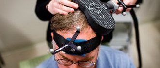
It is simple and painless. Electrodes, which are sometimes incorrectly called “sensors,” are placed on a person’s head in the area of the back of the head and forehead. Modulated pulses are sent through them from the device. Treatment lasts about 30 minutes, and the course is usually about 10 procedures. In some cases, electrical stimulation of the brain may cause a slight tingling sensation. This effect is painless, and many people even like it.
Indications for use
Transcranial electrical stimulation is a universal method of physiological therapy that is used for a wide variety of diseases and pathological conditions. The main indications for this type of electrotherapy are:
- chronic pain syndromes in neurology and internal medicine (treatment of neuropathic pain in postherpetic neuralgia, diabetes mellitus, oncology, phantom pain). It is known that its own endorphin is 20-30 times stronger than the analgesic effect of morphine;
- this method is indispensable in the complex treatment of depression, anxiety, PTSD, relief of various neuroses;
- stimulation of reparative processes in trophic ulcers and burns;
- borderline forms of arterial hypertension. Regulation of vascular tone leads to normalization of blood pressure;
- decreased immunity, chronic infections;
- in oncological practice, electrical brain stimulation can be used to slow down the rate of tumor growth;
- in pregnant women, TES is used in the treatment of toxicosis to prevent eclampsia;
- in the postmenopausal period to relieve neurotic conditions;
- in case of skin diseases - with atopic dermatitis, eczema, psoriasis, neurodermatitis;
- in the treatment of drug addiction and alcoholism (relief of withdrawal symptoms, physical and mental dependence);
- stimulation of various brain systems makes it possible to reduce the frequency of allergic manifestations, the dosage of antihistamines used and even hormones in the treatment of allergic diseases (asthma, hay fever, rhinitis, Quincke's edema, chronic urticaria);
- for endocrine diseases;
- to reduce the severity of inflammatory processes in autoimmune diseases (rheumatoid, psoriatic arthritis);
- for the treatment of insomnia disorders (insomnia).
It should be added that, despite the abundance of various indications for use and favorable reviews from patients, this method also has contraindications. We must not forget that any manipulations with electric current, be it treatment or electrodiagnostics, can generate magnetic fields and directly harm the operation of certain mechanisms.
Technique of TES therapy
Before using transcranial electrical stimulation to treat any disease, it is necessary to consult a physiotherapist and a neurologist. Therapy should not be performed immediately after eating; the interval between eating and the session should be at least two hours.
During TES therapy, the patient needs to lie down or sit, and the specialist places electrodes on the patient’s forehead, on the back of the head and on the nipples. Before applying the electrodes, the patient's skin is thoroughly cleaned and checked for damage, formations or rashes.
Ear jewelry should be removed during TES therapy. During the procedure, the patient may feel a slight vibration, tingling in the area where the electrodes are applied, and may also observe light flickering with their eyes closed. Electrodes do not leave marks on the skin.
To carry out the manipulation, special pulsed low-amplitude currents with a rectangular bipolar pulse shape are used. During the first procedure, the minimum current reading should be used, which should not be increased even if the patient does not have any third-party sensations. In subsequent sessions, the current levels are gradually increased until the patient begins to feel their presence. If discomfort is detected, the specialist should reduce the supplied current. Gradually, towards the end of the procedure, the current strength should be reduced to zero.
The patient should experience the above sensations, but they should not be unpleasant to him. In this way, the optimal current strength is regulated, and if it bears fruit in the form of positive results, then during further sessions the specialist will use exactly this current, without changing it upward.
If the patient has severe pain, then similar procedures can be performed several times a day with an interval of at least 4-6 hours. If the pain does not disappear, the patient is advised to undergo additional medical consultation and examination.
Currents affect the alpha rhythms of the human brain at a frequency of 70-80 hertz, stimulating the production of endogenous opioid peptides. After the session, those who suffer from blood pressure disorders are advised to rest for fifteen minutes and undergo a neck-collar massage. The duration of one procedure of transcranial electrical stimulation is 20 minutes, although in case of severe pain this time can be increased. The course of therapy usually includes about 15 sessions. In case of exacerbation of chronic diseases, TES therapy is aimed at relieving emerging symptoms, which can be done in 5 procedures. In 1 calendar year, it is permissible to conduct up to 60 sessions, repeating courses every 2-3 months.
Contraindications
Contraindications to this method are not an extensive list, but it must be remembered that transcranial electrical stimulation is contraindicated for:
- pathological lesions on the scalp at the site of electrode attachment (psoriasis, weeping eczema);
- the presence of space-occupying brain formations (tumors, parasitic cysts);
- history of epilepsy and episyndrome, or pathological delta rhythm according to EEG;
- crisis course of hypertension;
- presence of pheochromocytoma;
- increased thyroid function – hyperthyroidism in the stage of thyrotoxicosis;
- presence of a permanent pacemaker.
Finally, early childhood (up to 5–7 years) is also a contraindication to the procedure. But not at all because the child will be harmed. It’s just that the brain’s mediator systems have not fully matured, and for a young creature to be in a calm and relaxed state for half an hour is sheer torture.
Transcranial direct current stimulation
The key challenge of multielectrode transcranial electrical stimulation (TES) or transcranial direct current stimulation (tDCS) is to find the optimal stimulation pattern that delivers the required current density at the target and minimizes it in the rest of the brain, which can be mathematically defined as an optimization problem. Such optimization with least squares (LS) or Linearly Constrained Minimum Variance (LCMV) algorithms is typically expensive and requires multiple independent current sources.
Based on the principle of reciprocity in electroencephalography (EEG) and TES, optimal TES patterns can be quickly found. It is possible to define four different cortical targets in a detailed seven-objective finite element model and analyze the performance of different variants of TES methods considering reciprocity in terms of electrode density, targeting error, focality, intensity and directionality using LS and LCMV solutions as reference standards. The reciprocity algorithms are found to show good accuracy, comparable to LCMV and LS solutions. The use of higher electrode density improves focality, directionality and current intensity parameters.
Transcranial electrical stimulation (TES) is also known as transcranial direct current stimulation (tDCS) and transcranial alternating current stimulation (tACS). Because current levels are typically small (1-2 mA) and do not actually stimulate neurons, this technique is also called transcranial electrical neuromodulation (TEN). Even without neuronal stimulation, TES or TEN methods are capable of modifying the excitability of the cortex, as well as changing the rhythms of bioelectrical activity of the brain and the spatiotemporal activity of neural networks.
Compared to transcranial magnetic stimulation (TMS), TES is better tolerated, cost-effective, and an easy-to-use instrument. TES is a new therapy for the treatment of neuropsychiatric conditions such as depression, Parkinson's disease, anxiety and chronic pain.
Research has also shown that TES may be a valuable therapeutic tool for epilepsy, stroke rehabilitation, and other neurological and psychiatric disorders. It has also been suggested to improve cognitive skills such as memory or learning. This method may eventually become an alternative to the use of psychotropic drugs, since it does not affect the entire brain and has minimal side effects.
The requirement for specific targeting of neuronal regions of interest (ROI) is to use a methodology that minimizes, as much as possible, the current applied to brain regions unrelated to the target.
Despite these recent advances, debate continues regarding the clinical effectiveness of EFT, particularly the explanation for the variability in response to such therapy. About 36% of patients show the pattern of anodal excitatory/catadonic inhibitory effects that are commonly described in the literature. In addition, evidence of a nonlinear relationship between dosage and measured shock effects implies that agreement between treatment and bioelectrical activity recordings may be very sensitive to dosage accuracy.
Because the current flow of current cannot be completely focused, but rather follows the path of least resistance through the scalp tissue, an accurate model of electrode positions and conduction is required. Additionally, since current appears to show different effects when it is consistent with neuronal activity (normal to the cortical surface) versus crossing neurons (tangential flow), it is important to model human cortical geometry to the cortical surface using individual anatomical MRI, with the goal of calculating components of induced current density that are normal to the cortex than tangential currents.
In addition, there is increasing interest in electric shocks going beyond the traditional use of two large sponge electrodes, such as using a local high-resolution model of a single source (electrode) surrounded by four electrodes or massive dense electrode arrays to improve TES accuracy.
Refining the specification of the applied current density for each individual subject will therefore allow calculation of the effective dose individually, which may be important for understanding the large variability in responses observed among individuals.
In this regard, anatomically correct and head model-specific shock delivery to the TES becomes increasingly important in determining where to definitively test the clinical efficacy of TES in future clinical trials.
Several studies have been conducted to calculate dose for ROI and optimize electrode shape and size, as well as the total number of electrodes and their configuration. Estimation of dose to the original ROI is typically accomplished using finite element (FE) or finite difference (FD) current density modeling in detailed 3D head models. FE modeling has shown that high current densities can be observed in the most unexpected regions, due to the relatively high electrical conductivity of the cerebrospinal fluid (CSF) and the irregular shape of the cortex.
In addition to the need to model complex current application geometries, one of the uncertainties is the fact that the conductivity of human head tissue (in particular, the most resistant one, the skull bones) is not well known. Such conductance can be assessed using limited electrical impedance tomography (Beit) if such geometry is constructed accurately based on neuroimaging (structural MRI).
In general, TES uses two main types of electrodes: large anode and cathode patches (typically 5 by 7 cm) and significant smaller density EEG-like multiple electrodes. In the first, more traditional approach, a small number (usually two) of relatively large round or square electrodes are used in a bipolar configuration and in different mountings. Large electrode areas help reduce current density on the scalp. However, they do not allow for the reconfiguration of spatial electrode montages and hence stimulation patterns during the stimulation protocol on the time scale of brain dynamics (milliseconds). With advances in dense array EEG recording, it is now possible to analyze large electrode arrays for non-invasive neuromodulation. The use of a more flexible multi-electrode circuit allows for the implementation of much more universal and even closed circuits, the creation of dynamic stimulation protocols targeting several ROIs in one stimulation session, while simultaneously recording EEG for the purpose of neurophysical feedback and rapid adjustment of stimulation configuration templates using software. With a dense array of TES electrodes, the accuracy of focality and intensity of exposure to cortical targets also increases.
By changing the location of small electrodes (or a smaller cluster of electrodes approximating an area) on the scalp and comparing them with the level of exposure to a fixed current, it is possible to optimize the “delivery” of current to the ROI region of the cortex using a special algorithm. The problem of determining the required directional current density, without imposing additional restrictions on minimal impact on other areas of the brain, can be solved directly and accurately using the principle of reciprocity. Optimization of dense array TES is generally more difficult due to the much greater number of degrees of freedom than in two tDCS patch electrodes.
The main challenge is to find a current shock pattern describing the current (electrical source) or "sink" (output) levels for each electrode in a dense electrode array system that improves target accuracy in order to maximize current density at cortical ROI and minimize it in other brain regions. To meet safety constraints, limiting the current to the electrode requires type 1- constraints, which makes these algorithms iterative and therefore intensity calculations. In addition, these algorithms require an independent current source for each electrode, which increases hardware complexity and cost [with the exception of a small number of studies on dense tDCS arrays with fewer current sources and fewer electrodes.
The principle of reciprocity refers to the complementarity of the electric field at the cortical dipole location created by injecting current at the scalp, with the electrical potential at the scalp at the injection point caused by the same dipole. This method combines electroencephalography (EEG) and TES (FP) to effectively find the optimal solution by analyzing EEG and TES sources. A similar reciprocity relationship exists between magnetoencephalography (MEG) and TMS FP. This approach can be used as a guide to search for shock patterns both in a dense TES array with hardware and protection constraints, and in a dynamically reconfigurable multi-TMS.
With denser scalp coverage in a dense pole EEG array, the EEG topography for any cortical lead field is better approximated, and thus back current injection from these "pole" electrodes is expected to provide more accurate targeting. It remains unclear whether reciprocity-based targeting methods perform similarly or better than LS and LCMV methods, and whether using a very large number (256) of electrodes instead of (128) actually improves the performance of these methods. Preliminary results on using the reciprocity principle to obtain convenient current influence protocols using EEG networks with 128 and 256 high-density networks. The reciprocity method is optimal for maximizing the current density component at a target of the desired orientation. Four methods derived from the principle of reciprocity are described, empirically taking into account the additional requirements of minimizing the effects of TES on non-target brain regions and contrasting them with the LS and LCMV algorithms. Simulations on a detailed FE brain model using four representative cortical targets are possible to evaluate the performance of the methods in terms of targeting error (TE), focality, directionality, and shock intensity.
The first method, based on reciprocity, has a theoretical meaning where only one electrode introduces the total maximum current, and the remaining electrodes act as multiple "absorbers" to propagate back currents and minimize the impact of TES on non-target areas.
In the other three reciprocity methods, an additional constraint is considered: an upper limit on the current delivered by each electrode, which is usually considered in terms of a safety limitation in order to avoid skin irritation. These methods differ in the way they select "sinks" and provide the best optimization in terms of either total target intensity ("opposite" configuration) or focality index ("ring" configuration).
A soft tissue reference model for an adult subject could be obtained from T1-weighted MR images of the head using a 3T Allegra scanner (Siemens Healthcare, Erlangen, Germany). Bone structure was obtained from a CT scan of the same object recorded using a GE CT scanner (General Electric, Fairfield, United States). The registration matrix is 256 × 256 × 256 with a voxel size of 1 mm × 1 mm × 1 mm in both CT and T1 scans. To construct anatomically accurate model geometry, T1 MRI images are automatically segmented into seven tissue types (gray matter, white matter (WM), CSF, scalp, eyeballs, internal air, and skull). The TT volume is segmented into soft tissue, internal air components, and cranial bone. Typical electrode positions in EGI 128 and 256 high-density EGI sensor networks identified for the site in previous studies are conducted using a geodetic protogrammetry (GPS) system.
1.Electromagnetic stimulation
In modern therapy of neuropathologies and motor disorders, electromagnetic stimulation
finds wide application. In a number of clinical situations, this method has no equal in effectiveness and safety. The validity and effectiveness of this type of functional neurosurgery has been statistically confirmed. The essence of the technique is the constant or systematic impact on the nerve centers of electromagnetic pulses of a certain frequency and amplitude. The effectiveness of this effect has been proven, primarily for parkinsonism, dystonia, uncontrolled spasms and other disorders of muscle tone.
There are different forms of functional muscle pathology
: dystonia, uncontrolled movements, poor coordination, tremors, loss of the ability to walk and perform precise movements. In each specific case, diagnostics is aimed at identifying as accurately as possible the area of the brain that causes dysfunction. To do this, the patient’s electroencephalogram is carefully studied, which gives an idea of the required location of the electrode (localization and depth).
A must read! Help with treatment and hospitalization!
How to prepare for the procedure
No specific preparation is required, which allows transcranial stimulation to be performed on patients of various age categories.
During the preliminary conversation, the patient is told how the procedure will go. The course of treatment is usually 5-9 sessions.
The procedure is prescribed after examination by a doctor and certain studies. To exclude acute conditions, the following is performed:
- blood pressure measurement;
- temperature measurement;
- sometimes electrocardiography.
Depression
Waltre et al were the first to report the effects of transcranial magnetic stimulation on depression in three patients under 18 years of age. The children received daily treatment with 10 Hz TMS for two weeks. Two showed clinical improvement, but one of them complained of headache for two sessions. In another study, Loo et al tested the effects of 10 Hz TMS over six weeks on two 16-year-old teenage girls with depression and ADHD. As a result, there were no improvements in ADHD symptoms, but they reported a decrease in depressive symptoms. Despite the good results, all these data are preliminary because the studies were conducted without control groups. But they show that adolescents tolerate the study's intensive parameters well. No studies have been conducted using tDCS.
3.Preparation and performance of the operation
Each disorder is caused by disturbances in a specific area of the brain, so the installation of electrodes is carried out after a thorough diagnosis, which includes:
- electroencephalogram;
- MRI;
- CT;
- general diagnosis of the body's condition.
The procedure for installing electrodes and generator is carried out under local anesthesia
, since the brain itself has no pain receptors and only skin anesthesia is required. During surgical procedures, the patient is in full contact with the surgeon, which allows him to assess the condition and progress of the operation.
After operation
measures are taken to prevent infection of the affected areas, including a course of antibiotics. Discharge from the hospital occurs on days 3-5. After two weeks, a second visit to the neurosurgeon is necessary.
About our clinic Chistye Prudy metro station Medintercom page!