Structure of the spinal cord
Before talking about the components, you should understand the system itself. The spinal cord is located in the spinal canal, and is surrounded by three boundaries in the form of membranes:
- Soft
- Cobweb
- Solid
It is in constant interaction with internal organs and the brain. Its length is approximately 40 centimeters for women and 45 for men, weighing about 30 grams. It’s not for nothing that there is an expression: the spool is small, but expensive. The psychological state, the functioning of internal organs, reflexes and motor activity of a person depend on this small link.
The components of the spinal cord are gray and white matter. The location of the first of them is the central departments, the second is the periphery. In the embryo, the spinal cord occupies almost the entire spinal canal, and with subsequent growth it moves upward.
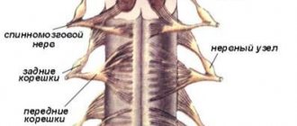
Looking at a cross-section of the human spinal cord (SC), you can see a central canal filled with brain fluid, gray matter in the shape of a butterfly (whose wings resemble horns) and white matter formed from paired cords - posterior, anterior and lateral.
Each of these sections performs a specific function that plays a vital role for a person. They ensure the passage and delivery of nerve impulses to various parts of the human body.
Structure and functions of the gray matter of the spinal cord. Rexed plates
The spinal cord is made up of gray and white matter . Gray matter consists of nerve cell bodies and nerve fibers—the processes of nerve cells. White matter is formed only by nerve fibers - processes of nerve cells (spinal cord and brain). Gray matter occupies a central position in the spinal cord. The central canal runs through the center of the gray matter. Outside the gray matter is the white matter of the spinal cord.
In each half of the spinal cord, gray matter forms gray columns. The right and left gray pillars are connected by a transverse plate - a gray commissure, in the center of which the opening of the central channel is visible. Anterior to the central canal is the anterior commissure of the spinal cord, and behind is the posterior commissure. On a cross section of the spinal cord, the gray columns, together with the gray commissure, have the shape of the letter “H” or a butterfly with spread wings (Fig. 2.5). The lateral projections of gray matter are called horns. There are paired, wider anterior horns and narrow, also paired posterior horns. The anterior horns of the spinal cord contain large nerve cells called motor neurons (motoneurons). Their axons form the bulk of the fibers of the anterior roots of the spinal nerves. The neurons located in each anterior horn form five nuclei: two medial and two lateral, as well as a central nucleus. The processes of the cells of these nuclei are directed to the skeletal muscles.
The posterior horn consists of interneurons, the processes of which (axons) are sent to the anterior horn, and also pass through the anterior white commissure to the opposite side of the spinal cord.
The nerve fibers (sensitive) of the dorsal roots, which are processes of nerve cells whose bodies are located in the spinal ganglia, end on the nerve cells of the nuclei of the dorsal horns. The peripheral part of the dorsal horns processes and conducts pain impulses. The average is associated with skin (tactile) sensitivity. The area at the base of the dorsal horn provides processing and conduction of muscle sensation.
The intermediate zone of gray matter of the spinal cord is located between the anterior and posterior horns. In this zone, from the VIII cervical to the II lumbar segment, there are projections of gray matter - the lateral horns. The lateral horns contain the centers of the sympathetic part of the autonomic nervous system in the form of groups of nerve cells united in the lateral (lateral) intermediate substance. The axons of these cells pass through the anterior horn and exit the spinal cord as part of the anterior roots of the spinal nerves. The intermedial nucleus (see Fig. 2.5) is the main “computing center” of the spinal cord. Here, sensory signals processed in the dorsal horn are compared with signals from the brain and a decision is made to initiate an autonomic or motor response. In the first case, trigger stimuli are sent to the lateral horn, in the second - to the anterior horn.
| Designations: 1. Spinal nerve fibers. 2. Afferent neurons of the spinal ganglion. 3. Posterior cord, funiculus dorsalis (posterior). 4. Posterolateral bundle, Lissauer tract (Lissauer, Heinrich, 1861-1891, German neurologist), fasciculus posterior lateralis. 5. Cervical spinothalamic tract (spinocervical tract, lateral tract of Morin, Morin D., 1955, 1964), tractus spinothalamicus cervicalis. 6. Posterior spinocerebellar tract, tractus spinocerebellaris dorsalis (posterior), Flechsig’s bundle (Paul Emil Flechsig, 1847-1929, German anatomist and neurologist). 7. Anterior spinocerebellar tract, tractus spinocerebellaris ventralis (anterior), Gowers bundle (William Richard Gowers, 1845-1915, British neurologist). 8. Lateral spinothalamic tracts, tractus spinothalamicus lateralis and anterior spinothalamic tracts, tractus spinothalamicus ventralis (anterior). |
The laminae of the gray matter of the spinal cord (Rexed's laminae) are heterogeneous anatomical structures of the gray matter of the spinal cord, identified on the basis of the morphology of their constituent neurons.
The gray matter of the spinal cord has “cytoarchitectural lamellarity,” that is, different areas (resembling longitudinally oriented plates) composed of neurons of homogeneous morphology (B. Rexed, 1952, Bror Rexed, 1914-2002, Swedish anatomist). In this regard, the gray matter of the spinal cord is divided into paired structures - plates (diagram). The plates are numbered with Roman numerals. Laminae I ÷ V form the posterior horns of the gray matter of the spinal cord. Plate VII forms the intermediate zone, which is the basis of all the horns of the spinal cord. Lamina IX consists of collections of large motor neurons called α-motoneurons and small motor neurons called gamma motor neurons. Axons of α-motoneurons innervate striated muscles. Axons of α-motoneurons innervate the contractile elements of muscle spindles. The axons of both α-motoneurons and α-motoneurons exit through the anterior (ventral) roots of the spinal nerves. Plates VII and VIII are very variable in structure. Plate VI is present only in the cervical and lumbosacral enlargements of the spinal cord. The collection of cells surrounding the central canal of the spinal cord along its entire length is often referred to as lamina X. All neurons that directly perform the function of a sensory receptor without an intermediary cell (primary sensory neurons) are located in the spinal ganglion, which is located in the intervertebral foramen. These neurons have two processes: peripheral and central. Peripheral processes transmit information from the periphery of the body from various receptors. The central processes of the same neurons form bundles of fibers that pass dorsolaterally into the spinal cord. These fibers transmit information about specific types of sensations to the central nervous system. This information follows further along special pathways to various parts of the nervous system and is used for the structural and functional organization (for control) of body systems. Information about pain and potentially harmful effects is transmitted mainly to the neurons of lamina I and lamina II of the gray matter. Information about tactile sensations is transmitted mainly to the cell bodies of lamina IV neurons or to the processes of these neurons. Information from muscle stretch receptors (from muscle spindles and tendon receptors) is transmitted along the nerve fibers of sensory neurons partially to the neurons of plates V, VI and VII. The collaterals of these nerve fibers, involved in the implementation of muscle stretch reflexes, are also directed to the neurons of plate IX.
In 1952, the Swedish anatomist Bror Rexed proposed dividing the gray matter into ten plates (layers), differing in the structure and functional significance of their constituent elements. This classification has received wide recognition and dissemination in the scientific world. Plates are usually designated by Roman numerals.
Laminae I to IV form the head of the dorsal horn, which is the primary sensory area.
Plate I is formed by many small neurons and large spindle-shaped cells lying parallel to the plate itself. It includes afferents from pain receptors, as well as axons of neurons in lamina II. The emerging processes contralaterally (that is, crosswise - the processes of the right posterior horn along the left cords and vice versa) carry information about pain and temperature sensitivity to the brain along the anterior and lateral cords (spinothalamic tract).
Plates II and III are formed by cells perpendicular to the edges of the plates. Corresponds to gelatinous substance. Both are affered by processes of the spinothalamic tract and transmit information downstream. Participate in the control of pain. The II plate also gives off processes to the I plate.
The IV plate corresponds to its own core. Receives information from plates II and III, the axons close the reflex arcs of the spinal cord on motor neurons and participate in the spinothalamic tract.
The V and VI plates form the neck of the posterior horn. Receive afferents from muscles. Plate VI corresponds to the Clarke nucleus. Receives afferents from muscles, tendons and ligaments, descending tracts from the brain. Two spinocerebellar tracts emerge from the plate: Fleschig's tract (variant: Flexig) (tractus spinocerebellaris dorsalis) - exits ipsilaterally (that is, into the cord of its own side) into the lateral cord Govers' tract (tractus spinocerebellaris ventralis) - exits contralaterally into the lateral cord.
VII occupies a significant part of the anterior horn. Almost all neurons of this plate are intercalary (with the exception of efferent neurons of Nucleus intermediolateralis. Receives afferentation from muscles and tendons, as well as many descending tracts. Axons go to plate IX.
Plate VIII is located in the ventromedial part of the anterior horn, around one of the parts of plate IX. Its neurons participate in propriospinal connections, that is, they connect different segments of the spinal cord with each other.
Plate IX is not unified in space; its parts lie inside plates VII and VIII. It corresponds to the motor nuclei, that is, it is the primary motor area, and contains motor neurons located somatotopically (that is, it represents a “map” of the body), for example, motor neurons of the flexor muscles usually lie above the motor neurons of the extensor muscles, neurons innervating the hand are more lateral, than those innervating the forearm, etc.
The X plate is located around the spinal canal, and is responsible for the commissural (between the left and right parts of the spinal cord) and other propriospinal connections.
EXAMINATION CARD No. 5 in Physiology of the Central Nervous System
What is gray matter?
Because the gray matter surrounds the central canal, it is called the intermediate canal. It is presented in the form of two vertical strands of irregular geometric shape, located on both sides of the central channel, called the left and right gray pillars. These elements are connected to each other by a thin plate (adhesion), surrounding and protecting the canal on all sides, adjacent from above to the fourth ventricle of the brain, ending below with the terminal ventricle called Krause.
The basic structure of the gray matter of the SC is clearly represented in the form of horns:
- The anterior ones are the location of the largest neurons, forming five main clusters or nuclei. These clusters provide motor activity of the human body, abdominal and thoracic vibrations during breathing.
- The structure of the posterior pillars (horns) is heterogeneous. It consists of axons and neurons, which are multi-polar cells. Each of them occupies a specific place on the territory of the CM. The processes of these neurons extend to the corresponding adjacent segments of the anterior layer.
- The lateral horns are responsible for the functionality of the autonomic nervous system (ANS) and have protrusions in the areas of the eighth cervical and second lumbar vertebrae. The upper (cervicothoracic) region regulates the functioning of the heart, digestive organs, sweat glands and blood vessels. Lower (lumbosacral) – defecation, urination, erection and ejaculation.
The nerve centers of the SC are segments with neurons located in them that affect all functioning receptors and internal organs. It is quite clear how important they are for the full life of the body.
What is the composition of gray matter?
Gray matter is formed from nerve cells, of which there are more than thirteen million (including glia and processes). All neurons grouped into nuclei or plexuses that make up the gray matter of the SC are divided into three main groups:
Internal cells with numerous processes that do not extend beyond the boundaries of the substance itself and provide interaction (forming synapses) with other neurons of the SC.
Root cells (the largest motto- and multi-neurons of the ANS), the neurons of which protrude beyond the boundaries of the gray matter and innervate skeletal muscles.
Tufted neurons forming many pathways leading to the brain. These cells got their name due to their ability to form bundles, which are a kind of switches connecting SC segments.
So, the spinal cord includes neuroglia, ganglion nerve cells, pulpal and non-pulpate nerve fibers. These cells are grouped into nuclei and are located in segments along the entire length of the spinal cord and its primary three-membered reflex arc.
Each segment of gray matter is separated by barriers in the form of Rexed plates that perform specific functions:
- Reception of impulses from the dorsal roots and their transmission to the spinothalamic tract.
- Accompaniment of impulses (afferents) of temperature and pain sensitivity.
- Processing information from the muscular system.
- Receiving and transmitting brain impulses.
- Innervation of the muscles of the limbs, body and internal organs.
Disturbances in the functioning of any of the links in this system lead to serious consequences, including paralysis, partial or complete.
Histology.RU
Material taken from the site www.hystology.ru
Anatomically, the spinal cord consists of two symmetrical halves, delimited from each other by the ventral median fissure and the dorsal median septum. The centrally located gray matter of the spinal cord contains multipolar nerve cells that form the nuclei of the spinal cord. Its peripheral part, the white matter, is represented by a collection of nerve fibers that are part of the various pathways of the central nervous system.
Gray matter of the spinal cord . Anatomically, the gray matter of the spinal cord consists of two halves connected by a commissure. Each of them has dorsal and ventral horns. In the thoracic and lumbar segments of the spinal cord, the superolateral section of the ventral horns can be distinguished as its lateral horns. In the gray commissure, which connects the two halves of the gray matter, there is the central canal of the spinal cord.
Gray matter is formed by multipolar neurons, unmyelinated and myelinated nerve fibers and neuroglia. Groups of nerve cells of the same functional significance form the nuclei of gray matter.
Based on morphological characteristics, localization, participation in nerve conduction in the gray matter of the spinal cord, the following types of cells can be distinguished: radicular cells - cells whose neurites leave the spinal cord as part of its ventral roots; internal cells, the neurites of which form synapses on the cells of the gray matter of the spinal cord brain, and tuft cells. Their neurites form separate bundles in the white matter that conduct nerve impulses from certain nuclei of the spinal cord to its other segments or to certain parts of the brain, forming the pathways of the central nervous system.
In the gray matter of the spinal cord, areas are distinguished that differ in neuronal composition, the nature of nerve fibers and neuroglia. Thus, in the dorsal horns of the gray matter, the spongy layer, gelatinous substance, the proper nucleus of the dorsal horn, its dorsal nucleus, or Clark’s nucleus should be distinguished.
The spongy layer of the dorsal horns of the gray matter contains small tufted cells embedded in a broadly looped glial framework.
The gelatinous substance is formed mainly by glial elements, which contain small amounts of small tuft cells.
The dorsal horn contains the dorsal horn nucleus, the thoracic nucleus (Clark's nucleus), and a significant number of diffusely scattered small multipolar interneurons.
The nucleus of the dorsal horn contains tufted cells, the axons of which pass through the anterior white commissure
Rice. 176. Schematic section of the spinal cord and spinal ganglion:
1 - 2 - reflex pathways of conscious proprioceptive sensations and touch; 3 and 4 - reflex pathways of proprioceptive impulses; 5 - reflex pathways of temperature and pain sensitivity; b — posterior own bundle; 7 - lateral own beam; 8 - anterior own bundle; 9 - posterior and 10 - anterior spinocerebellar tracts; 11 - spinothalamic tract; 12 - delicate bun; 13 - wedge-shaped bundle; 14 - rubro-spinal tract; 15 - thalamo-spinal tract; 16 - vestibulo-spinal tract; 17 - reticulospinal tract; 18 - tecto-spinal tract; 19 - corticospinal (pyramidal) lateral tract; 20 - corticospinal pyramidal anterior tract; 21 - own nucleus of the posterior horn; 22 - thoracic nucleus (Clark's nucleus); 23, 24 — nuclei of the intermediate zone; 25 - lateral nucleus (sympathetic); 26 - nuclei of the anterior horn.
to the opposite side of the spinal cord into the lateral cord of the white matter, where they form the ventral spinocerebellar and spinothalamic tracts (Fig. 176).
The dorsal horns contain a significant number of small, multipolar associative and commissural neurons, the neurites of which form synapses on the cells of the gray matter of the spinal cord of the same side (associative cells) or the opposite (commissural).
The dorsal nucleus is formed by large cells, their axons enter the lateral cord of the white matter on the same side and, as part of the dorsal spinocerebellar tract, enter the cerebellum.
The nerve cells of the intermediate zone of the gray matter form two nuclei: the medial intermediate nucleus, the neurites of which join the fibers of the ventral spinocerebellar tract of the same side, and the lateral intermediate nucleus, which contains associative cells of the sympathetic nervous system. The axons of these cells leave the spinal cord through the ventral roots of the spinal cord and form the white connecting branches of the sympathetic trunk.
The nuclei of the ventral horns of the gray matter are formed by the largest nerve cells of the spinal cord (100 - 140 µm in diameter). Their neurites form the bulk of the fibers of the ventral roots. Through the mixed spinal nerves they enter the skeletal muscles and end in the motor nerve endings. In the ventral horns of the gray matter of the spinal cord, two groups of motor cells are distinguished: the medial one, which innervates the muscles of the trunk, and the lateral one, which is characteristic of the region of the cervical and lumbar enlargements of the spinal cord. The lateral nucleus of the ventral horns contains neurocytes that innervate the muscles of the limbs.
Nerve cells are scattered in the gray matter, their axons in the white matter are divided into longer ascending and shorter descending branches. These fibers form their own (main) bundles of white matter adjacent to its gray matter. They give rise to many collaterals, ending in synapses on the motor cells of the anterior horns of 4–5 adjacent segments of the spinal cord. The spinal cord has three pairs of its own bundles.
The white matter of the spinal cord consists of myelinated nerve fibers and a supporting neuroglial framework. Nerve fibers in the white matter make up pathways (fiber complexes) - links of certain reflex arcs. Individual pathways are characterized by the position and functional identity of the cells whose processes are their fibers, their synaptic connections and position in the white matter of the spinal cord.
Among the conducting pathways, one should highlight: 1) the pathways of the spinal cord’s own reflex apparatus, 2) the pathways connecting the spinal cord and brain, 3) ascending (afferent) and 4) descending efferent (see anatomy).
Reviews (0)
Add a review
Secrets of his education
The gray matter of the SC is formed from many neurons with processes, united into nuclei and without a myelin sheath. To be more precise, it contains:
- Long neural processes
- Short neural processes
- Neuron cell bodies
- Connective and accompanying tissue
- Blood vessels
Each group of neurons is responsible for specific functions:
Sensitive ones provide the perception of impulses coming from tissues and organs. Their receptors are located throughout the human body and are concentrated on the skin and internal organs. Only their processes penetrate into the spinal cord, and the neurons themselves are located outside it.
Motor muscles are responsible for conducting impulses to the muscles from the centers of the spinal cord or brain.
Intercalary neurons have a complex structure and are located between the processes of sensory and motor neurons. Their main task is to receive and process all incoming information.
The anterior horn of the gray matter of the spinal cord is the largest and widest and houses motor neurons. The location of the sensitive nerve cells is the dorsal horn, which ensures the functioning of the internal glands and organs. The smallest and weakest middle horn is inhabited by intercalary neurons; it is here that the initial processing of all signals coming from various zones of the body takes place.
Most cells in the spinal cord are designed to control, analyze, receive and transmit pain impulses. They are multipolar, which distinguishes them from white matter neurons of the SC.
In a few words, gray matter can be characterized as a collection of nerve fibers and neurons with processes up to 0.1 millimeters in diameter and up to one and a half meters long. Each of these elements has its own purpose and different functionality.
Difference from white matter
This section is intended to calibrate concepts and answer the question of what gray and white matter of the brain are.
Gray matter
- Created by the nuclei of nerve cells and its relatives.
- It is located mainly in the central parts of the nervous system.
- Makes up no more than 40% of the total mass of the brain.
- Consumes about 3-5 ml of oxygen per minute.
- A structure that has a regulatory function.
White matter
- Formed by long myelinated axons.
- It is located primarily in the peripheral nervous system.
- Makes up more than 60% of the weight of the human brain.
- Consumes less than 1 ml of oxygen per minute.
- Responsible for conducting nerve impulses through the nervous system
It should be remembered that unlike the structure of the cerebral cortex, where the gray matter is a shell and covers the white substance, in the spinal cord the gray matter is surrounded by the white matter of the brain.
Functions and Features
Gray matter is located in the center of the spinal cord along almost the entire spinal column. At the levels of the lumbar and cervical regions it has a pronounced thickening, the presence of gray brain tissue is somewhat increased here. Thanks to this structure, human mobility and the ability to perform certain manipulations are ensured.
In addition, the SM performs a number of other important functions:
- Protective. The gray matter, like a membrane, is located around the central canal, protecting and circulating the cerebrospinal fluid (CSF), which delivers nutrients to tissues and nerve fibers.
- Regulatory. Most of the neurons and cells of the gray matter of the SC are capable of controlling many vital processes. They regulate the proper functioning of certain organs: kidneys, intestines, bladder, secretory glands and reproductive system.
- Conductor, which consists in ensuring information exchange between the brain and all systems, muscles, blood vessels and internal organs.
- Reflex. Many impulses received by the spinal cord are processed independently by it. At the same time, the load on the brain is reduced, responses occur without its participation and are called unconditioned reflexes. They play an important role, as they are sometimes created in an instant reaction (for example, to impulses of a warning and painful nature).
It turns out that the set of responsibilities assigned to the spinal cord is very large. This explains its complex structure and intricate structure.
