Positron emission tomography, or PET for short, is considered a doctor's test of the brain and other internal organs. Tomography is used mainly for diagnosing tumors. The method helps to find cancer in the latent stage. PET CT of the brain is used not only in oncology, but also in neurosurgery, psychiatry, cardiology, neurology and genetic testing.
PET CT scan of the brain is painless and absolutely safe. The procedure lasts no more than an hour. Unlike MRI and CT, which mainly study statistical indicators and the anatomical structure of the brain, PET/CT is aimed at a complete study of this vital organ in dynamics, that is, it directly examines the functions of the brain.
Indications for brain diagnostics using PET/CT
Experts recommend that certain people (those at particular risk) carry out this procedure regularly, without waiting for any signs of disease to appear. But there are indications when it is necessary to conduct PET/CT for everyone without exception.
Symptoms for which you are referred for a PET CT scan of the brain in Moscow:
- large and small convulsions (you should consult a specialist even if they appear in isolated cases);
- a disorder of consciousness in which a person loses the ability to perceive speech;
- loss of consciousness (especially if this occurs frequently);
- dizziness and chronic headache;
- feeling of lack of air, pain in the heart area, diarrhea and constipation, trembling limbs, decreased appetite;
- gag reflex and nausea;
- sharp darkening in the eyes, loss from the visual field, bifurcation of the visual picture.
Features of PET CT gallium
The radiopharmaceutical used in the scan is absorbed by all cells. Its versatility is due to its structure close to glucose. Cancerous formations absorb such a substance at a faster rate, which allows them to be detected during tomography.
In the picture, the tumor appears as a bright spot. This is because it absorbs more 18F-FDG. Based on the color saturation, number and location of these spots, the diagnostician can draw a conclusion about the stage and nature of the formation.
Another nuance should be taken into account by patients with diabetes. The presence of glucose in the radiopharmaceutical may reduce the reliability of the results or cause complications for the patient. Therefore, in this case, diagnosis is possible only after consultation with an endocrinologist.
Preparation for the brain PET/CT procedure
First of all, the day before a PET CT scan of the brain, it is forbidden to engage in physical exercise. You should not drink alcohol, caffeine or smoke cigarettes. This also applies to nicotine patches, hookah and chewing tobacco. You cannot ignore these recommendations, as you can harm your health, because the reaction can be the most unpredictable.
Before a PET CT scan of the brain, it is advisable to eat a light dinner, which should include fermented milk and cottage cheese products. 6-7 hours before the procedure, it is not advisable to eat, chewing gum or sweets. Drinking before PET CT scan of the brain is recommended; the patient should drink at least 0.5 liters of water or weak tea and coffee, without sugar or any sweeteners.
It is worth noting that the patient’s clothing should not contain metal buttons, fasteners, zippers, hooks or snaps. Clothing for a PET CT scan of the brain should be as comfortable and warm as possible. This is necessary for good muscle relaxation, and as a result, a high-quality image.
It is strictly prohibited to take watches, keys and other metal objects for a PET CT scan of the brain.
How is the research conducted?
- You must arrive at the PET center at the time specified when registering for the study;
- please turn off mobile phones and maintain silence in the premises of the PET center (especially in the waiting room for patients);
- after you change clothes, you will be taken to the waiting room and asked to provide your passport to complete the necessary documentation;
- then you will be invited to the office for a conversation with the doctor performing the study, where you will provide medical documentation;
- after a conversation with the doctor, you will be taken to the waiting room for patients, where you need to take a comfortable position, close your eyes and relax as much as possible;
- At the time appointed by the doctor, you will be invited to the treatment room and a radiopharmaceutical – 11C-Methionine – will be administered. The drug is administered intravenously through a thin catheter - the procedure does not cause discomfort during administration and cannot cause the development of allergic reactions;
- the patient returns to the waiting room and continues to relax, sitting in a comfortable position with his eyes closed;
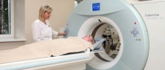
- 20–30 minutes after the introduction you will be invited for a scan, which lasts 25–30 minutes;
- scanning is performed in a supine position; during scanning you must lie still and not move (movements during scanning lead to the appearance of artifacts that make it difficult to evaluate the results of the study); The patient does not experience any sensations during scanning; the staff monitors the process and, if necessary, approaches the patient;
- for better relaxation, scanning is performed in a darkened room;
- After the scan is completed, the staff will escort you to the waiting room;
- After 15–20 minutes (the time required to reconstruct the diagnostic image), you will be informed about the completion of the examination and the approximate time for receiving the examination results.
Preparing for the diagnostic procedure
Before a PET CT scan of the brain, the patient is required to inform the doctor about existing chronic diseases, especially diabetes, and about medications taken.
Stages of PET CT scan of the brain with methionine.
- Preparation for PET CT scan of the brain. The patient will be asked to remove all jewelry, glasses, pins, dentures, hearing aids, hair clips, and wigs.
- Administration of a pharmacological agent. It contains active isotopes, and a PET CT scan of the brain with methionine is performed. Within an hour, the drug will spread through the blood vessels and accumulate in the tissues. The doctor will warn you that the medication may cause you to feel hot and will advise you to drink as much water as possible. During the action of isotopes during a PET CT scan of the brain in Moscow, the patient must lie down and not move. Talking is also prohibited, otherwise the image will be incorrect.
- Scanning. Done in half an hour to an hour.
- Rest for 15 minutes.
Whole body PET scan
It is the most common type of research in positron emission tomography. The main patient population is cancer patients. The principle of the method is based on the fact that the tumor cell, due to the fact that its metabolism is accelerated, absorbs the radiopharmaceutical much more intensively (compared to normal cells of the body). In addition, 18F-FDG, after entering the cell, does not participate in oxidation processes and, thus, ends up in the so-called metabolic trap.
As mentioned earlier, the object of research is the human organocomplex. As a rule, they look in the range from the earlobe to the middle third of the thigh.
The main diseases that are studied using 18F-FDG PET-CT are lymphomas, breast cancer, thyroid cancer, esophageal cancer, colorectal cancer, uterine cancer, ovarian cancer and many others.
It is important to say that 18F-FDG PET should not be used for fibroadenomas, neuroendocrine tumors, prostate cancer, and some types of renal cell carcinoma, since these types of tumors do not accumulate 18F-FDG radiopharmaceuticals.
With 18F-FDG PET, the extent of the tumor process (primary tumor, regional metastases, distant metastases) is primarily assessed. It should be noted that in some cases, with a widespread tumor process and an unperformed biopsy with morphological (histological) examination, it is not possible to establish the primary tumor.
Whole body PET-CT with 18F-FDG focus of pathological accumulation of radiopharmaceuticals
The 18F-FDG PET method also makes it possible to evaluate cancer treatment, i.e. say whether the treatment helps or not. But it is important to know that in order to conduct a high-quality study and eliminate errors, it is necessary to observe certain periods between the treatment session and the PET study: a) 3 months after surgery for an adequate assessment of the surgical area, b) 4 weeks after the last radiation therapy session, c) at least 3 weeks after the last administration of chemotherapy.
Preparation for PET examination:
- On the day of the study, the patient must be hungry.
- 6 hours before the test you can eat 2 boiled eggs.
- You can drink plain unsweetened and still water without restrictions, tea, coffee without sugar and without milk (this is also possible).
- In patients with diabetes mellitus, glucose-lowering medications are discontinued on the day of the study. IMPORTANT: the blood glucose level should not be higher than 11 mmol/l, otherwise the study will be refused.
The patient should expect that he will spend at least 4 hours in the department (usually 4-6 hours). Of this time, 30 minutes are allocated for measuring the patient’s height and weight, as well as for filling out documents: a contract for the provision of medical services, informed consent for the study, etc. After registration, the patient is taken to the treatment room, where he is administered the 18F-FDG radiopharmaceutical. After administration of the radiopharmaceutical, the patient is given 1 liter of water and sent to the waiting room for 1 hour 30 minutes (minimum). After the designated time has passed, the patient is invited to a tomograph scan, which lasts on average 30 minutes. After the scan, the patient either remains to wait for the report or goes home to pick up the report the next day.
Some aspects of interpretation of the conclusion
The main concept used in the description of PET studies with 18F-FDG in the conclusions is the “focus of pathological accumulation of radiopharmaceuticals” as well as its characteristic such as Standard Uptake Value or SUV, in addition, you can often find such concepts as focal hypermetabolism, zone of hypermetabolism etc. Let's look at these concepts in more detail.
The focus of pathological accumulation of radiopharmaceuticals is an area of living tissue with a high accumulation of radiopharmaceuticals. As a rule, these are tumor nodes, metastases, and foci of inflammation.
Standard Uptake Value is a dimensionless value that quantitatively characterizes the level of radiopharmaceutical accumulation in the area of interest (usually in the focus of pathological accumulation of radiopharmaceuticals, but normal tissues can also be assessed, for example, in lymphomas). It is important to note that different tomographs calculate this indicator differently. The author of these lines knows at least 5 formulas for its calculation. Therefore, dynamic monitoring (repeated PET study) should be performed on the same tomograph, and ideally by the same doctor.
Diagnostic results
How informative is PET CT for brain tumors?
- The tumor site in the brain is being identified.
- The dynamics of the tumor and its relapse are observed.
- The extent of the tumor is determined.
- Metastases are visible in the lymph nodes.
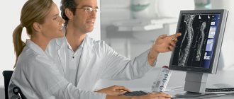
The doctor makes an accurate decoding of the PET CT scan of the brain. It allows you to see pathologies and identify serious diseases. In cases of mental illness, the difference between the brain of a healthy person and one with depression is clearly visible.
For PET CT scans of the brain, decoding is necessary to make a correct diagnosis and timely treatment.
When else is a PET scan scheduled?
As already mentioned, positron emission tomography is a highly effective technique for the early diagnosis of cancer. By introducing indicator substances into the body, research is carried out on almost all systems and organs. PET is much more accurate than MRI in assessing cancers such as head and neck lymphoma, lung cancer, prostate cancer or colon cancer. Tracer substances make it possible to determine the site of cancer and the spread of metastases.
In cases where treatment has already been prescribed, PET makes it possible to ensure that the fight against the disease is being carried out effectively, and the prescribed methods/drugs are producing results. Also, positron emission tomography allows you to make a final decision about whether surgical intervention to remove a cancerous tumor is necessary and possible in principle.
PET CT with gallium PSMA
This diagnostic method is necessary for men with suspected prostate tumors. PSMA is a prostate specific membrane antigen. It is able to attach to cancer cells in the prostate gland. In order for the particles to be visualized on the tomograph monitor during the study, they are charged with gallium 68. But the diagnostician can detect not only the presence of oncology, but also assess its size and structure.
In this case, positron emission tomography and computed tomography are indicated not only for patients who are suspected of having cancer, but also for those who have already undergone prostatectomy or hormonal therapy. A rising PSA makes it possible to notice a relapse of the disease even in the initial stages. Thanks to this, local irradiation can be used for treatment, avoiding chemotherapy and taking hormonal drugs.
