Histology.RU
Material taken from the site www.hystology.ru
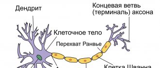
The processes of nerve cells together with the neuroglial cells covering them form nerve fibers. The processes of nerve cells located in them (dendrites or neurites) are called axial cylinders, and the oligodendroglopy cells covering them are called neurolemmocytes (lemmocytes, Schwann cells). In accordance with the composition of nerve fibers and the morphological features of their structure, myelinated and non-myelinated nerve fibers are distinguished.
Unmyelinated (non-myelinated) nerve fibers are characteristic of the autonomic nervous system. Lemmocytes - oligodendroglial cells - as part of the non-myelin fiber are tightly adjacent to each other, forming continuous strands. With light microscopy, the boundaries of glial cells in the fiber are not visible and the cells collectively look like a continuous ribbon containing their characteristic oval nuclei. The soft nerve fiber contains several axial cylinders, that is, processes of various nerve cells that can freely leave it and pass into adjacent fibers. During the formation of nerve fibers, the plasmalemma of the glial
Rice. 159. Unmyelinated nerve fibers (according to Cajal).
Rice. 160. Scheme of the structure of an unmyelinated nerve fiber:
1 - nucleus and 2 - cytoplasm of the lemmocyte; 3 - 4 - axons of nerve cells immersed in the cytoplasm of the lemmocyte (3 - completely, 4 - partially); 5 - mitochondria; 6 - rough enoplasmic seta; 7 - microtubules; 8 - collagen fibers of the endoneurium; 9 - basement membrane; 10 — mesaxon
Rice. 161. Electron micrograph of a cross section of unmyelinated nerve fibers of the rat mesentery:
1 - lemmocyte nucleus; 2 — lemmocyte cytoplasm; 3 — axial cylinders; 4 — mesaxon (according to Faucett).
the cells bend, forming more or less deep folds containing processes of nerve cells - axial cylinders. The areas of the plasmalemma of the lemmocyte, brought together in the area of the fold, form a double membrane - mesaxon, on which the axial cylinder is suspended. With light microscopy, the lemmocyte plasmalemma and mesaxons are not detected, which gives the impression that the axial cylinders are immersed directly in the cytoplasm of glial cells (Fig. 159, 160, 161).
Myelinated (meaty) nerve fibers . The diameter of myelin fibers ranges from 1 to 20 µm. They contain one axial cylinder - a dendrite or neurite of a nerve cell, covered with a membrane formed by successively located neuroglial cells - lemmocytes. In the sheath, the fibers are distinguished
Rice. 162. Myelinated nerve fibers from the frog sciatic nerve treated with osmic acid:
1 - axial cylinder; 2 - lemmocyte; 3 - myelin sheath; 4 - connective tissue; 5 — notches; 6 - interception.
two layers: the inner - myelin, thicker and the outer - thin, containing the cytoplasm and nuclei of lemmocytes.
At the border of two lemmocytes, the sheath of the myelin fiber becomes thinner, and a narrowing of the fiber is formed - a nodal interception (interception of Ranvier). The section of nerve fiber between two nodes is called the internodal segment. Its shell corresponds to one lemmocyte (Fig. 162). During the development of the myelinated nerve fiber, the axial cylinder, plunging into the cytoplasm of the lemmocyte, brings with it its plasmalemma. Here, as in an unmyelinated fiber, a mesaxon is formed, which, successively lengthening and concentrically layering on the axial cylinder, forms a zone of densely located membranes - the myelin sheath of the fiber (Fig. 163, 164).
Electron microscopy shows that mesaxon membranes form alternating light lines (8 - 12 nm), corresponding to their lipid layers, and dark thin lines formed by protein molecules (Fig. 165).
The outer layer of the myelin fiber sheath - neurilemma (Schwann membrane) - corresponds to the cytoplasm of lemmocytes with their nuclei pushed to the periphery. Densely located turns of mesaxon in the corresponding sections of the fiber, deviating from the orientation parallel to the axial cylinder, disperse and form obliquely oriented zones of the shell, richer in cytoplasm. When the fibers are osminated, they stand out as light lines—myelin notches.
In the interception area, layers of myelin sheath membranes are in contact with the axial cylinder of the fiber. In accordance with the sequence of growth of the axial cylinder and the formation of mesaxon layers, the deeper layers of the latter are shorter than the superficial ones and are located further from the interception.
Adjacent lemmocytes of the fiber sheath interact with each other through a system of more or less pronounced finger-like processes, which, intertwining, form their contacts and are observed in different sections on sections. The surface of the myelin fiber is covered with a basement membrane connected to strands of collagen fibers of the surrounding connective tissue.
Rice. 163. Scheme of development of myelin fiber:
1 — contact of the axolemma and the membrane of the lemmocyte; 2 - slot; 3 - axolemma and membranes of the lemmocyte; 4 — lemmocyte cytoplasm; 5 — mesaxon.
Rice. 164. Scheme of the structure of myelin fiber:
1 - axon; 2 — mesaxon; 3 - notches; 4 - interception; 5 — lemmocyte protoplasm; 6 — lemmocyte nucleus; 7 - neurilemma; S—endoneurium.
Rice. 165. Electron micrograph of a cross-section of myelin fiber from the cranial cervical ganglion of cattle (Kozlov’s preparation):
1 - lemmocyte nucleus; 2 — lemmocyte cytoplasm; 3 - plates of the myelin sheath; 4 - neurofilaments in the cytoplasm of the axon.
The axial cylinder of nerve fibers consists of neuroplasm - the cytoplasm of nerve cells containing longitudinally oriented neurofilaments and neurotubules. The presence of various organelles and their localization are specific to the axial cylinders of fibers of various functional significance.
Reviews (0)
Add a review
NERVOUS SYSTEM
NERVOUS SYSTEM, a set of organs and structures in the body of animals and humans, formed by elements of nervous tissue; perceives irritations coming from the external environment and from the organs and tissues of the body itself, analyzes, processes and stores information, regulates and coordinates the functions of the body, ensures the integration of the whole organism and an appropriate response to external influences. Activities of N. s. is based on two physiological. processes: excitation and inhibition. The body's response to external or internal irritations - reflexes that consist of sequential reactions: perception of irritations (receiving information), conducting excitation (transmitting information) to the associative centers of the nervous system; analyzing and storing received information; transmitting a signal to effector organs (for example, muscles, glands), the activity of which needs to be changed in accordance with changes in external conditions and the state of the body. The set of nerve structures involved in this sequence of reactions is called a reflex arc. It includes: receptors, afferent (centripetal) nerve fibers, nerve centers, efferent (centrifugal) nerve fibers and effector organs. There are two categories of reflex reactions of the body to external influences: stereotypical, hereditarily fixed unconditioned reflexes and conditioned reflexes, which are acquired during the life of the organism and allow more subtle and perfect adaptation to changing external conditions.
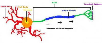
Basic structural and functional unit of N. s. - a neuron that has a number of processes: a long axon that conducts nerve impulses centrifugally, and several short branching dendrites that conduct impulses centripetally. The terminal branches of the axon are connected to the dendrites of neighboring neurons; in contact zones - synapses, with the participation of mediators, the transmission and transformation of nerve impulses occurs.
During the phylogenesis of N. s. first appears in coelenterates. Their diffuse N. s. represented by neurons evenly distributed throughout the body and connected to each other by processes. They form a common nerve network, developing in both the ectodermal and endodermal layers of the body wall and located directly under the epithelium. Excitement in such a N. s. carried out from any point of irritation in all directions; there are no specific ones. reactions to different irritants. In diffuse nodular N. s. Some manifestations of the concentration of nerve elements are observed in the form of nodular accumulations of nerve cells around the mouth opening (in coral polyps) and along the edge of the umbrella (in jellyfish). In ctenophores, ganglion-like clusters of neurons form near the mouth, along rows of ciliated pectal plates, and near the balance organ. In this type of N. s. directional conduction of excitation becomes possible. During the evolution of animals, with an intensification of the lifestyle and an increase in the variety of movements, differentiation of the elements of the receptor apparatus occurs with the development of specializations. sensory organs (see Sensory organs) and the concentration of neurons near the anterior (in terms of movement) end of the body. In this case, to one degree or another, the neurons are immersed deep in the body. A new type of N.S. organization is being formed. in the form of a complex of nerve trunks or cords in which the cell bodies of neurons and some of their processes are located; The longitudinal nerve trunks are connected to each other by transverse commissures. This level of organization corresponds to N. s. flat and roundworms, as well as echinoderms and hemichordates. The efferent processes of neurons located in the nerve trunks are sent to the effector organs, forming the peripheral. nerves. In the anterior part of the animal's body, the longitudinal trunks are connected to each other by the peripharyngeal nerve ring. More massive clusters of nerve cells are located here - the cerebral or cerebral ganglia. That. the formation of central and peripheral begins. departments of N. s. Further concentration of N.'s elements. During phylogenesis, it led to the development of the ganglionic type of neurological system, characterized by an accumulation of neurons in ganglia connected to each other by longitudinal and transverse connections (connectives and commissures) formed by processes of nerve cells. In annelids and arthropods, ganglia are located under the intestine in the form of an abdominal paired nerve chain (the so-called neural scala; there are two ganglia in each body segment); the anterior pair of ganglia of the abdominal nerve chain is connected by a ring commissure with a pair of suprapharyngeal ganglia. Mollusks are characterized by a scattered-nodular type of N., in which the ganglia are distributed in different parts of the body.
The main types of structure of the nervous system: 1 – diffuse nervous system of the hydra; 2 – system of nerve cords (orthogon) of the trematode; 3 – ganglion nervous system of the earthworm; 4 &ndas…
Differentiation diff. organ systems was accompanied by their integration with the help of N. with. The process of head formation (cephalization) is associated with increased development and differentiation of the suprapharyngeal ganglia, forming complex ganglion complexes (the so-called brain). The ganglia of the abdominal “nervous ladder” merge with each other in pairs (“the ladder” is transformed into a chain); in some groups of arthropods, successive ganglia also merge in the longitudinal direction with the formation of complex nerve ganglia, up to the union of all ganglia of the abdominal chain.
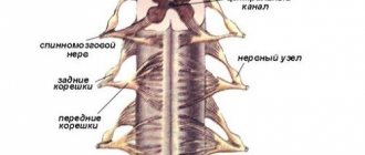
General type of organization N. s. chordates generally correspond to the principle of the structure of nerve trunks in which various components are distributed. neurons and pathways. At the same time, it has a number of unique features. In the embryos of chordates, the rudiment of N. s. (neural plate) arises on the dorsal side of the body above the notochord rudiment; During neurulation, the neural plate is transformed into a neural tube with a cavity located inside (neurocoel). In this regard, N.'s device with. Chordates are classified into a special tubular type. The organization of scientific research achieves the greatest complexity and perfection. in vertebrates: it is characterized by the highest degree of centralization, differentiation and integration. Improvement of N. s. during the evolution of vertebrates is associated with the development of adaptations to the general activation of the lifestyle. Divisions of the central nervous system (CNS) are formed. In ontogenesis in vertebrates, the head section of the neural tube, due to the uneven growth of its different parts, forms several. bubble-like swellings (the so-called brain vesicles), from which the parts of the brain are further differentiated. The brain continues behind the spinal cord, which outwardly retains the poorly differentiated structure of the neural tube, in which the cell bodies of neurons are grouped around the neurocoel, forming the gray matter of the spinal cord. Around it is white matter, consisting of the axons of nerve cells. Here are the pathways through which information is transmitted within the central nervous system. In the brain, gray matter forms various. clusters (nuclei) surrounded by white matter of the pathways. In some parts of the brain, gray matter forms the superficial layer (cortex). The neurocoel forms the ventricular system of the brain. The functions of different parts of the central nervous system are complexly intertwined; most of them are provided by the interaction of neurons located in the nuclei of different parts of the brain and forming complexly organized systems. Peripheral N. s. in vertebrates it is also difficult to differentiate and includes the cranial and spinal nerves, their ganglia and the sympathetic nervous system. The latter is part of the so-called. autonomous vegetative N. system, controlling the functions of the body’s autonomic systems (circulatory, respiratory, digestive, excretory and reproductive). See also Metasympathetic Nervous System, Parasympathetic Nervous System, Sympathetic Nervous System, Neurophysiology.