Nervous system
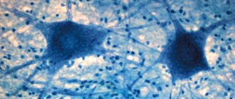
Most cells in the body have a similar structure. They have a compact shape enclosed in a shell. Inside there is a nucleus and a set of organelles that perform the synthesis and metabolism of necessary substances. However, the structure and functions of the neuron are different. It is a structural unit of nervous tissue. These cells provide communication between all body systems.
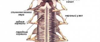
The basis of the central nervous system is the brain and spinal cord. These two centers secrete gray and white matter. The differences are related to the functions performed. One part receives a signal from the stimulus and processes it, while the other is responsible for carrying out the necessary response command. Outside the main centers, the nervous tissue forms bundles of clusters (nodes or ganglia). They branch, spreading a signal-conducting network throughout the body (peripheral nervous system).
Nerve cells
To provide multiple connections, the neuron has a special structure. In addition to the body, in which the main organelles are concentrated, there are processes. Some of them are short (dendrites), usually there are several of them, the other (axon) is one, and its length in individual structures can reach 1 meter.
The structure of the nerve cell of the neuron is designed in such a way as to ensure the best interchange of information. Dendrites are highly branched (like the crown of a tree). With their endings they interact with the processes of other cells. The place where they meet is called a synapse. This is where the impulse is received and transmitted. Its direction: receptor – dendrite – cell body (soma) – axon – reacting organ or tissue.
The internal structure of a neuron is similar in composition to organelles to other structural units of tissue. It contains a nucleus and cytoplasm bounded by a membrane. Inside there are mitochondria and ribosomes, microtubules, the endoplasmic reticulum, and the Golgi apparatus.
Neurons
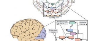
A neuron is a complex, highly specialized cell with processes capable of generating, perceiving, transforming and transmitting electrical signals, and also capable of forming functional contacts and exchanging information with other cells.
Each neuron has only 1 axon, the length of which can reach several tens of centimeters. Sometimes lateral processes extend from the axon - collaterals . Axon endings typically branch and are called terminals . The place where the axon extends from the cell soma is called axonal hillock .
In relation to the processes, the soma of the neuron performs a trophic function, regulating metabolism. A neuron has characteristics common to all cells: it has a membrane, a nucleus and a cytoplasm in which organelles are located (endoplasmic reticulum, Golgi apparatus, mitochondria, lysosomes, ribosomes, etc.).
In addition, the neuroplasm contains special-purpose organelles: microtubules and microfilaments , which differ in size and structure. Microfilaments represent the internal skeleton of the neuroplasm and are located in the soma. Microtubules stretch along the axon along the internal cavities from the soma to the end of the axon. Biologically active substances spread through them.
In addition, a distinctive feature of neurons is the presence of mitochondria in the axon as an additional source of energy. Adult neurons are not capable of division.
Types of neurons
There are several classifications of neurons based on different characteristics: the shape of the soma, the number of processes, the functions and effects that the neuron has on other cells.
Depending on the shape of the soma, they are distinguished: 1. Granular (ganglionic) neurons , in which the soma has a round shape; 2. Pyramidal neurons of different sizes - large and small pyramids; 3. Stellate neurons ; 4. Fusiform neurons .
Based on the number of processes (by structure) there are: 1. Unipolar neurons (single-processed) , having one process extending from the cell soma, are practically never found in the human nervous system; 2. Pseudounipolar neurons (false unipolar), such neurons have a T-shaped branching process, these are cells of general sensitivity (pain, temperature changes and touch); 3. Bipolar neurons (bipolar) having one dendrite and one axon (i.e. 2 processes), these are cells of special sensitivity (vision, smell, taste, hearing and vestibular stimulation); 4. Multipolar neurons (multi-process) , which have many dendrites and one axon (i.e. many processes); small multipolar neurons are associative; medium and large multipolar, pyramidal neurons - motor, effector.
Unipolar cells (without dendrites) are not typical for adults and are observed only during embryogenesis. Instead, in the human body there are pseudounipolar cells, in which a single axon divides into 2 branches immediately after leaving the cell body. Bipolar neurons are present in the retina and transmit excitation from photoreceptors to ganglion cells that form the optic nerve. Multipolar neurons make up the majority of cells in the nervous system.
According to the functions they perform, neurons are: 1. Afferent (receptor, sensory) neurons - sensory (pseudo-unipolar), their somas are located outside the central nervous system in ganglia (spinal or cranial). Along sensory neurons, nerve impulses move from the periphery to the center.
The shape of the soma is granular. Afferent neurons have one dendrite that connects to receptors (skin, muscle, tendon, etc.). Through dendrites, information about the properties of stimuli is transmitted to the soma of the neuron and along the axon to the central nervous system.
Example of sensory neurons: a neuron that responds to skin stimulation.
2. Efferent (effector, secretory, motor) neurons regulate the work of effectors (muscles, glands, etc.). Those. they can send orders to the muscles and glands. These are multipolar neurons, their somas have a stellate or pyramidal shape. They lie in the spinal cord or brain or in the ganglia of the autonomic nervous system.
Short, abundantly branching dendrites receive impulses from other neurons, and long axons extend beyond the central nervous system and, as part of the nerve, go to effectors (working organs), for example, to skeletal muscle.
Example of motor neurons: spinal cord motor neuron.
The cell bodies of sensory neurons lie outside the spinal cord, while motor neurons lie in the anterior horn of the spinal cord.
3. The betting neurons (contact, interneurons, associative, closing) make up the bulk of the brain. They communicate between afferent and efferent neurons and process information coming from receptors to the central nervous system.
These are mainly multipolar stellate-shaped neurons. Among interneurons, neurons with long and short axons are distinguished.
Example of interneurons: neuron of the olfactory bulb, pyramidal cell of the cerebral cortex.
The chain of neurons consisting of sensory, intercalary and efferent neurons is called the reflex arc. All activities of the nervous system, as defined by I.M. Sechenov, is of a reflex nature (“reflex” means reflection).
According to the effect that neurons have on other cells: 1. Excitatory neurons have an activating effect, increasing the excitability of the cells with which they are connected. 2. Inhibitory neurons reduce cell excitability, causing an inhibitory effect.
Nerve fibers and nerves
Nerve fibers are processes of nerve cells covered with a glial sheath that carry out nerve impulses. Through them, nerve impulses can be transmitted over long distances (up to a meter).
The classification of nerve fibers is based on morphological and functional characteristics.
Based on morphological characteristics, they are distinguished: 1. M myelinated (meat) nerve fibers are nerve fibers that have a myelin sheath; 2. Non -myelinated (non-myelinated) nerve fibers are fibers that do not have a myelin sheath.
According to functional characteristics, they are distinguished: 1. Afferent (sensitive) nerve fibers; 2. Efferent (motor) nerve fibers.
Nerve fibers extending beyond the nervous system form nerves. A nerve is a collection of nerve fibers. Each nerve has a sheath and a blood supply.
There are spinal nerves connected to the spinal cord (31 pairs) and cranial nerves (12 pairs) connected to the brain. Depending on the quantitative ratio of afferent and efferent fibers within one nerve, sensory, motor and mixed nerves are distinguished (see table below).
In sensory nerves, afferent fibers predominate, in motor nerves, efferent fibers predominate, in mixed nerves, the quantitative ratio of afferent and efferent fibers is approximately equal. All spinal nerves are mixed nerves. Among the cranial nerves, there are three types of nerves listed above.
List of cranial nerves with dominant fiber designation
I pair - olfactory nerves (sensitive); II pair - optic nerves (sensitive); III pair - oculomotor (motor); IV pair - trochlear nerves (motor); V pair - trigeminal nerves (mixed); VI pair - abducens nerves (motor); VII pair - facial nerves (mixed); VIII pair - vestibulo-cochlear nerves (sensitive); IX pair - glossopharyngeal nerves (mixed); X pair - vagus nerves (sensitive); XI pair - accessory nerves (motor); XII pair - hypoglossal nerves (motor).
Structure and types of neurons
In most cases, several thick branches (dendrites) extend from the cell soma (base). They do not have a clear boundary with the body and are covered with a common membrane. As they move away, the trunks become thinner and branch out. As a result, their thinnest parts look like pointed threads.
The special structure of the neuron (thin and long axon) implies the need to protect its fiber along its entire length. Therefore, on top it is covered with a sheath of Schwann cells that form myelin, with nodes of Ranvier between them. This structure provides additional protection, isolates passing impulses, and additionally nourishes and supports the threads.
The axon originates from a characteristic hill (mound). The process eventually also branches, but this does not occur along its entire length, but closer to the end, at the points of connection with other neurons or tissues.
Main types of neurons and their functions
The main functions of the central nervous system are:
- unification of all parts of the body into a single whole and their regulation;
- management of the state and behavior of the body in accordance with environmental conditions and its needs.
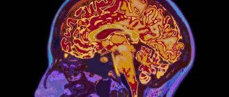
In humans, the leading part of the central nervous system is the cerebral cortex. It controls the most complex functions in human life - mental processes (consciousness, thinking, memory, speech, etc.).
The main methods for studying the functions of the central nervous system are methods of removal and stimulation, recording electrical phenomena, the method of conditioned reflexes, computed tomography, thermal imaging, and nuclear magnetic resonance.
The main functions of neurons are:
- perception of external stimuli - receptor function,
- processing is an integrative function
- transmission of nerve influences to other neurons or working organs is an effector function.
The body of the neuron is called the soma, where information processing processes take place.
The dendrites of neurons serve as inputs to the neuron. The output of a neuron is an axon; it transmits the signal further - to another nerve cell or working organ (muscle, gland).
The initial part of the axon and the extension at the point of its exit from the cell body - the axon hillock - are particularly highly excitable. This is where the nerve impulse originates.
Neurons are divided into three main types:
- afferent (sensitive, or centripetal) transmit information from receptors to the central nervous system. The bodies of these neurons are located outside the central nervous system - in the spinal ganglia and in the ganglia of the cranial nerves. Afferent neurons have a long process - a dendrite, which contacts the receptor at the periphery or forms a receptor itself, as well as a second process - an axon - which enters the spinal cord through the dorsal horns.
- Efferent neurons (motor, centrifugal) are associated with the transmission of descending influences from the upper levels of the nervous system to the underlying ones or from the central nervous system to the working organs. Efferent neurons are characterized by a branched network of short processes - dendrites and one long process - an axon.
- Intermediate (associative, interneurons, interneurons) are smaller cells that communicate between afferent and efferent neurons. They transmit nerve influences horizontally and vertically (above and below) directions.
The interaction of neurons with each other and with organs occurs through special formations - synapses (contact).
They are formed by the terminal branches of neurons on the body or processes of another neuron. The more synapses there are on a nerve cell, the more it perceives various irritations and the wider the sphere of influence on its activity and the possibility of participation in the body’s reactions.
There are 3 elements in the structure of the synapse:
1) presynaptic membrane formed by thickening of the membrane of the terminal branch of the axon;
2) synaptic cleft
3) postsynaptic membrane - thickening of the adjacent surface of the next neuron.
Impulse transmission is carried out in 2 ways: chemical and physical. Chemical pathway - using a mediator, which can be excitatory (acetylcholine, norepinephrine) or inhibitory (gamma-aminobutyric acid)
The first causes depolarization of the postsynaptic membrane and the formation of an excitatory postsynaptic potential (EPSP). To excite a neuron, the EPSP must reach a threshold level (10 mV). The effect of the mediator is short-lived (1-2ms), after which it is broken down into choline and acetic acid or absorbed back. In inhibitory synapses, potassium ions intensively enter the postsynaptic membrane and increase the polarization of the membrane. In this case, inhibitory postsynaptic potential (IPSP) is recorded. As a result, the cell becomes inhibited. It is more difficult to arouse her than in the original state.
Classification
Neurons are divided into types depending on the type of mediator (mediator of the conductive impulse) released at the axon terminals. This can be choline, adrenaline, etc. Depending on their location in the parts of the central nervous system, they can relate to somatic neurons or autonomic ones. There are receptive cells (afferent) and transmitting feedback signals (efferent) in response to irritation. Between them there may be interneurons responsible for the exchange of information within the central nervous system. Depending on the type of response, cells can inhibit excitation or, conversely, increase it.
According to their state of readiness, they are distinguished: “silent”, which begin to act (transmit an impulse) only in the presence of a certain type of irritation, and background, which constantly monitor (continuous generation of signals). Depending on the type of information perceived from the sensors, the structure of the neuron also changes. In this regard, they are classified into bimodal, with a relatively simple response to irritation (two interrelated types of sensation: a prick and, as a result, pain, and polymodal. This is a more complex structure - polymodal neurons (specific and ambiguous reaction).
Neuron structure
Neurons, or neurocytes, of different parts of the nervous system differ significantly from each other in functional significance and morphological features.
Depending on their function, neurons are divided into:
- receptor (sensitive, afferent) - generate a nerve impulse under the influence of various influences from the external or internal environment of the body;
- intercalary (associative) - carry out various connections between neurons;
- effector (efferent, motor) - transmit excitation to the tissues of the working organs, prompting them to action.
A characteristic feature of all mature neurons is the presence of processes.
These processes ensure the conduction of a nerve impulse through the human body from one part to another, sometimes very distant, and therefore their length varies widely - from several micrometers to 1-1.5 m.
Based on their functional significance, neuronal processes are divided into two types. Some perform the function of removing nerve impulses, usually from the bodies of neurons, and are called axons or neurites.
The neurite ends with the terminal apparatus or on another neuron, or on the tissues of the working organ, muscles, glands.
The second type of nerve cell extensions is called dendrites. In most cases, they are highly branched, which determines their name. Dendrites conduct impulses to the neuron body.
Based on the number of processes, neurons are divided into three groups:
- unipolar - cells with one process;
- bipolar - cells with two processes;
- multipolar - cells with three or more processes.
Multipolar cells are most common in mammals and humans.
Of the many processes of such a neuron, one is represented by a neurite, while all the others are dendrites.
Bipolar cells have two processes - a neurite and a dendrite. True bipolar cells are rare in the human body. These include some of the cells of the retina, the spiral ganglion of the inner ear and some others. However, based on the essence of their structure, a large group of afferent, so-called pseudounipolar neurons of the cranial and spinal nerve ganglia should be classified as bipolar cells.
They are called pseudounipolar because the neurite and dendrite of these cells begin with a common outgrowth of the body, creating the impression of a single process, followed by a T-shaped division.
There are no true unipolar cells, that is, cells with one process - a neurite, in the human body.
The vast majority of human neurons contain one nucleus located in the center, less often - eccentrically.
Binuclear neurons, and especially multinucleated ones, are extremely rare, for example: neurons in the prostate gland and cervix. The shape of the neuron nuclei is round. In accordance with the high metabolic activity, the chromatin in their nuclei is dispersed. The nucleus has 1, and sometimes 2 and 3 large nucleoli.
In accordance with the high specificity of the functional activity of neurons, they have a specialized plasmalemma, their cytoplasm is rich in organelles.
The cytoplasm has a well-developed endoplasmic reticulum, ribosomes, mitochondria, Golgi complex, lysosomes, neurotubules and neurofilaments.
The plasmalemma of neurons, in addition to the function typical of the cytolemma of any cell, is characterized by the ability to conduct excitation. The essence of this process comes down to the rapid movement of local depolarization of the plasmalemma along its dendrites to the perikaryon and axon.
The abundance of granular endoplasmic reticulum in neurocytes corresponds to a high level of synthetic processes in the cytoplasm and, in particular, the synthesis of proteins necessary to maintain the mass of their perikarya and processes.
Axons that do not have protein-synthesizing organelles are characterized by a constant flow of cytoplasm from the perikarya to the terminals at a speed of 1-3 mm per day. This is a slow current carrying proteins, in particular enzymes necessary for the synthesis of mediators in axon terminals.
In addition, there is a fast current (5-10 mm per hour) transporting mainly components necessary for synaptic function. In addition to the flow of substances from the perikaryon to the terminals of axons and dendrites, a reverse (retrograde) current is also observed, through which a number of cytoplasmic components return from the terminals to the cell body.
The transport of substances along the processes of neurocytes involves the endoplasmic reticulum, membrane-bound vesicles and granules, microtubules and the actinomyosin cytoskeletal system.
The Golgi complex in nerve cells is defined as a cluster of rings, twisted threads, and grains of various shapes.
The cell center is often located between the nucleus and dendrites. Mitochondria are located both in the body of the neuron and in all processes. The cytoplasm of neurocytes in the terminal apparatus of processes, in particular in the area of synapses, is especially rich in mitochondria.
Neurofibrils
When nervous tissue is impregnated with silver, neurofibrils are revealed in the cytoplasm of neurons, forming a dense network in the perikaryon of the cell and oriented in parallel within the dendrites and axons, including their finest terminal branches.
Using electron microscopy, it was established that neurofibrils correspond to bundles of neurofilaments with a diameter of 6-10 nm and neurotubules (neurotubes) with a diameter of 20-30 nm, located in the perikaryon and dendrites between chromatophilic clumps and oriented parallel to the axon.
Secretory neurons
The ability to synthesize and secrete biologically active substances, in particular mediators, is characteristic of all neurocytes.
However, there are neurocytes specialized primarily to perform this function - secretory neurons, for example, cells of the neurosecretory nuclei of the hypothalamic region of the brain. Secretory neurons have a number of specific morphological characteristics:
- secretory neurons are large neurons;
- in the cytoplasm of neurons and in axons there are secretion granules of varying sizes - neurosecretion, containing protein, and in some cases lipids and polysaccharides;
- many secretory neurons have nuclei of irregular shape, which indicates their high functional activity.
Features, structure and functions of a neuron
The surface of the neuron membrane is covered with small projections (spikes) to increase the contact area. In total, they can occupy up to 40% of the cell area. The nucleus of a neuron, like that of other types of cells, carries hereditary information. Nerve cells do not divide by mitosis. If the connection between the axon and the body is broken, the process dies. However, if the soma has not been damaged, it is able to generate and grow a new axon.
The fragile structure of the neuron suggests the presence of additional “care”. Protective, supporting, secretory and trophic (nutrition) functions are provided by neuroglia. Its cells fill all the space around. To a certain extent, it helps restore broken connections, and also fights infections and generally “takes care” of neurons.
Cell membrane
This element provides a barrier function, separating the internal environment from the neuroglia located outside. The thinnest film consists of two layers of protein molecules and phospholipids located between them. The structure of the neuron membrane suggests the presence in its structure of specific receptors responsible for recognizing stimuli. They have selective sensitivity and, if necessary, “turn on” in the presence of a counterparty. The connection between the internal and external environments occurs through tubules that allow calcium or potassium ions to pass through. At the same time, they open or close under the influence of protein receptors.
Thanks to the membrane, the cell has its potential. When it is transmitted along the chain, excitable tissue is innervated. Contact between the membranes of neighboring neurons occurs at synapses. Maintaining a constant internal environment is an important component of the life of any cell. And the membrane subtly regulates the concentration of molecules and charged ions in the cytoplasm. At the same time, they are transported in the required quantities for metabolic reactions to occur at an optimal level.