Neurons: structure, types and types
Nervous tissue contains nerve cells and neuroglia (Fig. 1) . The tissue forms the brain and spinal cord, nerve fibers and nodes. The nervous system is responsible for the coordinated functioning of organs and organ systems and ensures the body’s connection with the environment.
Rice. 1. Horse retinal nerve cells
A neuron is a basic, highly specialized cell of nervous tissue. It receives, processes and transmits information. It consists of a body or soma, which contains the nucleus with the bulk of the cytoplasm, and processes. The diameter of the nerve cell body is 15–150 μm or 0.001 mm.
Types of neurons by number of processes (Fig. 2) :
- bipolar;
- unipolar;
- multipolar;
- pseudounipolar.
Rice. 2. Types of neurons
Neuronal cell bodies are concentrated mainly in the gray matter of the brain and spinal cord. Long processes stretch long distances from the place where the nerve cells with the nucleus are located. The length of the axon can reach 1 m or more.
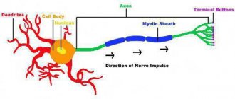
Components of a motor (multipolar) neuron (Fig. 3) :
- The body of a nerve cell with a centrally located nucleus.
- Short branching processes called dendrites that carry information to the cell body.
- A long cellular extension, an axon, that carries information from the neuron body.
- Insulating myelin sheath of the axon from Schwann cells (part of neuroglia).
- The nodes of Ranvier are narrow gaps separating Schwann cells.
- Sensory endings are receptors.
Rice. 3. Structure of a neuron
Types of neurons depending on the function performed
| Main title | Additional titles | Functions |
| Sensitive | Afferent, sensory | They carry information about sensation (impulse) from the surface of the body and internal organs to the brain. |
| Insert | Associative, connecting, switching | They make up about 99% of all nerve cells, process, analyze information, and develop decisions. |
| Motor | Effector, efferent | They carry impulses from the brain and spinal cord to the executive organs. |
Demyelinating diseases
Diseases that lead to the destruction of the myelin sheath are most often of an autoimmune nature; another cause may be treatment with antipsychotics or a hereditary predisposition. The destruction of the lipid layer causes a decrease in the speed of irritation impulses.
Diseases are divided into those that affect the central nervous system and pathologies that damage the peripheral network. Diseases that affect the functioning of the central nervous system:
- Myelopathy of the spinal cord occurs as a result of compression of myelin fibers by intervertebral hernias, tumors, bone fragments, after a spinal cord stroke. In patients, sensitivity and muscle strength in the affected area decrease, paresis of the arms or legs occurs, the functioning of the intestines and urinary system is disrupted, and muscle atrophy of the lower extremities develops.
- Leukodystrophy of the brain causes damage to the white matter. Patients have poor coordination of movements and cannot maintain balance. Muscle weakness develops, involuntary spasms and nervous tics appear. Memory, intellectual abilities, vision and hearing gradually deteriorate. In later stages, blindness, deafness, complete paralysis, and difficulty swallowing food occur.
- Small focal leukoencephalopathy of the brain most often affects men over 60 years of age. The main reasons are arterial hypertension and hereditary predisposition. Patients experience memory and attention deterioration, lethargy, and difficulty speaking. The gait slows down, coordination of movements is impaired, urinary incontinence appears, and the patient has difficulty swallowing food.
- Osmotic demyelination syndrome is characterized by the breakdown of myelin sheaths in brain tissue. Patients have a disorder of the speech apparatus, a constant feeling of drowsiness, depression or increased excitability, mutism, paresis of all extremities. In the early stages of the disease, the demyelination process is reversible.
- Multiple sclerosis is manifested by numbness of one or two limbs, partial or complete loss of vision, pain when moving the eyes, dizziness, fatigue, tremors of the limbs, poor coordination of movements, tingling in various parts of the body.
- Devic's disease is an inflammatory autoimmune disease that affects the optic nerve and spinal cord. Symptoms include varying degrees of visual impairment, including blindness, paraparesis, tetraparesis, and impaired functioning of the pelvic organs.
Symptoms of the disease depend on the area of damage to the myelin fibers. The process of demyelination can be detected using computed tomography and magnetic resonance therapy. Signs of damage to the peripheral nervous system are detected on electromyography.
Neuroglia
Neuroglial cells lie between neurons and act as support, protection, and nutrition for nervous tissue. They participate in the formation of the myelin sheath of nerve fibers (nerves). The membrane consists of Schwann cells filled with a fat-like substance.
Neuroglia include astrocytes that are stellate in shape and small in size. They have numerous processes, are part of the gray matter of the brain, and participate in the formation of the blood-brain barrier.
Oligodendrocytes are responsible for performing the main functions of neuroglia - support, nutrition, insulation and regeneration. Microglia are cells with 2 processes capable of phagocytosis. Such cellular elements of nervous tissue protect neurons from foreign substances and bodies and remove decay products.
Neuroglia differ from neurons in a number of properties. Auxiliary cells multiply, but are not able to be excited, do not form or conduct impulses. The formation of myelin sheaths by Schwann cells occurs gradually during the first 3–10 years of life.
Main structure
Nervous tissue consists of neurons, receptor cells and neuroglia, which are intercellular substances.
Neurons are unique excitable fibers, consisting of a number of elements and units: nuclei, enveloped in a membrane of cytoplasm and receptors that transport various elements and impulses. In addition, they participate in the process of division, movement and synthesis.
Receptor cells are processes of different sizes that transmit impulses throughout the human body. They are divided into dendrites - small, thick nerve endings and axons - thin and long in structure.
Neuroglia fill the entire intercellular space between tissue components. They create uniform and constant nutrition, division and movement of impulses along the ligaments and systems of the body. They are located in the part of the central nervous system where the largest concentration of neurons is located. They are characterized by a functional regime in the body - performing immunological, barrier, support and other functions.
Functions
Main functions of nervous tissue:
- Construction Due to its structure, nervous tissue participates in the formation of the brain, the central nervous system, in particular fibers, nodes, processes and the elements connecting them. It is capable of forming an entire system and ensuring its harmonious functioning.
- Data processing. With the help of cell neurons, our body perceives information coming from the outside, processes it, analyzes it and then transforms it into specific impulses that are transmitted to the brain and central nervous system. Histology studies specifically the ability of nervous tissue to produce signals that enter the brain.
- Regulating the interaction of systems. Adaptation to various circumstances and conditions occurs. It is able to unite all the vital support systems of the body, competently managing them and regulating their work.
Functions of nervous tissue
There are three main functions in the actions of the central nervous system:
System construction
Nervous tissue forms a single system with balanced, constant functioning of all organs. It forms the tissue of the brain, nervous system, and participates in the creation of all elements of connective tissue with its processes, fibers and other elements of the neuron.
Informing
Thanks to neurons, the human body receives information from external factors, internal impulses, processes it, extracts the necessary responses and transmits information to one or another organ. The ability of the central nervous system to timely transmit an impulse to the brain to a particular stimulus is considered to be moderately studied at this stage.
Interaction of systems
Nervous tissue adapts and interacts at various cellular levels with all systems and departments of the body. It is able to control, change and improve the functioning of organs and facilitate their interaction. It collects all incoming information, stores it, changes it and regulates responses.
Structural classification of neurons
Taking into account their structure and components, the following types can be distinguished:
- unipolar, consisting of one process (found in animals, but not in humans);
- pseudounipolar, consisting of one dendrite with two branches (located in the spinal ganglia);
- bipolar, consisting of two units - an axon and a dendrite and are located in sensory organs (eye, ear, nose);
- multipolar, consisting of a large number of dendrites and one axon. They are the largest in the tissue structure and are located in the central nervous system.
The human body consists of many tissues and cells, among which nervous tissue is represented by only two important specific components. The characteristics and features of these components are determined by their physiological functions and definitions. The main feature of nervous tissue is to receive irritating and exciting factors and transmit response impulses and reactions to the brain. Accordingly, it transmits signals through the nerve plates throughout the body, to tissues and skin cells.
Nervous tissue reactions
Excitability is the ability to respond to various irritating and provoking factors by the entire body system as a whole and by cells individually. It has two thresholds: the lower one, which reacts to small point impulses (cold, heat, injection) and the upper one, which is a reaction in the form of a pain syndrome to sharp irritating factors from the outside.
It has two processes:
- excitation or reaction to the influence of a stimulus, expressed in the modification of metabolic cellular processes of all tissues;
- inhibition or response to the excitation process.
The first is characterized by a change in metabolism in neurons, which is accompanied by the penetration of differently charged ions through the plasma membrane of proteins and lipids. They affect the movement of ions and the transmission of impulses. The difference in the characteristics of the fields at rest is small, but it affects the ion density of the intracellular and extracellular spaces of the cell. Therefore, excitability is characterized by migration and freedom of movement both from one cell to another and within it.
The second one stops, inhibits or blocks any activity in nerve tissues and its cells. Some of the nerve endings react to all excitation responses, and some to inhibition, which creates a harmonious interaction of the entire vital system of the human body as a whole.
Both reactions constitute a complex work of ions, replacing each other during the response to an irritating effect. Considering that all changes occur as a consequence of various reactions in the body, energy expenditure in irritating situations, it can be noted that they are the harmonious state of the neuron itself.
Conductivity is the ability to transmit various impulses. This stage looks like the transfer of information from one cell to another and its movement to any part of the nervous tissue and the entire system. Moving to different areas of tissue, a neuron charged with an impulse changes the ion density throughout the entire area of movement.
Irritability is the ability of tissue to transition from a resting state to an active phase, thanks to receptors. This condition is provoked by incoming impulses, both from external stimuli and from internal reactions of the body. For example:
- skin reacts to burn or touch;
- eye - for flash or dust;
- ear - to a sharp cry or whisper.
When any of these processes are disrupted, an imbalance occurs in the human body. So, during loss of consciousness, action on irritating factors stops, the functioning of all psychomotor manifestations stops. The receptor endings no longer send impulses to the brain. It, in turn, does not transmit any responses to the aggregate cells and organs.
Chemical component of nervous tissue
- Water – its content in nerve tissue and fibers is insignificant, but necessary. It prevents sticking and ensures ion mobility.
- Protein compounds – albumin, globulin and others. These substances are found in axons and nerve processes in small quantities. A significant content of proteins in the form of amylase, maltase and other mediators is found throughout the nervous tissue.
- Carbohydrates - glucose and sugar-like substances are included in the chemical composition of cells in small quantities, but are necessary for the balanced functioning of receptors.
- Fats - phospholipids, cholesterol, cerebrosides increase in volume and improve during human development.
- Macro and microelements - iron, magnesium, copper, sodium, potassium and other substances are contained in proportion throughout the entire nervous tissue. The presence of these elements is very important in the development and normal functioning of all body systems.
Nervous tissue
(The study of the histological structure of nervous tissue is associated with the names of outstanding Russian scientists - A. S. Dogel, A. I. Babukhin, A. N. Mislavsky, A. A. Zavarzin, B. I. Lavrentiev, etc.
)
Nervous tissue, together with its special supporting tissue - neuroglia, which develops from a common rudiment with nerve elements, and connective tissue forms the nervous system of the body - the brain and spinal cord, numerous peripheral nerves and specifically differentiated nerve endings, scattered in large numbers in various tissues and organs bodies.
The main properties of nervous tissue are excitability and conductivity. Nervous tissue is able to perceive irritations from the external environment and from internal organs and tissues and transmit them through its fibers to other tissues and organs of the body. Nervous tissue generally develops from the outer germ layer (ectoderm).
The perception of irritation, its conduction and transmission - these most important functions of nervous tissue - in the simplest unicellular organisms occur in one cell. In multicellular organisms, already during their embryonic development, a special perceptive and conducting system is formed, consisting of specifically differentiated nervous elements.
The presence of special nerve cells in multicellular organisms, which play such an important role in the life of the organism, led to the formation of special tissues that protect the nervous tissue from external irritations. The increase in the volume of the body and the complication of its structure entailed a significant complication and expansion of the conducting nervous system. In vertebrates, there is almost no area on the surface or inside the body where the apparatus of the nervous system does not perceive irritations.
The nervous system unites all parts of the body with their various structures and functions into one harmonious whole. One part of the nerve fibers - centripetal - transmits irritations from the periphery to the nerve centers located in the brain and spinal cord. From the nerve centers to all parts of the body there is another part of the nerve fibers - centrifugal, the endings of which transmit impulses from the centers of the brain and spinal cord to the organs and tissues of the body (skin, muscles, glands, etc.).
The nervous system positively regulates the functioning of all organs and systems of the body. By regulating metabolism in the body, it also carries out a trophic function, influencing the degree of absorption of nutrients by all tissues of the body, maintaining a constant chemical composition, and metabolism.
The nervous system, with the help of devices that perceive external irritation, communicates between a person and the external environment. Finally, all the richness and diversity of a person’s mental life, all of his higher nervous activity are directly related to the nervous system.
No other system of the body can so completely and quickly realize the influences of the external and internal environment and provide certain functional and structural changes in the interests of the whole organism, like the nervous system. The main significance of the nervous system for the body is to respond (react) appropriately to everything that affects it from the outside and inside, and to establish a certain level of body functions that ensures the highest optimum of its vital activity as a whole.
The higher the organism is developed, the more developed and the more important its nervous system is, the more complex the connections and interaction of the organism with the environment, as well as the interaction between parts of the body.
The body's reactions carried out by the central nervous system in response to a particular irritation emanating from the external or internal environment are called reflexes. The basis of all nervous activity carried out by nervous tissue is a reflex.
Neuroglia
. Nervous tissue consists of neuroglia and the nervous tissue itself. Neuroglia* is a tissue involved in the formation of the nervous system. It makes up the stroma, in which the more delicate nerve elements are located. This tissue also develops from the ectoderm, but it is unable to perceive and conduct excitation. The sheath of neuroglia, located between the connective and nervous tissue, plays not only a mechanical support role, but also retains extraneous irritations and, like a kind of cover, protects it. But mainly neuroglia regulates the metabolism of nervous tissue, carries out a trophic function, being an intermediary between nervous tissue and the internal environment of the body. It also plays the role of removing decay products. In addition to neuroglia, connective tissue also performs a supporting and nutritional function in the nervous system. Such supporting trophic elements, for example, include one of the types of neuroglia that appears earliest in embryonic development, namely ependyma - the thinnest layer of cells that covers the walls of the brain cavities from the inside.
* (From the Greek word "glia" - glue, putty.
)
Neuroglia cannot be considered as a simple substitute for supporting connective tissue: it not only has the same origin as nervous tissue, but also continues to remain in the closest connection with it.
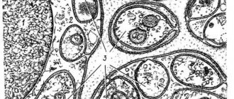
It is customary to distinguish between two main types of neuroglia: macroglia and microglia. The shape of macroglial cells is very diverse, but process (star-shaped) cells with a rounded body and many processes of different lengths predominate. These cells are called astrocytes (Fig. 32); their long processes are densely intertwined with the processes of neighboring astrocytes, forming something like felt. Another type of macroglial cell, oligodendroglia, has tree-like branching processes. This type of matfoglia is most often found around nerve fibers. Star-shaped macroglial cells are interconnected through thin, never-interrupted thread-like processes into a continuous syncytial network in which the nerve elements of the brain and spinal cord are located. Neuroglia, a thin layer everywhere, separates the nervous tissue itself from the connective tissue membranes and blood vessels.
Rice. 32. Astrocytic macroglia from the spinal cord (according to Zavarzin). 1 - capillary; 2 - processes of astrocytes (stellate cells); 3 - astrocyte bodies
Microglia, according to some scientists, are formed from mesenchyme; its cells are also diverse and represent modified macrophages. It penetrates the nervous system along with the vessels and also has supporting-trophic and protective functions. The main function of its elements is phagocytosis.
Neuroglia plays an important role in all pathological processes. For example, with various injuries, neuroglia grow and replace the area of dead nervous tissue.
Nerve cells
. The main elements of nervous tissue are nerve cells, which have a variety of shapes, in most cases polygonal. Their size ranges from 4 to 150 μ, but some of them can reach large sizes and are visible even with the naked eye, such as nerve cells in the spinal cord of a bull. A nerve cell usually consists of a more or less protoplasm-rich body containing a large vesicular, chromatin-poor nucleus with clearly visible nucleoli inside. The cytoplasm of a nerve cell has a very complex structure. One of the features of nerve cells is the absence of centrosomes in them. Along with the usual components, there are special formations in the cytoplasm. The most important elements in the structure of protoplasm are special Nissl bodies. These bodies have a round or angular shape of granular lumps. Due to their resemblance to the spots on the skin of a tiger, they are also called tigroid.
With any irritation of a nerve cell, the first thing that occurs is changes in the structure and location of these bodies. What Nissl bodies essentially are and what their function is has not yet been clarified with precision. It was only noticed that the number of clumps is significantly less in cells that have been subjected to irritation for a long time, that is, in tired cells. Having disappeared when the cells are overstrained from excessive work, the little bodies are restored again during rest. For example, in birds, Nissl bodies disappear during flight and reappear during rest. They are present in dendrites and absent in neurites (see below).
In many cells of the nerve ganglia, pigments are often deposited in the form of round inclusions of varying sizes - the dark pigment melanin and the yellow pigment, which accumulates throughout life in almost all ganglion cells. The latter is also called wear pigment.
From the body of the nerve cell (Fig. 33) processes of unequal sizes extend. A nerve cell with its processes is called a neuron (or neuron).
Rice. 33. Nervous tissue. A - types of nerve cells. The first one on the left is a single-process nerve cell; the second is a bi-process nerve cell; the third is a multi-processed nerve cell. 1 - dendrites; 2-3 - neurites. B - structure of a neuron. 1 - cell body; 2 - core; 3 - dendrites; 4 - neurite (axon); 5 - branched end of the neurite; 6 - neurilemma; 7 - myelin; 8 — axial cylinder; 9 - Ranvier interceptions. C - Nissl bodies (tigroid) in the protoplasm of a nerve cell. D - neurofibrils of a nerve cell
The nerve cell is a vital neurodynamic and trophic center for the entire neuron.
The cell body and its processes are penetrated by a dense network of the finest fibers intertwined in different directions - neurofibrils. Most scientists consider neurofibrils to be conductors of nervous excitation. However, according to other researchers, the conductor of nervous excitation is the liquid part of the cytoplasia, and neurofibrils represent the supporting apparatus of the cell.
The longer of the processes of a nerve cell (Fig. 34), which conducts irritation to other nerve cells or to peripheral organs, is called a neurite, while all the other processes that receive irritations from other nerve cells or from the periphery are called dendrites *. Dendrites divide repeatedly near the cell body and can form rich branches in the form of a dense bush. But there are a special type of dendrites - dendrites of sensitive cells of the spinal ganglia; they are long, reaching the periphery. The dendrites of a nerve cell enormously increase its surface, increasing both the possibility of nutrition and the perception of nerve stimuli emanating from the terminal branches of the neurites of other nerve cells.
* (From the Greek word "dendron" - tree. Most dendrites have the appearance of a tree with branches.
)
Rice. 34. A - unipolar motor nerve cell of insects. 1 - cell body; 2 - cell process; 3 - neuritis; 4 — dendrites (according to Zavarzin). B - bipolar nerve cell from dragonfly skin. 1 - unbranched process - neuritis; 2 - tree-like process - dendrite. B - multipolar motor nerve cell. 1 - neuritis; 2 - dendrites
A neurite is the main process of a nerve cell; it is called a nerve fiber, or axon*, and consists of a sheath and an axial cylinder. All nerve fibers are processes of nerve cells. Connecting into bundles, they form nerves.
* (From the Latin word axis - axis.
)
Having departed from the nerve cell in the form of a cone-shaped protrusion, the neurite does not branch at the beginning of its path, maintaining a diameter of several microns. It can stretch over enormous distances. The length of a nerve fiber is measured by the distance from its nerve cell to the final branching at the periphery in any organ (muscle, gland, etc.). For example, nerve fibers heading from the spinal cord to the peripheral part of the lower limb are up to 1 m long. Along the way, the neurite gives off thin branches (collaterals)*.
* (From the Latin word collateralis - lateral.
)
Along their entire length, nerve fibers are accompanied by neuroglia, which forms their sheath and separates them from the surrounding connective tissue. The glial sheath of the nerve fiber is called neurilemma. It consists of a thin outer layer of the Schwann membrane and an inner layer in the form of a delicate coupling, impregnated with a special fat-like substance, myelin. The myelin sheath has a shiny white appearance, and the fibers possessing it are called myelin, or pulp; fibers that do not contain myelin in their sheath are called pulpless (remakovic).
The very substance of the nerve substance, consisting of a long process of a nerve cell (i.e., a neurite), runs in the very center along the axis of the nerve fiber, covered with a neurilemma, and is called the axial cylinder. It is the most important part of the nerve fiber. The myelin sheath is interrupted in places, forming the so-called nodes of Ranvier (Fig. 35). At the site of interception, the Schwann membrane is adjacent directly to the axial cylinder (neurite). The interceptions externally divide the nerve fiber into a number of segments. It is assumed that necessary nutrients penetrate through them to the nerve fiber. The myelin sheath serves to protect as well as nourish the delicate nerve process, so distant from the cell body. The thickness of the shell varies; the longest axial cylinders also have the thickest fleshy covers. The caliber of axons is of significant importance in the physiology of peripheral nerves: it turns out that, unlike somatic nerve fibers, the autonomic nervous system is 2-5 times thinner; the diameter of somatic fibers is 12-14 μ, and vegetative - from 2 to 7 μ. Impulses travel through somatic nerve fibers much faster than through fibers of the autonomic system.
Rice. 35. Scheme of the structure of the pulpal nerve fiber. 1 - Ranvier interception; 2 - Schwann membrane; 3 - Schwann cell; 4 - axial cylinder; 5 - myelin sheath
The nerve fiber also has a system of lymphatic slits, which under certain circumstances can serve as a path for the spread in the ascending direction, up to the central nervous system and meninges, of various types of harmful, toxic and infectious principles (tetanus, anaerobic bacteria, etc.).
The nerve fibers of the brain and spinal cord are connected into groups that make up the so-called pathways or tracts; they form the white matter of the brain. Outside the brain, nerve fibers join together in bundles to form nerves. Some fibers are partly located within the brain, partly outside it; the part located in the brain is called the central nerve fiber, the part located outside the brain is called the peripheral nerve fiber. Peripheral nerve fibers stretch parallel to each other and, connecting into bundles, form spinal or cranial peripheral nerves covered with a connective tissue sheath.
The nerve is a roundish, often flattened, whitish cord. Inside the nerve, between the individual bundles of nerve fibers, there is a connective tissue endoneurium containing the vessels of the nerve.
Nerve fibers end either near other irregular cells (in the brain, in the ganglia of the autonomic nervous system) or on the periphery in organs and tissues. Ending at the periphery, they lose their membranes, remain “naked” and form nerve endings of various sizes and shapes. Moreover, both in the neurite itself and in its terminal branches, neurofibrils follow everywhere; since peripheral nerves are divided into centrifugal, carrying impulses from the central nervous system to muscles, glands, etc., and centripetal, carrying sensitive impulses from the periphery of the body (from the skin, mucous membranes, blood vessels, etc.) to the central nervous system, then the endings their neurites are sharply different from each other.
At the periphery, the neurite of the centripetal (sensitive) fiber branches and forms either a complex nerve ending, the so-called sensitive “encapsulated body,” or penetrates the surface of the skin with separate thin fibers devoid of a myelin sheath, which form small swellings among the epithelial cells - sensitive nerve endings. Centrifugal fibers, for example motor fibers, when approaching the striated muscle (Fig. 36), lose their shells, and the bare axial cylinders disintegrate into terminal branches in the shape of “deer antlers” or form tiny loops or ears submerged under the sarcolemma.
Rice. 36. A - motor ending on a muscle fiber. 1 - nerve fiber; 2 - muscle fiber. B - free nerve endings in the epithelium, 1 - free nerve endings; 2 - epithelial cells
* * *
Consequently, tissues consist of cells and intercellular substance, individual organs are built from a complex of various tissues. The normal functioning of the body depends on the condition of all its organs, tissues and cells.
To carry out their functions, tissues are combined into organs. Organs represent a collection of specific elements (cells) not only of a given (for example, muscle) tissue; they include a number of other auxiliary tissues and, above all, certain elements of connective tissue that unite the functional elements of a given organ anatomically.
Further, the organ includes blood vessels, lymphatic vessels, nerves, etc.
A set of organs, built from the same tissues and having the same function and development, regardless of their location in the body, constitute a system of these organs: for example, the skeletal, muscular system, vascular system.
A set of organs of different structures, but intended for some general basic function of the body, makes up the apparatus. For example, the musculoskeletal system consists of bones, joints, and muscles. The digestive apparatus includes a wide variety of organs: tongue, esophagus, stomach, liver, intestines, etc. The reproductive apparatus includes internal and external genital organs: glands, organs for conducting and storing reproductive products, embryo development, copulation organs, etc.
However, strict differentiation in the application of this terminology is not observed, and different authors apply it in their own way: for example, Rauber identifies the concept of apparatus and system. Lysenkov and other anatomists talk about the locomotor system, but about the digestive, genitourinary system, although the latter includes two different, most important functions of the body: excretion and reproduction. On the other hand, it is impossible to limit functions, for example, breathing or excretion, to the respiratory or urinary organs alone, since these functions are also performed by other organs (skin, etc.).
Most guides talk about the digestive system, respiratory system, etc.
In humans, the following organ systems are distinguished: skeletal system; muscular system; nervous system; sensory system; circulatory and lymphatic system; respiratory system; digestive system; genitourinary system; endocrine system (endocrine glands).
It must be emphasized that not a single tissue, not a single organ or individual system in the body functions in isolation. It would be absolutely wrong to imagine a living organism composed of cells, tissues and organs independent of each other. The organism is not a machine, but a complex living complex that is a single whole. All its tissues and organs are not just summed up, but continuously interact and are united (integrated) by a close internal connection into a single mass, functioning as one integral system, constantly and continuously interacting with the external environment and being in the process of mobile, fluid, constantly fluctuating, changeable equilibrium . The unification of the body is achieved through the central nervous system directly and through the liquid internal environment of the body under its influence (neuro-humoral regulation).
Based on the enormous scientific material accumulated over the past two decades, it can be argued that there is hardly any function in the body that, in its manifestation, would not be controlled by the higher part of the central nervous system. The more complex the organism, the more specialized systems and parts it contains, the stronger their interconnection is manifested. In the process of historical development of the organism, not only the specialization of the parts of the whole increased, but also the ways of their interconnection. The more specialized a given part of the body is, the more dependent it is on other parts. Consequently, specialization is inevitably associated with the closest subordination of parts united into a whole organism by the nervous system.
So, an organism is a very complex living system that functions as a single whole. Therefore, when highlighting individual systems for convenience of study, one must always remember that none of them functions without unification and connection with others, being an inseparable part only in the general life activity of the whole organism.