The white matter of the spinal cord is its most important element, as it ensures the conduction of signals to different parts of the body. When examining the structure of the spinal cord in cross-section, it is clear that the white matter envelops the gray matter. Despite the fact that the functions of the spinal cord and its organization have been studied by medical science for a very long time, certain subtleties of the formation and functioning of white matter still conceal many mysteries. It is precisely because of the complexity of the organization of the spinal cord, as well as the processes occurring in the neurons of that area, that not in all cases, when herbs appear in this area, doctors can completely eliminate their consequences and restore the mobility of the limbs or simply a violation of the sensitivity of individual areas bodies.
Why is white matter needed?
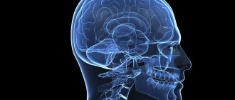
The structure of white and gray matter in the brain
The white and gray matter have a close relationship, which is designed to ensure the necessary level of transmission of nerve impulses from the central nervous system to the peripheral nerves. The central nervous system, that is, the brain, is in close interaction with the spinal cord, so most doctors do not separate these two components of the main nervous organization in the human body.
So, the main task of white matter is the transmission of nerve impulses to the central nervous system and, conversely, the transmission of impulses coming from the brain to the peripheral nerves. Peripheral nerves are a set of nerve fibers that provide innervation to all organs and tissues present in the human body. Disturbance in the conduction of nerve impulses inevitably leads to loss of sensitivity and control over certain organs and tissues.
The main task of white matter is its conductive function, which regulates the functioning of all parts of the nervous system. Signals that the white matter receives through the gray matter horns coming from the central nervous system, and in addition, those that go through the white matter nerve bundles from the central nervous system are transmitted along the descending pathways of the white matter. All signals received from peripheral nerves are transmitted to the gray matter and through some white matter fascicles via ascending pathways. The white matter consists of myelinated processes.
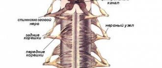
Spinal cord and its pathways
Despite the fact that when cut, the white and gray matter of the spinal cord look approximately the same and differ only in shade, in fact these parts of the spinal cord perform completely different functions and have different structures. How exactly the columns of gray matter of the spinal cord function is still largely a mystery, but it is believed that this part is the most ancient, and its main function is the transformation and transmission of information to the central nervous system.
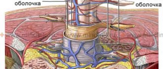
In the center of the spinal cord is the central canal, which, during normal functioning, is filled with cerebrospinal fluid, which is necessary to ensure the water-salt balance of the spinal cord tissue. The white matter on one side is in contact with the gray, and on the other it is covered with soft, arachnoid and hard shells.
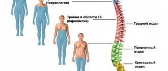
Considering that the entire spinal cord is located in the spinal canal of the spine, it itself is divided into 5 segments, which belong to and have the same names as the sections of the spine.
Total information
The spinal cord, like the brain, is a vital organ in our body.
It lies in the spinal canal, which is formed by the roots of the vertebrae. At the level of the pyramidal fibers, the spinal cord begins, not the brain, and at the top the organ passes into the medulla oblongata. From below it continues to the first or second lumbar vertebra. Inside the organ there is a cavity or central canal, unlike the head. The protective function is performed by the pia mater, arachnoid and dura mater, just like the brain. Cerebrospinal fluid fills the spaces between the membranes and the spinal canal in humans. The epidural space is filled with adipose tissue and a venous network; the space is located between the bone of the vertebrae and the outer hard shell of the substance.
The diameter of the organ varies along its entire length (thickenings intumescentia cervicalis and intumescentia lumbalis are noticeable in the cervical and lumbar regions). The segmental structure of the organ, the nervous system, is responsible for the thickenings that are formed due to the fusion of the reflex arcs of the upper and lower extremities.
Thirty-two parts are distinguished throughout the entire organ, the nervous system: eight cervical, twelve thoracic, five lumbar, five sacral and one or two coccygeal. The length of the spinal cord in men and women is different; in representatives of the stronger sex the length reaches 45 centimeters, in the weak it is only 41 centimeters.
Anatomical features
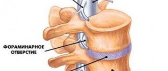
The medullary canal is located inside the spine
A section of the spinal cord reveals that the gray matter has significantly less mass than the white matter. Studies have revealed that the gray matter of the spinal cord has a mass approximately 12 times less than the mass of the white matter. White matter has a complex anatomical structure.
The white matter of the spinal cord is formed by several types of nerve cells, which have very different origins. Individual cells are processes of the gray. Other cells come from the cells of the sensory ganglia, which, although not structural elements of the spinal cord, are directly related to it. The third type of cells comes from the ganglion cells of the central nervous system.
Considering the specificity of nerve cells, we can conclude that the white matter serves to connect nerve cells located in different parts of the body. This is very important, because during movement, muscles in different parts of the body are used, so such a nervous organization allows you to connect the activities of all tissues.
The white matter has pronounced segmentation. Thus, the posterior, anterior and lateral grooves are separators that form the so-called cords:
- Anterior cord. Anatomically, the anterior columns are located between the anterior horn of the gray matter and the anterior median fissure. This area contains descending pathways through which signals pass from the cortex, and in addition, from the midbrain to all important organs and tissues of the body.
- Posterior cord. Anatomically, the posterior cords are localized between the posterior and anterior horns of the gray matter of the spinal cord. The posterior funiculi contain delicate, wedge-shaped and ascending fascicles. These bundles are separated from each other, and the posterior intermediate grooves serve as a separator. The wedge-shaped bundle of nerves contained in the posterior region of this cord carries nerve impulses from the upper limbs to the brain. The delicate bundle transmits impulses to the brain from the lower extremities.
- Lateral cord. Anatomically, it is located between the posterior and anterior horn. This cord contains both ascending and descending pathways.
The structure of white matter includes a complex system of varying length and thickness of non-pulp and pulpal nerve fibers in combination with supporting tissue, which is designated neuroglia. The white matter also contains small blood vessels that have almost no connective tissue.
Anatomically, the white matter of one half is connected to the white of the other half by a commissure, and in the area of the central spinal canal extending transversely in front there is a white commissure. Different fibers are tied into bundles. It is worth considering in more detail the bundles that conduct nerve impulses and their functions.
Structure of gray matter
Substantia grisea is an ideal structure consisting of:
- neurons;
- dendrites;
- unmyelinated axons;
- glial cells;
- thin capillaries.
The latter color the bark brown and, contrary to popular belief, the substance is not gray, but grey-brown.
Numerous labyrinth-like depressions and bulges form convolutions known as cerebral gyri. The main function of gray matter is to ensure communication between the human body and the outside world, as well as to regulate reflexes and ensure higher mental functions. And if the substantia grisea consists of neurons, then the substantia alba appears in the form of myelin-covered axons (neuron processes), which act as conductors and serve to transmit signals and provide communication between the hemispheres and nerve centers. The myelin sheath gives the substance its characteristic white color.
The gray substance in the spinal structure resembles the contours of the letter H or the wings of a butterfly. Depending on their location and functions, gray pillars are divided into: rear, front and side. The lateral parts of the dorsal region, in turn, are divided into:
- Posterior - consist of intermediate nerve cells. Receive signals from ganglia.
- The anterior ones consist of motor neurons. The main function is to ensure muscle tone.
- Lateral - consist of sensory and visceral neurons. Responsible for motor functions.
Main ascending paths
Ascending pathways serve to transmit impulses from peripheral nerves to the brain. Most of the ascending pathways deliver nerve impulses to the cerebellar and cortical regions of the central nervous system. Some ascending white matter tracts are so fused together that they simply cannot be considered separately. It is possible to distinguish 6 independent and interconnected ascending bundles located in the white matter.
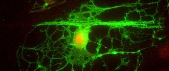
How is the connection between two human brain organs
- Gaulle's thin bundle and Burdach's wedge-shaped bundle. These bundles are formed from special cells of the spinal ganglia. A thin beam is formed from 19 lower segments. The wedge-shaped bundle is formed from 12 upper segments. The fibers of both of these bundles are integrated into the spinal cord through the dorsal roots and transmit collaterals to special neurons. Axons reach the nuclei of the same name.
- Ventral and lateral pathways. Considering what each path consists of, the sensory cells of the spinal ganglia, which are integrated into the dorsal horns, are immediately isolated. The cells included in these bundles move to the gray and touch the switching nuclei located in the thalamus.
- Ventral spinocerebellar tract of Gowers. Contains special neurons of the spinal ganglia that pass into the area of Clarke's nucleus. The axons ascend to the upper parts of the central nervous system, where they enter the ipsilateral half of the cerebellum through its superior peduncles.
- Dorsal spinocerebellar tract of Flexing. Contains neurons of the spinal ganglia at the very beginning, and then switches to nuclear cells in the intermediate zone of the gray matter. The axons reach the longitudinal medulla by passing through the inferior cerebellar peduncle and then pass into the ipsilateral region of the cerebellum.
These are not all the ascending pathways that run in the white matter of the spinal cord, but at present the nerve bundles presented above are the most studied.
Histology
The spinal cord consists of two symmetrical halves, delimited from each other in front by a deep median fissure, and behind by a connective tissue septum. In fresh preparations of the spinal cord, the naked eye can see that its substance is heterogeneous. The inner part of the organ is darker - this is its gray matter (lat. substantia grisea). At the periphery of the spinal cord there is a lighter white matter (lat. substantia alba). Gray matter is shaped like an "H" or a butterfly in a cross-section of the brain. The projections of gray matter are commonly called horns. There are anterior, or ventral, posterior, or dorsal, and lateral, or lateral, horns[3].
Along the spinal cord, the relationship between gray and white matter changes. Gray matter is represented by the smallest number of cells in the thoracic region. The largest is in the lumbar region.
Gray matter
The gray matter of the spinal cord consists of neuronal cell bodies, unmyelinated and thin myelinated fibers, and neuroglia. The main component of gray matter, distinguishing it from white matter, are multipolar neurons [3].
Cells similar in size, fine structure and functional significance lie in the gray matter in groups called nuclei. Among the neurons of the spinal cord, the following types of cells can be distinguished:
- radicular cells (lat. neurocytus radiculatus), the axons of which leave the spinal cord as part of its anterior roots;
- internal cells (lat. neurocytus internus), the processes of which end in synapses within the gray matter of the spinal cord;
- tuft cells (lat. neurocytus funicularis), the axons of which pass through the white matter in separate bundles of fibers, carrying nerve impulses from certain nuclei of the spinal cord to its other segments or to the corresponding parts of the brain, forming pathways.
Individual areas of the gray matter of the spinal cord differ significantly from each other in the composition of neurons, nerve fibers and neuroglia[3].
In the posterior horns there is a spongy layer
,
gelatinous substance
,
dorsal horn nucleus proper
and
thoracic nucleus
. Between the posterior and lateral horns, the gray matter protrudes into the white matter in strands, as a result of which a network-like loosening is formed, called the reticular formation [3].
Spongy layer
The dorsal horn is characterized by a broadly looped glial skeleton, which contains a large number of small interneurons [3].
In gelatinous substance
glial elements predominate. The nerve cells here are small and their number is insignificant.
The posterior horns are rich in diffusely located intercalary cells
. These are small multipolar association and commissural cells, the axons of which end within the gray matter of the spinal cord on the same side (association cells) or the opposite side (commissural cells).
Neurons of the spongy zone, gelatinous substance and intercalary cells communicate between the sensory cells of the spinal ganglia and the motor cells of the anterior horns, closing local reflex arcs. The nucleus of the posterior horn is located in the middle of the
. It consists of interneurons, the axons of which pass through the anterior white commissure to the opposite side of the spinal cord into the lateral funiculus of the white matter, where they are part of the ventral spinocerebellar and spinothalamic tracts and are sent to the cerebellum and thalamus [3].
Thoracic core
(Clark's nucleus) consists of large interneurons with highly branched dendrites. Their axons exit into the lateral cord of the white matter on the same side and, as part of the posterior spinocerebellar tract (Flexig's tract), ascend to the cerebellum [3].
In the intermediate zone, the medial intermediate nucleus
, the axons of the cells of which join the anterior spinal-cerebellar tract (Gowers tract) of the same side, and
the lateral intermediate nucleus
, located in the lateral horns and representing a group of associative cells of the sympathetic reflex arc. The axons of these cells leave the brain along with somatic motor fibers as part of the anterior roots and are separated from them in the form of white connecting branches of the sympathetic trunk [3].
The anterior horns contain the largest neurons of the spinal cord, which have a body diameter of 100-150 μm and form nuclei of significant volume. This is the same as the neurons of the lateral horn nuclei, the root cells, since their axons make up the bulk of the fibers of the anterior roots. As part of the mixed spinal nerves, they enter the periphery and form motor endings in the skeletal muscles. Thus, these nuclei represent motor somatic centers. In the anterior horns, the medial and lateral groups of motor cells are most pronounced. The first innervates the muscles of the trunk and is well developed throughout the spinal cord. The second is located in the area of the cervical and lumbar thickenings and innervates the muscles of the limbs [3].
Motor neurons provide efferent information to skeletal striated muscles and are large cells (100-150 µm in diameter). In the axon terminals there are synaptic vesicles with acetylcholine, on the neuron body and dendrites there are numerous synapses - up to 1000 or more axosomatic terminals. Motor neurons are combined into 5 groups of motor nuclei - lateral (anterior and posterior), medial (anterior and posterior), central. In the nuclei, neurons form columns[3].
The gray matter of the spinal cord contains many scattered tufted neurons. The axons of these cells exit into the white matter and immediately divide into longer ascending and shorter descending branches. Collectively, these fibers form their own, or main, bundles of white matter, directly adjacent to the gray matter. Along their course, they give rise to many collaterals, which, like the branches themselves, end with synapses on the motor cells of the anterior horns of 4-5 adjacent segments of the spinal cord [3].
Spinal cord gray matter nuclei (right) and Rexed plates (left)
Layers of gray matter according to Rexed
In 1952, the Swedish anatomist Bror Rexed proposed dividing the gray matter into ten plates (layers), differing in the structure and functional significance of their constituent elements. This classification has received wide recognition and dissemination in the scientific world. Plates are usually designated by Roman numerals.
Laminae I to IV form the head of the dorsal horn, which is the primary sensory area.
I plate
formed by many small neurons and large spindle-shaped cells lying parallel to the plate itself. It includes afferents from pain receptors, as well as axons of neurons in lamina II. The emerging processes contralaterally (that is, crosswise - the processes of the right posterior horn along the left cords and vice versa) carry information about pain and temperature sensitivity to the brain along the anterior and lateral cords (spinothalamic tract).
II and III plates
formed by cells perpendicular to the edges of the plates. Corresponds to gelatinous substance. Both are affered by processes of the spinothalamic tract and transmit information downstream. Participate in the control of pain. The II plate also gives off processes to the I plate.
IV plate
corresponds to its own core. Receives information from plates II and III, the axons close the reflex arcs of the spinal cord on motor neurons and participate in the spinothalamic tract.
V and VI plates
form the neck of the posterior horn. Receive afferents from muscles. Plate VI corresponds to the Clarke nucleus. Receives afferents from muscles, tendons and ligaments, descending tracts from the brain. Two spinocerebellar tracts emerge from the plate:
- Fleschig's tract (option: Flexig) (lat. tractus spinocerebellaris dorsalis) - exits ipsilaterally (that is, into the cord of its own side) into the lateral cord
- Govers' tract (lat. tractus spinocerebellaris ventralis) - exits contralaterally into the lateral cord
VII
occupies a significant part of the anterior horn. Almost all the neurons of this plate are intercalary (with the exception of the efferent neurons of the Latin Nucleus intermediolateralis). Receives afferentation from muscles and tendons, as well as many descending tracts. Axons go into lamina IX.
VIII plate
located in the ventromedial part of the anterior horn, around one of the parts of plate IX. Its neurons participate in propriospinal connections, that is, they connect different segments of the spinal cord with each other.
Plate IX
is not united in space, its parts lie inside the VII and VIII plates. It corresponds to the motor nuclei, that is, it is the primary motor area, and contains motor neurons located somatotopically (that is, it represents a “map” of the body), for example, motor neurons of the flexor muscles usually lie above the motor neurons of the extensor muscles, neurons innervating the hand are more lateral, than those innervating the forearm, etc.
X plate
located around the spinal canal, and is responsible for commissural (between the left and right parts of the spinal cord) and other propriospinal connections.
White matter
White matter surrounds gray matter. The grooves of the spinal cord divide it into cords (lat. funiculi): anterior, lateral and posterior. The cords are nerve tracts that connect the spinal cord to the brain.
The widest and deepest groove is the lat. Fissura medianus anterior (anterior median fissure), dividing the white matter between the anterior horns of the gray matter. Opposite her is lat. Sulcus medianus posterior (posterior median groove).
A pair of lateral grooves (lat. Sulcus lateralis posterior & anterior) go, respectively, to the posterior and anterior horns of the gray matter.
The posterior cord is divided (lat. Sulcus intermedia posterior), forming two ascending tracts: the one closest to the posterior median sulcus of the lat. Fasciculus gracilis (delicate or thin tuft), and more lateral lat. Fasciculus cuneatus (wedge-shaped fascicle). The internal bundle, thin, rises from the lowest parts of the spinal cord, while the wedge-shaped one is formed only at the level of the thoracic region.
Spinal nerves
Scheme.
Fibers in the spinal nerve (from the 8th cervical to 2-3 lumbar segments). Sensory fibers are indicated in blue, motor (somatic) in red, preganglionic sympathetic in dark green, postganglionic sympathetic in light green.
The anterior and posterior roots fuse to form the spinal (spinal) nerve.
Spinal nerves contain four functional components:
- GSA (general somatic afferent) - receive sensory fibers from the surface of the body;
- GVA (general vegetative afferent) - receive sensory fibers from visceral organs;
- GSE (general somatic efferent) - innervate skeletal muscles;
- GVE (general vegetative efferent) - innervate the autonomous (vegetative) ganglia.
The areas of skin served by various spinal nerves are called dermatomes.
The main descending tracts of the spinal cord
The descending pathways are closely connected with the gray matter region and ganglia. These bundles transmit nerve electrical impulses that emanate from the central nervous system and are sent to the periphery. Descending pathways are currently even less studied than ascending pathways. Descending paths, like ascending ones, often intertwine with each other, forming almost monolithic structures, so some of them should be considered without dividing into separate paths:
- Ventral and lateral corticospinal tracts. They originate from pyramidal neurons of the lowest layers of the motor zone of the cerebral cortex. Next, the fibers cross the cerebral hemispheres, the base of the midbrain, and then move along the ventral sections of the so-called Varoliev and medulla oblongata, reaching the spinal cord.
- Tectospinal. It originates from cells in the quadrigeminal region of the midbrain and ends with a connection in the region of mononeurons of the anterior horns.
- Rubrospinal. The base of the path is the cells located in the region of the red nuclei of the central nervous system, there are crossings of the midbrain region, and the endings of the nerve fibers of this path lie in the region of the neurons of the intermediate zone.
- Vestibulospinal tracts. This is a collective concept that reflects several types of bundles, which originate from the vestibular nuclei located in the medulla oblongata and end in the anterior cells of the anterior horns.
- Olivospinal. It is formed by the axons of olivary cells localized in the longitudinal medulla and ends in the region of mononeurons.
- Reticulospinal. It is the connector between the spinal cord and the reticular formation.
These are the main pathways that are most studied at present. However, it should be noted that there are also local bundles that also perform a conducting function, but at the same time connect different segments located at different levels of the spinal cord.
What is the danger of track damage?
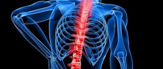
Despite the fact that the white matter is hidden under three membranes that protect the entire spinal cord from damage, and is located in the hard frame of the spine, cases of damage to the spinal cord due to injury are not uncommon. The second cause of conduction disturbance is infection, but it is not so common. As a rule, with spinal injuries, it is the white matter that is first affected, since it lies close to the surface of the spinal canal of the spine.
The degree of dysfunction may depend on the characteristics of the injury or damage, so in some cases the dysfunction will be reversible, in others it will be partially reversible, and in others there may be irreversible consequences.
As a rule, irreversible consequences due to damage to the spinal cord are observed when a large rupture occurs. In this case, the conduction function is disrupted. If there is a spinal bruise in which the spinal cord is compressed, there are several options for damage to the connections between the nerve cells of the white matter with different consequences.
In some cases, certain fibers are torn, but there is a possibility of their healing and restoration of the transmission of nerve impulses. The complete restoration of a damaged bundle may require considerable time, since nerve fibers grow together extremely difficult, and the possibility of conducting nerve impulses through them depends on their integrity. In other cases, there may be a partial restoration of the conduction of electrical impulses through damaged nerve fibers, then sensitivity in certain parts of the body may be restored, but not fully.
The degree of traumatization is not the only thing that affects the possibilities of rehabilitation, because... much depends on how quickly first aid was provided and how professionally further resuscitation was carried out. In order for the nerves to begin conducting electrical impulses, they need to be retrained to do so. The regeneration process is also influenced by other characteristics of the human body, including age, metabolic rate, chronic diseases, etc.
How gray matter influences some human abilities
The gray tissue of the brain, regulating the processing of signals from the outside and generating effector impulses, is not only responsible for the functioning of the entire human nervous system, but also affects his abilities: mental, cognitive, physical, etc.
Various experiments by scientists have shown that a person’s abilities depend on the volume of the gray substance, while changes in the amount of white substance did not show any noticeable changes.
Experiments by British scientists have shown that the thinner the cerebral cortex, therefore, the smaller the volume of gray substance, the worse a person copes with solving logical problems, the less various abilities he has, and also with a low volume of substance, subjects often had problems with reaction speed , speech dysfunctions, memory problems and poor intellectual abilities.
At the same time, studies have shown that learning foreign languages, memorizing poetry, scientific or artistic works, and playing music affect the enlargement of the cerebral cortex. The longer and more intense the learning process, the greater the volume of gray substance becomes, therefore, the more abilities, including mental ones, a person displays.
A decrease in the amount of gray matter is affected by:
- a person’s lifestyle is a sedentary, inert, inactive, from a physical and mental point of view, way of life;
- abuse of bad habits - alcohol, drug addiction and smoking reduce the volume of gray substance.
For example: those suffering from alcoholism experience a significant decrease in the amount of brain tissue, which is reflected in behavior and mental functions: incoherent speech, problems with memory and perception, inhibition of thought processes.