Diagnostic methods
Electroneurography (ENMG) is a procedure in which the nerve is stimulated with weak electrical impulses. In other places on the nerve, the speed at which these impulses reach these points is measured. To carry out the procedure, special electrodes are placed on the passage of the facial nerve. The examination takes approximately 40 minutes, allowing you to identify the following manifestations:
- inflammation of the nerve (with a decrease in the speed of impulse transmission);
- rupture of the nerve fiber (the signal is not transmitted);
- possible muscle atrophy (not all muscle fibers receive impulse);
- disturbance of the conductivity of the nerve column (muscles react poorly to signals).
The electromyography method involves studying natural impulses without the use of electric current. Needle electrodes are inserted into some areas of the facial muscles, which will measure the propagation of impulses along the fibers. The study is carried out in 2 stages. First, impulses are measured in a relaxed state, and then when trying to use facial muscles. The procedure takes from 40 minutes to an hour and can reveal the following disorders:
- slowing down the passage of impulses through muscle fibers;
- reduction in the number of fibers responding to the signal.
Inflammation therapy
The recovery process lasts on average 8–10 weeks. During this time, the causes of the development of neuritis are clarified (if possible), inflammation of the facial nerve is relieved, measures are taken to preserve the eye and prevent muscle strain on the affected side in case of paralysis.
For the latter purpose, the following procedures can be carried out.
- Fixation of tissues with adhesive tape on the diseased half of the face; It can also be used to cover the eyelid to prevent the eyeball from drying out. This event is possible with mild paralysis.
- Stitching the upper and lower eyelids together is indicated if the weakening of the muscles is severe and the upper eyelid does not close at all.
- For the same purpose, implants are inserted into the upper eyelid.
- In modern medicine, preference is also given to botulinum toxin for the eyelids. Subsequently, preparations with this substance can also be used on the affected side of the face as an aesthetic correction and to prevent complications (tightening and contraction of muscles).
Drug treatment of facial nerve neuritis involves taking the following medications:
- corticosteroids – relieve inflammation and swelling, promote the conduction of impulses along nerve fibers;
- painkillers – used for severe pain;
- diuretics - indicated to relieve swelling of the nerve and tissues;
- antiviral - given in cases where inflammation of the facial nerve is caused by herpes simplex and varicella-zoster viruses (acyclovir);
- antispasmodics – relieve pain, vasospasm, improve blood circulation;
- neurotropic drugs - prescribed for muscle contractions;
- B vitamins - to restore metabolism in damaged areas.
Neuritis of the facial nerve can also be treated with auxiliary methods: massage, mud and paraffin applications, electrophoresis, UV irradiation, ultra-high frequency therapy (UHF) and the like.
Their widespread use is currently quite controversial - due to unproven positive effects and recorded complications during or after completion of the course.
The basis should be drug treatment with corticosteroids - they are the ones that in most cases allow you to get rid of the lesion and prevent contractures.
httpv://www.youtube.com/watch?v=embed/TXMg6KbR-_w
Neuritis of the facial nerve is a disease that is characterized by the appearance of pain in places where the nerve trunk is damaged and its paralysis. In most cases, with timely consultation with a doctor, the disease is cured, but complications and relapses are possible. Sometimes there are other variants of neuritis, for which symptomatic therapy is also indicated
It is important to carry out treatment with a doctor, and not on your own - this will avoid unpleasant irreversible consequences
mozgius.ru
Manifestation of the disease
The main symptoms of neuritis of the facial nerve are pronounced:
- pain in various parts of the face, in the back of the head, ear and eyeball, on the lips, gums, tongue, which can intensify with hypothermia or touch;
- paralysis, manifested by weakening of the muscles of one half of the face.
In addition, you may experience:
- hyperacusis (increased sensitivity to sounds), hearing loss;
- decreased or complete loss of taste sensitivity.
The paralysis that characterizes facial neuritis can be easily diagnosed visually. The patient has:
- the folds on the forehead of the affected side are smoothed out;
- there is a distortion of the mouth;
- it is impossible to close the eye on the affected side, and it rolls back when trying to look up (Bell's symptom).
A person needs to urgently consult a doctor if he cannot frown his forehead, puff out his cheeks, wrinkle his nose, whistle or blow briefly, draw water into his mouth, blink his eyes alternately, or easily close both eyes. The sooner facial neuritis is diagnosed and treated, the higher the likelihood that the paralysis will disappear .
What a person’s face looks like when paralysis appears can be seen in the photo below.
Signs of paralysis can vary in severity, from mild to severe. A first-degree lesion may, at first glance, not be noticeable and be diagnosed only upon careful examination (the patient’s eye has difficulty closing, and slight asymmetry of the mouth is detected). Subsequent degrees are characterized by an increase in the severity of paralysis: moderate weakness, severe, severe and complete.
As a result of paralysis, a person may begin to leak saliva from the corner of the mouth on the affected side, bite the inside of the cheek while eating, and swell when talking (sailing symptom). The person speaks with difficulty and indistinctly. He may suffer from constant dry mouth and thirst.
If a patient exhibits signs of sudden weakening of the facial muscles without pain, then only paralysis is diagnosed (ICD-10 code - G51.0). However, both cases are treated according to the same scheme.
Severe symptoms of facial neuritis suggest that even in sleep a person cannot cover the eyeball with his eyelid. In the first days this leads to dryness and redness, and in subsequent days it threatens the development of keratopathy and complete blindness.
How to treat
Recovery occurs after 2-2.5 months. During this period, the inflammatory process of the nerve of the facial zone passes; it is necessary to prevent overextension of the paralyzed area of the face. To do this you need:
- Secure the affected facial part with an adhesive plaster, cover the eyelid to avoid drying out the conjunctiva of the eye. This procedure is performed for mild neuritis, when paralysis is mild;
- sew the lower eyelid with the upper eyelid if there is severe weakness and it is impossible to close the eyes with the upper eyelid; implantation is also indicated for this purpose;
- In medical practice, botulinum toxin has recently been used for eyelid paralysis. In the future, products with this toxin are used for neuritis to correct the aesthetic appearance of the face in order to prevent complicated conditions.
Treatment of facial paralysis with drugs involves the use of:
- corticosteroid drugs (prednisolone), which relieve swelling and help normalize impulse conduction along the nerves of the face;
- analgesic group of drugs for severe pain;
- diuretics necessary to relieve swelling near the nerve (furosemide);
- antiviral drugs if neuritis of the facial nerve is caused by herpes simplex, Varicella-Zoster virus (gerpevir);
- antispasmodics, relieving pain syndromes, vascular spasms, improving microcirculation;
- neurotropic drugs prescribed for muscle twitching;
- anticholinesterase drugs (proserin, galantamine), indicated in the third week of the disease;
- vitamin group B, necessary to restore the metabolic processes of the affected area.
Facial neuropathy is auxiliary treated with physiotherapeutic procedures, including classical and acupressure massage, paraffin and therapeutic mud applications, electrophoresis administration of medication, and ultra-high-frequency exposure to currents.
Massage with physical therapy is indicated from the beginning of the second week with a gradual increase in load. In the third week, phonophoresis with hydrocortisone is indicated; ultrasound will not be superfluous.
The basis of treatment is the use of corticosteroids that relieve inflammation and prevent contracture manifestations.
It should be remembered that early diagnosis of facial paralysis facilitates timely treatment until the manifestations of neuropathy completely disappear. Basically, with timely treatment, the disease is completely cured, however, relapses are possible. There are other manifestations of types of neuritis of the facial nerve, where symptomatic treatment is also indicated. Self-medication will not bring anything good except harm and complications.
Complications and prevention
It is believed that with idiopathic etiology, complete recovery almost always occurs, whereas after injury or infection, it is not possible to restore the function of the trigeminal nerve.
The most serious consequence of the lesion is contracture of the facial muscles, when the patient feels numbness in one half of the face. In fact, part of the face does not move, it literally cramps.
Other complications include blepharospasm (twitching), muscle atrophy, facial synkinesis, for example, tears suddenly flow when chewing, conjunctivitis due to partial closure of the eyelids.
Relapse of the disease is much more difficult to treat, so it is better to prevent it by adhering to preventive measures:
- treat all viral diseases in a timely manner, do not suffer them “on your feet”;
- try not to get too cold;
- take vitamin complexes to avoid nutritional deficiencies;
- carry out hardening procedures;
- Avoid nervous tension and stress.
umozg.ru
Complications and consequences
The disease is dangerous due to its complications . The most common residual effects include the following:
- weakness of facial muscles;
- partial facial asymmetry;
- uncontrolled involuntary movements of facial muscles during conversation, laughter, smiling;
- muscle paresis (narrowing of the palpebral fissure (half-closed eye), deepening of the nasolabial fold, appearance of a dimple on the chin, raised cheek, drooping corner of the mouth).
- soreness in certain areas;
- excessive tearing;
- increased muscle tone.
Comments
(function(w, d, n, s, t) { w = w || []; w.push(function() { Ya.Context.AdvManager.render({ blockId: 'RA-324974-4', renderTo : 'yandex_rtb_R-A-324974-4', async: true }); }); t = d.getElementsByTagName('script'); s = d.createElement('script'); s.type = 'text/javascript '; s.src = '//an.yandex.ru/system/context.js'; s.async = true; t.parentNode.insertBefore(s, t); })(this, this.document, 'yandexContextAsyncCallbacks ');»+»ipt>
- Automatic gates
- Wooden gates and fences
- Hedge
- Brick fences
- Fence and gate structures
- Metal fences and gates
- Fence and gate installation
- Answers on questions
- Finishing the fence and gate
- Fence and gate repair
- Advertising on the website
- Our specialists
Establishing diagnosis
Neuritis affects both children and adults equally. The course of the disease is divided into three stages:
- Acute – lasts up to 14 days.
- Subacute – takes four weeks.
- Chronic – more than a month.
Treatment results depend on the depth of the neuropathy, how quickly the diagnosis was made, and the etiology of the lesion. The doctor rarely has doubts about the nature of the disease, since the symptoms are too obvious.
The main thing is that you can quickly contact a specialist who will determine the cause of the disease. For this purpose, an MRI is prescribed to exclude tumors in the brain. Electroneuromyography is used to establish the source of inflammation, its nature and to find out at what stage the deviation is now.
Neuralgia and neuritis, unspecified
ICD-10 Category: M79.2
Definition and general information
Synonyms: Alcock syndrome, groin pain, pudendal neuralgia
Inguinal neuralgia is a syndrome of chronic neuropathic pain that is aggravated by sitting and which has no corresponding organic cause. Inguinal neuralgia is often associated with the presence of pelvic organ dysfunction.
The estimated prevalence in France is 1/6000.
Etiology and pathogenesis
The causes of inguinal neuralgia remain unclear. There are several possible sites of nerve compression: the area of the posterior pelvic ligaments (sacrotuberous and sacrospinal ligaments), in Alcock's canal (due to splitting of the aponeurosis of the obturator muscle) and the falciform part of the sacrotuberous ligament. There is also the possibility of proximal nerve entrapment at the level of the suprapiriform canal and distal entrapment of the dorsal clitoral/penis nerve at the level of the suprapubic canal. Inguinal neuralgia can also be caused by cycling, childbirth, and scarring from surgery or radiation therapy.
Clinical manifestations
The disease usually appears between the ages of 50 and 70 years. Inguinal neuralgia manifests itself as neuropathic pain of varying intensity in the perineal area. The pain is described as intense, sharp, with a burning sensation, and sometimes numbness. A sensation of a foreign body in the rectum or vagina (sympathalgia) has also been reported. The pain is unilateral or often midline, more intense during the day when sitting or wearing tight clothing. Pain is often associated with increased sensitivity in the pelvic area, with dysfunction of the bladder (pollakiuria, dysuria), anorectal area (dysgesia, increased pain during peristalsis) and the genital area (dyspareunia, intolerance to contact with the vulva, postcoital exacerbation of pain, constant genital arousal, erectile dysfunction). dysfunction), as well as myofascial pain in the buttocks. The co-occurrence of symptoms of stem radiculitis is common. There are several forms of inguinal neuralgia: benign, regressive, with exacerbations, stable and severe with progressive worsening of symptoms.
Neuralgia and neuritis, unspecified: Diagnosis
Diagnostic criteria include the presence of pain in the area of innervation of the pudendal nerve, which is aggravated in a sitting position, the absence of sensory disturbances, pain does not cause awakening during sleep and is relieved by blockade of the pudendal nerve using anesthetics. MRI allows you to classify inguinal neuralgia based on the location of the entrapment: Type I - in the sciatic notch; type II - ischial spine and sacroischial ligament; type IIIA - obturator internus muscle; type IIIB - obturator internus and piriformis muscles and type IV - distal branches of the pudendal nerve.
Normal MRI results do not exclude the diagnosis of inguinal neuralgia.
Differential diagnosis
Differential diagnosis includes tumor compression of nervous tissue (neurofibromas, schwannomas), external compression (endometriosis, bone metastases), infections or dermatological diseases (chronic urethritis, lichen sclerosus), other sacral neuropathies (herpes zoster, herpes), transient proctalgia, postpartum perineal neuropathy, complex regional pain syndromes, perineal neuropathy involving the inferior gluteal nerve, sciatic bursitis, piriformis syndrome and vulvodynia.
Neuralgia and neuritis, unspecified: Treatment
Management includes treatment for neuropathic pain (gabapentin, pregabalin, duloxetine, amitriptyline), transcutaneous posterior tibial nerve stimulation, physical therapy, osteopathy, and short-term psychotherapy.
The effect of infiltration of the pudendal nerve with anesthetics is limited. In refractory forms, transglutinal decompression of the pudendal nerve was effective. In cases where surgical intervention is ineffective, implantation of a neurostimulator at the level of the conus terminalis can be proposed.
Inguinal neuralgia significantly affects quality of life, but has no effect on life expectancy.
wikimed.pro
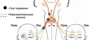
Neuritis of the facial nerve on the right ICD 10 – MedPage
In ICD-10, lesions of the facial nerve are coded in category 051.
| 1 | 2 | 3 |
| C51.0 | Bell's Palsy Facial Palsy | OFD. Neuropathy of the facial nerve PRFD. Acute neuropathy of the left facial nerve with damage to it |
distal area against the background of arterial hypertension with moderate paresis of facial muscles, recovery phase
Note.
Bell's palsy is an acute neuropathy of the facial nerve, usually caused by compression of the facial nerve in a bony canal due to swelling or inflammation. Neuropathy is often idiopathic in nature, but can be triggered by a viral infection (including herpes).
Predisposes to the development of neuropathy: arterial hypertension, diabetes mellitus, pregnancy. Diagnosis requires the exclusion of other conditions that cause damage to the facial nerve (brucellosis, syphilis, otitis, damage to the parotid glands, tumors, leukemic infiltration, sarcoidosis, multiple sclerosis, etc.).
Idiopathic neuropathy tends to recover spontaneously. Some patients develop irreversible residual effects in the form of paresis, synkinesis, contracture of facial muscles, “crocodile tears” syndrome, dysacusia, dysgeusia, and chronic pain syndrome.
When formulating a diagnosis, one should indicate the phase (progression, stabilization, recovery), the degree of weakness of the facial muscles (mild paresis is characterized by the loss of only emotional movements, while movements performed on command remain relatively intact; with severe paresis, voluntary movements are lost, but tone remains Relatively preserved in plegia, movements are completely lost and muscle hypotonia occurs). It is also advisable to indicate the level of nerve damage (proximal - characterized by weakened tear secretion and dry eye, and distal - characterized by lacrimation), since with proximal damage, recovery is slower. After 12 months after the development of neuropathy, it is recommended to use the term “consequences of neuropathy” indicating the nature and severity of residual effects
051.1 Inflammation of the knee joint OFD. Ganglionitis of the geniculate ganglion Excluded: postherpeti-
logical lesion of the knee joint (B02.2)
Note.
It is characterized by a combination of acutely developing unilateral paralysis of the facial muscles with pain in the ear and parotid region, signs of involvement of the cochleovestibular nerve (dizziness, hearing loss) and sometimes the trigeminal nerve.
Herpetic lesion of the node (Ramsay Hunt syndrome) is coded in another category
| 1 | 2 | 3 |
| 051.2 | Rossolimo-Melkersson syndrome Rossolimo-Melkersson-Rosenthal syndrome | OFD. The same as in ICD-10 PRFD. Idiopathic bilateral recurrent neuropathy of the facial nerve (Rossolimo-Melkersson-Rosenthal syndrome), acute phase |
| Note. Rossolimo-Melkersson-Rosenthal syndrome is a condition characterized by recurrent, often bilateral damage to the facial nerve, which is accompanied by granulomatous cheilitis, tongue folding and angioedema of the face. Other causes of bilateral facial nerve damage should be excluded (neuroborreliosis, syphilis, infectious mononucleosis, sarcoidosis, idiopathic cranial pachymeningitis, meningeal carcinomatosis, Paget’s disease, etc.) | ||
| 051.3 | Clonic hemifacial spasm | OFD. Facial hemispasm PRFD. Facial hemispasm on the right due to compression of the nerve by the loop of the superior cerebellar artery, clonic form, compensation during treatment with botulinum toxin |
| Note. Facial hemispasm is characterized by paroxysms of involuntary contractions of muscles innervated by the facial nerve. In the intervals between spasms, the face remains symmetrical or slight weakness of the facial muscles on the side of the spasm is detected. In most cases, it is associated with compression of the nerve by a small artery or vein (most often branches of the posterior inferior cerebellar artery), less often - a tumor or arteriovenous malformation. Sometimes occurs as a result of acute neuropathy or nerve injury. When formulating a diagnosis in the presence of MRI (or MR angiography) data, the source of compression of the facial nerve should be indicated | ||
| 051.4 | Facial myokymia | OFD. The same as in ICD-10 PRFD. Idiopathic facial myokymia with predominant involvement of the eyelids of the right eye, remitting course, exacerbation phase |
| Note. Facial myokymia is manifested by wave-like contractions of groups of fibers of facial muscles, in which neighboring groups of fibers contract in turn. Facial myokymia is associated with increased excitability of peripheral motor neurons and may be a sign |
| 1 | 2 | 3 |
| damage to the brain stem caused by a tumor, stroke or multiple sclerosis. However, this subcategory only codes for idiopathic facial myokymia, which most often involves the eyelids and is usually a benign condition, often occurring in healthy individuals due to overwork | ||
| C51.8 | Other facial nerve lesions | OFD - see note |
| Note. This subheading codes other diseases of the facial nerve that are not included in the previous headings. Facial hemiatrophy (Parry-Romberg disease) is coded under p67.4 | ||
| C51.9 | Facial nerve lesion, unspecified | Code for statistical recording of unspecified cases of facial nerve lesions |
1.6.
Source: //zakon.today/nevrologiya_1068/porajenie-litsevogo-vii-125187.html
Consilium Medicum No. 09 2021 – Trigeminal neuralgia
For citation Hide list Trigeminal neuralgia. Consilium Medicum.
2016; 09: Trigeminal neuralgia - NTN (trigeminal neuralgia, Fothergill's disease, painful tic) - the most common form of paroxysmal facial pain, a chronic disease that occurs with remissions and exacerbations, characterized by attacks of extremely intense shooting, usually unilateral, pain in innervation zones II, III or extremely rarely the first branch of the trigeminal nerve. The prevalence of NTN is quite high and amounts to up to 30–50 patients per 100 thousand population, and the incidence in the world, according to WHO, is in the range of 2–4 people per 100 thousand population [2, 3]. Women are more susceptible - 5 per 100 thousand population, men - 2.7 per 100 thousand. Typical NTN debuts at the 50th anniversary of life, occurring more often on the right - 70%, on the left - 28%, in rare cases it can be bilateral - 2% [4].
VI Diseases of the nervous system (G00–G99) Lesions of individual nerves, nerve roots and plexuses (G50–G59) G50.0 Paroxysmal facial pain syndrome, painful tic G53.0* Neuralgia after herpes zoster (B02.2+) Postherpetic : inflammation of the genu ganglion, trigeminal neuralgia
International classification of headaches [5]:
• primary (idiopathic) NTN (caused by compression of the trigeminal root) • secondary (symptomatic) NTN (manifestation of other central nervous system diseases: multiple sclerosis, tumors, brainstem stroke, etc.)
Etiology
The most common cause of NTN is compression of the trigeminal nerve at the extra- or intracranial level. Intracranial factors: dilated convoluted cerebellar arteries, aneurysms of the basilar artery, space-occupying processes in the posterior cranial fossa, tumors of the cerebellopontine angle.
Extracranial factors: compression in the bone canal through which the nerve passes, associated with its congenital narrowness or acquired as a result of a chronic inflammatory process in adjacent areas (caries, sinusitis), local odontogenic or rhinogenic inflammatory processes.
The development of NTN can be promoted by infectious processes, vascular, endocrine-metabolic and allergic disorders, and much less frequently by demyelination of the trigeminal nerve root in multiple sclerosis [2, 6–8].
Pathogenesis
From the perspective of the “collar pain control” theory of Melzack and Wall (Wall, Melzack.
Gate control theory, 1965), with neuralgia of the V and IX pairs of the cranial nerve, caused by compression of their roots at the entrance to the brain stem, demyelination of fast-conducting (antinociceptive) type A fibers occurs with the appearance of many additional voltage-dependent sodium channels in the demyelinated areas and the formation of contacts of these areas with unmyelinated (nociceptive) type C fibers. This leads to the formation of prolonged and high-amplitude activity of pathologically altered A fibers, which is manifested by painful paroxysms in the face and oral cavity [3].
Typical signs of pain during an attack of NTN:
• Character: paroxysmal, shooting, comparable to an electric current discharge • Localization: definite, not changing for many years, within the zones of innervation of the trigeminal nerve • Direction: pain comes from one area and reaches another • Duration: attack does not exceed 2 minutes ( usually 10–15 s) • Presence of: – refractory period – “light” interval between two separate attacks – trigger zones (areas on the skin of the face and in the oral cavity, mild irritation of which causes paroxysm) – trigger factors (actions that cause pain attacks: washing, brushing teeth, chewing, talking)
• Typical behavior: patients freeze in the position in which they were caught by an attack
Neuralgia of individual branches of the trigeminal nerve:
• Nasociliary (Charlene's neuralgia) – stabbing pain radiating to the central part of the forehead when touching the outer surface of the nostril • Supraorbital – paroxysmal or constant pain in the area of the supraorbital notch and the medial part of the forehead • Lingual nerve – pain in half of the tongue, aggravated by eating and talking • Infraorbital nerve – pain of low intensity, feeling of numbness of the mucous membrane of the upper jaw and infraorbital region
Treatment
Modern treatment tactics for NTN imply an integrated approach, taking into account the genesis and form of the disease (predominance of central or peripheral components), evidence-based medicine, and the individual characteristics of a particular patient. It includes medications, physiotherapeutic and surgical methods [2].
*According to the recommendations of the European Federation of Neurological Societies (2009). **Level of evidence. List of Spanish literature Hide list 1. Tovazhnyanskaya E.L. Trigeminal neuralgia: modern aspects of complex therapy. Intl. neurol. magazine 2010. 2. Gritsai N.N., Kobzistaya N.A.
Classic trigeminal neuralgia and odontogenic pain syndrome. News of medicine and pharmacy. 2009; 299:23–5. 3. Diagnostic evaluation and treatment of trigeminal neuralgia. NeuroNEWS. 2009; 3:21–6. 4. Neurology: national guidelines. Edited by E.I. Gusev, A.N. Konovalov, V.I. Skvortsova, A.B. Gekht. M.: GEOTAR-Media, 2009. 5.
International classification of headaches. Ed. 2nd. M.: GlaxoSmithKline Trading, 2003. 6. Reveguk E.A., Karpov S.M. Relevance of the problem of trigeminal neuralgia in neurology. Advances of modern natural science. 2013; 9: 127–8. 7. Sapon N.A. Questions of the pathogenesis of trigeminal neuralgia (postulates, contradictions and new approaches). Ukr. neurosurgical journal.
2005; 2:54–9. 8. Stepanchenko A.V. Typical trigeminal neuralgia. M.: Publishing house. group "VKhM", 1994. 9. Lutsky I.S., Lyutikova L.V., Lutsky E.I. B vitamins in neurological practice. Intl. neurol. magazine 2008; 2: 89–93. 10. Ba A. Metabolic and structural role of thiamine in nervous tissues. Cell Mol Neurobiol 2008; 28:923–1. 11. Gibson GE, Blass JP.
Thiamine-dependent processes and treatment strategies in neurodegeneration. Antioxid Redox Signal 2007; 9:1605–19. 12. Mamchur V.I., Dronov S.N., Zhiluk V.I. Clinical and pharmacological aspects of the use of B vitamin complexes in the treatment of vertebrogenic pain syndromes. Health of Ukraine. 2009; 9. 13. Wilson RG, Davis RE. Clinical chemistry of vitamin B6.
Adv Clin Chem 1983; 23:1–68.
14. Solomon LR. Disorders of cobalamin (vitamin B12) metabolism emerging: concept in pathophysiology, diagnosis and treatment. Blood Rev 2007; 21: 113–30.
Source: //con-med.ru/magazines/consilium_medicum/232910/233015/
Source: https://medstranica.ru/nevrit-liczevogo-nerva-sprava-mkb-10.html
Folk remedies
An inflamed facial nerve can be treated with folk remedies. A doctor's consultation is required in advance so as not to aggravate the disease and achieve the desired result. The best folk remedies are:
- heating with salt and sand;
- mumiyo solution;
- rubbing with white acacia tincture;
- compresses and chamomile tea;
- rubbing ointment from black poplar buds;
- red rose tea.
To warm it up, you can take a glass of salt or regular sand, heat it in a frying pan or in the microwave. After this, you need to put everything in a cloth bag and apply it to the affected area before going to bed for 30 minutes for a month. This procedure is acceptable 7 days after the onset of symptoms. Its main benefit is warmth, which promotes recovery.
A ready-made 10% solution of mumiyo is sold at the pharmacy. A cotton swab soaked in the solution is used to rub the affected part of the face for 5 minutes. A small dose of mumiyo is taken orally (0.2 g) with milk. This should be done for about 2 weeks. This will further strengthen the peripheral nerves (if neuralgia is observed) and the immune system.
To rub with white acacia tincture, you need to take 4 tbsp. l. its flowers, infuse in a glass of vodka for 1 week. You need to rub it 2 times a day for about a month. Chamomile has a good effect. You need to take 3 bags and brew with 1 cup of water, leaving for 15 minutes. The resulting tea should be drunk, and the bags themselves should be used for compresses. They should be applied to the skin of the face and covered with oilcloth and cloth on top. Chamomile flowers have an anti-inflammatory effect, relieve spasm and swelling. The procedure is carried out throughout the course of treatment of the disease.
Fresh or dried black poplar buds are ground in butter. The finished mixture must be applied to the affected areas once a day for 2 weeks immediately after warming up. This remedy helps relieve inflammation and relieves pain. The noticeable effect occurs after about 5-7 days.
Rose petals poured with boiling water must be left to steep for 10 minutes. Then drink 200 ml of the resulting drink. Therapy lasts a month. This tea contains essential oils and other essential substances that benefit the nervous system.
Symptoms of facial nerve neuropathy
The main source of unpleasant symptoms is muscle weakness or paralysis. Visually, there is a distortion of facial expressions and asymmetry.
Symptoms of facial neuropathy form a complex that includes:
- Acute pain in the affected half of the face.
- Headache.
- Inability to wet the eyelids, the outer corner of the eye and corner of the mouth are drooping.
- Smoothing the folds of the nasolabial triangle and wrinkles on the forehead.
- Tearfulness, which is replaced by dry eyes, a feeling of “sand” in the eyes.
- Increased intraocular pressure.
- Drooling (in some cases).
- Intolerance to noise, bright light.
- Slurred speech.
- Loss of taste.
The first signs may appear a day before neuritis, usually a dull pain in the temporal bone, which intensifies with active facial expressions. Then the pain becomes acute, which is felt regardless of muscle tension. At the same time, headaches and inadequate reactions to external stimuli such as light and noise occur.
Peripheral neuropathy of the facial nerve, resulting from otitis or mastoiditis, can begin suddenly against a background of pain and discomfort in the ear area.
Pathogenesis
The pathogenetic mechanism for the development of Bell's palsy depends on the cause.
Ischemic neuropathy of the facial nerve occurs due to vascular ischemia, which causes damage to the motor neurons of the facial nerve. This condition is often observed during a hypertensive crisis.
Compressive ischemic neuropathy of the facial nerve develops as a result of compression of the nerve by nearby structures with subsequent circulatory impairment. Bone fragments, hematomas, hemostasis and lymphostasis can negatively affect the branches of the facial nerve and disrupt normal functioning.
[21], [22], [23], [24], [25], [26], [27], [28], [29], [30], [31]
Signs of the disease
Trigeminal neuralgia produces fairly clear symptoms, so diagnosing the disease does not cause difficulties. The disease is characterized by the appearance of a sharp, burning pain in the face that occurs suddenly. The painful attack does not last long, for a maximum of 2 minutes or seconds (10-20), after which it goes away on its own. As we wrote above, pain occurs in the area where one of the three branches of the nerve is affected. Patients who suffer most are those in whom all three branches of the trigeminal nerve are affected.
The pain always occurs on one side of the face. Sometimes it can be transient - affecting one branch of the nerve, then another. The pain radiates to the eye, ear, neck, and occipital region; patients call this pain shooting and compare it to an electric discharge.
An attack of neuralgia is accompanied by convulsive contractions of the facial (facial, chewing) muscles, while the patient does not scream or cry, but tries to minimize movements. Patients experience increased salivation, lacrimation, and sweating (see causes of excessive sweating). The skin turns red and signs of rhinitis may appear.
Pain occurs both for no apparent reason and with additional irritations: talking, shaving, chewing. During the periods between painful attacks, no signs of the disease can be detected. Sometimes there is mild pain if you press on the exit points of the facial nerve.
Typically, the location of the pain remains unchanged for several years. Since patients with such neuralgia more often chew food on the healthy side, over time, muscle thickening, dystrophy of the masticatory muscles, and decreased sensitivity may occur on the affected part of the face.
The disease is characterized by excruciating pain. When collecting anamnesis and examination, doctors note that patients talk with horror about the pain they suffered, trying not to touch the area of the face where the attack occurred. Patients are usually tense and anxious in anticipation of exacerbation of neuralgia
This must be taken into account when choosing a treatment method for trigeminal neuralgia - it is very important to reassure the patient, instill in him confidence that the treatment will be effective and the pain will not return
Which doctor should I contact? Having experienced an attack of severe facial pain, especially if it manifests itself in the area of the jaws and teeth, most patients associate its occurrence with dental pathologies. This is not true. Your road lies to a doctor - a neurologist.
Coding of facial nerve neuritis in the ICD. Facial nerve neuropathy ICD 10
According to the nature of the course, there are 3 types of trigeminal neuralgia:
- Spicy . Characterized by frequent attacks of unbearable pain. The number of attacks per day in rare cases can reach 300. The intensity of pain in acute trigeminal neuralgia intensifies when touching trigger points (nose, chin, temple).
- Subacute . Attacks of pain are present, but their intensity and frequency are reduced.
- Chronic . The disease occurs cyclically: periods of exacerbation are followed by periods of remission. During remission, the patient does not experience acute attacks of pain - the sensations have a less pronounced aching character. A new exacerbation usually occurs unexpectedly without obvious reasons.
A separate point is made about the atypical nature of the course of the disease . In the atypical form of trigeminal neuralgia, pain does not occur in attacks, but is constant.
Severe aching or throbbing pain, burning, itching are signs of atypical trigeminal neuralgia.
Due to the occurrence
Because of their occurrence, doctors distinguish between primary and secondary neuralgia:
- Primary or true neuralgia is caused by a direct effect on the trigeminal nerve - compression, irritation or disruption of blood flow.
Primary neuralgia occurs regardless of any other diseases. - Secondary neuralgia (symptomatic) is a pathology that develops against the background of other diseases (tumors, infections, inflammations).
By localization
Types of trigeminal neuralgia according to localization:
- 1st branch – the areas of the eyeballs and eyelids, forehead and upper part of the back of the nose are affected;
- 2nd branch – pain syndrome affects the lower eyelids, cheekbones, tip of the nose, upper jaw;
- 3rd branch – the lower jaw and oral cavity are vulnerable.
Most often, pathology of the 2nd and 3rd branches is diagnosed.
Signs and symptoms of facial nerve neurosis
The main symptom is very severe pain that occurs in any part of the nerve trunk: in the area of the cheekbones, eyebrows, forehead or jaw. As the intensity decreases, a burning sensation occurs in the affected area. The pain goes away during sleep.
Paralysis of facial muscles occurs. Most often, the facial muscles are affected on one side. Since the muscles responsible for chewing are also affected, it becomes difficult to chew food, and speech changes. It is not possible to hold liquid food in the mouth.
Often a person bites his cheek while eating.
Dry mouth may occur if the endings in the salivary gland are damaged. It becomes difficult to close your eyes, which causes the mucous membrane to dry out. The patient rarely blinks. The tip of the tongue cannot taste food. Hearing decreases, and it becomes especially difficult to distinguish low sounds. Possible sleep disturbances.
The patient's face changes. Due to the fact that one side is paralyzed, symmetry is greatly impaired. The eye on the affected side is open too wide, the nasolabial fold is smoothed out, the corner of the lips is drooping.
Based on the detected symptoms, treatment should be selected by a doctor.
Treatment of lesions of the facial or trigeminal nerve
The first thing to do (if the cause of the lesion is not known) is to undergo a full diagnosis and find out why this problem occurred. This will allow the doctor to choose the right treatment tactics.
It is very important to consult a doctor as soon as possible if you experience the first symptoms of complications after tooth extraction or implant installation.
If treatment is started promptly, complete restoration of muscle functionality can be achieved.
It is also important that if numbness persists for 3 months and no measures have been taken, it will most likely not be possible to restore the affected nerve, since persistent degenerative changes develop in it.
Diagnostics
- a blood test to check for a viral or bacterial infection that could cause nerve damage,
- CT or MRI of the skull and brain to determine the affected area,
- electromyography, which involves direct impact on nerve endings to determine the level of disturbance in the passage of impulses along the nerve.
Contraindications
Depending on the type of exposure to a physical factor, contraindications for physiotherapy may be different. But basically, the development of complications and the presence of systemic diseases serves as a basis for stopping the physiotherapy procedure.
- Development of contracture.
- Pathological synkinesis.
- Excessive excitability of the nerve fiber.
- Tendency to bleed.
- Heart diseases.
- Malignant neoplasms.
- High pressure.
- Mental illnesses.
- The presence of metal objects inside the body (during magnetic therapy).
CEREBRAL PALSY AND OTHER PARALYTIC SYNDROMES G80-G83
G80 Cerebral palsy
Included: Little's disease Excluded: hereditary spastic paraplegia (G11.4)
G80.0 Spastic cerebral palsy. Congenital spastic palsy (cerebral) G80.1 Spastic diplegia G80.2 Infantile hemiplegia G80.3 Dyskinetic cerebral palsy. Athetoid cerebral palsy G80.4 Ataxic cerebral palsy G80.8 Another type of cerebral palsy. Mixed syndromes of cerebral palsy G80.9 Cerebral palsy, unspecified. Cerebral palsy NOS
G81 Hemiplegia
Note• For initial coding, this category should be used only when hemiplegia (complete) (incomplete) is reported without further specification or is stated to be established or long-term, but its cause is not specified. This category is also used for coding for multiple reasons to identify types of hemiplegia due to any cause. Excludes: congenital and cerebral palsy (G80. -) G81.0 Flaccid hemiplegia G81.1 Spastic hemiplegia G81.9 Hemiplegia, unspecified
G82 Paraplegia and tetraplegia
Note• For primary coding, this rubric should be used only when the listed conditions are reported without further clarification or they are stated to have been established for a long time or have existed for a long time, but their cause is not specified• This rubric is also used when coding for multiple reasons for identifying these conditions due to any cause. Excludes: congenital or cerebral palsy (G80.-)
G82.0 Flaccid paraplegia G82.1 Spastic paraplegia G82.2 Paraplegia, unspecified. Paralysis of both lower limbs NOS. Paraplegia (lower) NOS G82.3 Flaccid tetraplegia G82.4 Spastic tetraplegia G82.5 Tetraplegia, unspecified. Quadriplegia NOS
G83 Other paralytic syndromes
Note• For primary coding, this rubric should be used only when the listed conditions are reported without further clarification or they are stated to have been established for a long time or have existed for a long time, but their cause is not specified• This rubric is also used when coding for multiple reasons for identifying these conditions due to any cause. Includes: paralysis (complete) (incomplete), except as specified in sections G80 - G82
G83.0 Diplegia of the upper limbs. Diplegia (upper). Paralysis of both upper limbs G83.1 Monoplegia of the lower limb. Paralysis of the lower limb G83.2 Monoplegia of the upper limb. Paralysis of the upper limb G83.3 Monoplegia, unspecified G83.4 Cauda equina syndrome. Neurogenic bladder associated with cauda equina syndrome Excludes: spinal bladder NOS (G95.8) G83.8 Other specified paralytic syndromes. Todd's palsy (post-epileptic) G83.9 Paralytic syndrome, unspecified
Complications and prevention
Failure to comply with doctor's instructions, skipping procedures and taking medications, self-medication and untimely initiation of therapy can lead to serious consequences of inflammation of the facial nerve:
- amyotrophy;
- muscle contracture (tightening and loss of elasticity);
- involuntary contractions of facial muscles (facial hemispasm and blepharospasm);
- facial synkinesis (improper distribution of nerve impulses);
- inflammation of the cornea (conjunctivitis) and others.
ARVE Error: id and provider shortcodes attributes are mandatory for old shortcodes. It is recommended to switch to new shortcodes that need only url
In some cases, the disease does not go away forever. Sometimes neuritis returns to the same half of the face. Relapses of the disease are much more difficult to treat, and recovery does not always occur. To prevent the disease from reappearing, it is necessary to follow preventive measures:
- Do not overcool your face, avoiding even small drafts and being under air conditioning. You cannot go out into the air with wet hair and sit on public transport near an open window. In cold weather, you must wear a hat.
- Treat viral diseases in a timely manner.
- Avoid extreme stress.
- If possible, go for health treatment to sanatoriums located in dry and hot places.
- Eat properly and balanced.
- Take vitamins.
- Strengthen the body by hardening.
- Carry out self-massage sessions for the face.
Risk factors
The likelihood of neuritis increases in healthy people if they work in cold conditions, near refrigeration units, air conditioners, or in a draft.
Another equally important factor is facial piercing, namely the eyebrow, lower lip or the front third of the tongue. Firstly, an unprofessional puncture can cause nerve injury. Secondly, even a correct puncture does not guarantee complete safety, since inadequate care of the puncture site, contact with insufficiently sterile instruments, dust and dirt can lead to infection with pathogenic microorganisms and cause an acute inflammatory process.
Facial nerve neuropathy in children occurs when there is a family history of this disease. If one of the parents has a history of neuritis, then there is a possibility of it occurring in the child.
, , , , ,
Forecast
With proper treatment and compliance with the recommendations of the attending physician, the prognosis for neuropathy of the facial nerve is favorable. It is important to remember that repeated cases of neuropathy are much worse tolerated by the body and cause irreversible processes in the nerve and muscle tissue. To avoid relapse of the disease, it is important to adhere to prevention.
[44], [45], [46], [47]
RCHR (Republican Center for Health Development of the Ministry of Health of the Republic of Kazakhstan) Version: Archive - Clinical protocols of the Ministry of Health of the Republic of Kazakhstan - 2010 (Order No. 239)
Prevention
General strengthening procedures, a balanced diet, moderate physical activity and gradual hardening can increase the body's resistance.
It is necessary to avoid sudden changes in temperature, not to overcool, avoid drafts and long stays near the air conditioner in the summer.
Another mandatory point is timely treatment of ear, nose and throat diseases. Treatment must be comprehensive and effective, otherwise a partially cured disease may take a chronic form. Maintaining bed rest during illness will not only speed up recovery, but also prevent the occurrence of complications.
, , , , , , ,
How does facial neuritis manifest?
The symptomatic picture is bright, expressed in:
- pain in different facial areas, in the occipital region, ear, eye area; lips, gums, and tongue also hurt. The pain worsens when touching a hypersensitive area or exposure to cold;
- paralyzed state of one side of the facial area with muscle laxity.
The patient is hypersensitive to sound effects, he hears poorly, perceives taste poorly or does not feel it at all.
Facial paralysis, characteristic of this neuropathy, is easily diagnosed. The patient has:
- smoothness of the frontal fold of the inflamed facial area;
- skewed oral area;
- a symptom in which the patient cannot completely close the eyelids of the paralyzed area of the face, but when he looks up, the eyeball is observed to roll back.
The patient must urgently receive medical help if he is unable to move his eyebrows, puff out his cheeks, whistle or blow sharply, fill the mouth with water, blink one and the other eye, or close his eyes completely.
The disease is expressed in different degrees of severity. In its mild form, it is almost invisible visually, only a thorough medical examination, during which the doctor detects a difficult-to-close eye and oral asymmetry. The moderate and severe form is expressed by aggravation of the condition in the form of moderate, pronounced, severe, absolute weakness.
With an inflamed facial nerve, the patient may drool from the corner of the mouth in the paralyzed area, bite the inside of the cheek when chewing, and the cheek swells during a conversation. The patient's speech is slurred and it is difficult for him to speak. His mouth is dry and he is thirsty.
Facial neuritis: symptoms, treatment, photos, consequences
Facial neuritis ( synonym: Bell's palsy
) is a disease from the group of mononeuropathies, characterized by an acute onset, rapid development of clinical manifestations, and probable persistent consequences.
- Causes
- Symptoms
- Diagnostics
- Treatment
- Photo
- author's material
- Consequences
This condition involves different parts of the facial nerve, including different branches, which can lead to slightly different symptoms. It should also be noted that a vivid clinical picture with “terrible” manifestations in the form of sagging half of the face often serves as a reason to suspect a stroke or other severe pathologies of the brain.
In fact, neuritis of the facial nerve is quite easily cured and, as a rule, leaves no consequences if therapy is started in a timely manner.
Information for doctors: according to ICD 10, neuritis of the facial nerve is encrypted under code G51.0. You should also indicate the cause of the disease (infection, injury, etc.), phase (exacerbation, incomplete remission, etc.), damage to the proximal or distal parts, severity of paresis and other symptoms.
Causes
There are many reasons, it is often impossible to clearly differentiate a specific factor, which is when we talk about idiopathic Bell's palsy. Often the cause is an infectious-allergic lesion. It develops in the second or third week after an acute viral infection.
Hypothermia may also be an additional cause. Neuritis often develops as a result of traumatic lesions, sometimes during plastic surgery, surgery on the salivary glands, and surgery for sinusitis.
In this case there is a clear connection with trauma; Treatment usually does not give good results.
Symptoms
The symptoms are quite numerous. Depending on the degree of damage, localization of the main lesion, phase of the disease:
- Weakness of the facial and chewing muscles on one side, manifested in facial asymmetry and sagging cheeks.
- Leakage of food from the mouth.
- Lagophthalmos is the inability to completely close the eye, leaving a small strip of white.
- Impaired sensitivity of the tongue.
- Watery eyes or, conversely, dry eyes.
- Numbness of the cheek, soft palate and tongue.
- Impaired facial skin sensitivity.
- Pain in the parotid region is especially common with infectious-allergic lesions.
Long-term untreated neuritis can lead to persistent pathological manifestations. The clinic in this case will be as follows:
- Persistent paresis of the facial muscles.
- Facial sensitivity disorders.
- Pathological twitching - synkinesis - in the cheek, eyebrows, etc. Often accompanied by severe unpleasant or even painful sensations.
Diagnostics
Diagnosis is usually not difficult for neurologists. The diagnosis is established based on medical history and neurological manifestations. In case of an old process, it makes sense to conduct ENMG of the facial nerve, which reveals a decrease in the excitability of nerve fibers and a decrease in the speed of impulse conduction.
Treatment
Treatment of neuritis of the facial nerve, especially with acutely developed manifestations, should begin immediately. Therapy includes drug treatment, physiotherapy techniques, exercise therapy, taping therapy (adhesive plaster traction).
- Drug treatment necessarily includes the prescription of neuroprotective drugs (primarily B vitamins), vascular therapy (Trental as the drug of choice), antioxidant treatment (Mexidol, vitamin E can be used), and hormonal treatment. Prednisolone for neuritis of the facial nerve is prescribed in the case of an infectious-allergic nature of the lesion and only in case of early treatment by the patient (on the first to fourth day after the onset of the disease). The hormone is prescribed in a decreasing pattern. Typically, on the first day, 50-70 mg is prescribed (10-14 tablets, in two or three doses), the dose is accumulated for the first three days, then there is a gradual (5 mg per day) reduction in the dosage until it is completely discontinued.
- Various techniques are used in physiotherapy. Electrophoresis with proserine and magnetic fields are often used. Prescription of procedures occurs only after consultation with a physiotherapist, identification of contraindications and the best method of treatment. In the recovery phase, as well as in case of long-term consequences, massage can be prescribed. Facial massage is done very carefully, using an activating technique on the affected side. Massage is an excellent procedure for preventing the development of synkinesis.
- The exercise therapy complex consists of exercises that allow for early muscle activation. When performing exercises, the healthy side is usually supported by the hand. This is necessary so that further redistribution of tone in favor of healthy muscles is not aggravated.
- Taping therapy involves applying adhesive tape to the weak muscles of the affected area. Sometimes it is during taping therapy that it is advisable to engage in physical therapy exercises. Depending on the number of muscles affected, a different number of adhesive tapes are applied. The time for one procedure is 1-2 hours.
Consequences
The development of persistent consequences of the disease occurs in approximately 10% of cases, while more than half are cases with untimely treatment.
Among the consequences of neuritis of the facial nerve, contractures (persistent spasm of the muscles of the affected side), the already mentioned synkinesis, as well as paresis of the facial muscles, which develops more often with a traumatic lesion, should be highlighted.
Treatment of the consequences is symptomatic; in case of severe synkinesis, botulinum toxin injections are possible.
Alexey Borisov (neurologist)
Practicing neurologist. Graduated from Irkutsk State Medical University. Works in the faculty clinic of nervous diseases. Read more…
Surgery
In some situations, the above treatment methods do not bring results. If there is no noticeable improvement in the condition within 8-10 months, then the patient needs surgery. In no case should you delay with it, since it is effective only in 1 year of illness. After this period, the processes occurring in the muscles can no longer be stopped.
In most cases, surgery is performed for ischemic neuritis. With this form of the disease, the nerve is pinched in a narrow canal. Long-term inflammatory processes in the ears and skull injuries lead to this condition. You cannot do without surgery for neuritis, which is caused by nerve ruptures due to injury.
At the beginning of the operation, a small incision is made near the auricle, and the site of the nerve rupture is cleaned. The most favorable option is the possibility of suturing the facial nerve. Sometimes it is necessary to create another channel for the nerve if its length is not enough for direct suturing. Nerve transplantation from the femur is permitted. The latter method involves stitching in 2 places at once, which leads to disruption of impulse conduction.
Diagnostics
Diagnostic criteria
Neuropathy of the facial nerve occurs as an idiopathic disease or as a consequence of various intra- and extracranial processes (meningitis, tumors of the cerebellopontine angle, aneurysms of the basilar artery, multiple sclerosis, Guillain-Barré syndrome, diabetes mellitus, tumors of the parotid gland, leukemic infiltration of the nerve in the canal of the temporal bone) . Idiopathic forms of facial neuropathy (Bell's palsy) are one of the most common types of neurological pathology. Paralysis often occurs after cooling. It is assumed that the basis of the disease is ischemia, leading to swelling of the nerve and its pinching in the facial canal.
Complaints and anamnesis: facial asymmetry, weakness of the facial muscles, inability to raise an eyebrow, close an eye, or puff out a cheek. The anamnesis reveals what was the cause - hypothermia, injury, infection, either against the background of complete health, or from birth as a result of a birth injury.
Etiology of the disease
Scientists have not established the exact cause of the disease. According to the international classification ICD 10, the disease belongs to class 6 - diseases of the nervous system and has code G51. The following factors lead to inflammation of the facial nerve:
- Hypothermia of the body. In this condition, a person’s immunity sharply decreases; the blood vessels of the hypothermic area (cheek) narrow greatly, causing spasm of muscle tissue and blood vessels, which leads to subsequent disruption of nerve nutrition.
- Herpetic infection. Most people have it in their bodies without even realizing it. The favorite place for reproduction is nerve cells.
- Alcohol abuse. Alcohol in large quantities tends to cause inflammation of nerves throughout the body, including the facial one.
- Hypertension (high blood pressure) often causes increased pressure inside the skull and stroke. If the vessel rupture and hemorrhage occur near the facial nerve, it will cause disease.
- Pregnancy (especially the first 3 months). During this period, a serious hormonal change occurs in a woman’s body, which also affects the nervous system.
- Neoplasms of the brain. There are cases when a tumor in a certain place presses on a nerve.
- Atherosclerosis. This disease causes blockage of the blood vessels that supply the nerve. Without receiving the necessary substances, neurons die.
- Diabetes. Due to metabolic disorders, foci of inflammation may appear in the area of the facial nerve.
- Multiple sclerosis. One of the manifestations of the disease is the destruction of nerve sheaths and the formation of plaques.
- Otitis and sinusitis. The infection can spread to surrounding tissues, including the facial nerve.
- Traumatic brain injuries, ear injuries. The impact can damage and rupture nerve fibers, causing swelling and inflammation.
- Frequent stress and depression that negatively affect the entire nervous system.
- Dental problems. Since the facial nerve runs very close to the oral cavity, diseases of the teeth and jaw can lead to complications associated with its activity.
Many of these reasons cause spasm of blood vessels, expansion of capillaries in which blood stagnates. Penetrating through the walls of capillaries, blood fluid collects in the spaces between the cells. This causes tissue swelling, compression of veins and lymphatic vessels, and obstruction of lymph outflow. In some cases, neuritis is not inflammatory in nature (neuropathy).
What is facial neuritis and disease code according to ICD-10
Neuritis (inflammation, neuropathy) of the facial nerve (Bell's palsy, facial paralysis) is the most common idiopathic form of paralysis.
The disease is characterized by degenerative changes in the nerve, leading to disruption of the innervation of facial muscles . Complete or partial damage to the nerve is possible.
In the International Classification of Diseases (ICD-10), Bell's palsy is coded G51.0 and is a subdivision of G51 (Facial nerve lesions).
According to WHO, the prevalence of facial neuritis is 13-24 cases per 100,000 population and ranks second among pathologies of the peripheral nervous system after vertebrogenic diseases.
In children, LPN is the most common form of damage to the peripheral nervous system. It accounts for more than 90% of all mononeuropathies occurring in childhood.
Most often, people aged 25-60 years suffer from inflammation of the facial nerve.
There are two types of neuritis of the facial nerve:
- primary;
- secondary.
Application of electrophoresis
Electrophoresis with Dibazol, Proserin, Nivalin, Potassium and vitamin B1 is prescribed for inflammation of nerve fibers, impaired cell metabolism and muscle atrophy. The essence of the procedure is that medicinal substances are introduced into the human body through a direct electric current of low voltage and strength. The physiotherapist increases the intensity of the current until a slight tingling sensation is felt on the skin. Up to 20 procedures lasting from 10 minutes to half an hour may be needed.
In case of paralysis of the facial muscles, their contracture, pain, or damage to nerve fibers, diadynamic therapy may be prescribed. During the procedure, cloth pads with electrodes soaked in warm water are placed over the areas where the nerves pass, and then electrical impulses are applied through them. They enter the muscles through the skin and contract them, dispersing fluid, fighting inflammation, and promoting nerve fiber restoration. 1 procedure lasts up to 20 minutes and is repeated from 10 to 30 times.
In the subacute stage of nerve inflammation and muscle paralysis, paraffin and ozokerite applications are indicated. Thanks to the thermal, mechanical and chemical action of the applications, the regeneration of affected nerve fibers is accelerated and the consequences of inflammation are prevented. A thin layer of heated paraffin or ozokerite is applied to the facial skin, wait until it hardens, and apply several more layers. After this, everything is covered with cellophane and cloth. The duration of the manipulation is about 40 minutes. Approximately 10 sessions are required. After any of the physical procedures described above, it is forbidden to go out into the cold.
A week after inflammation of the facial nerve, a special massage can be performed. To do this, you need to contact a specialist, since this massage has a number of features. Immediately before the massage, you need to warm up the neck muscles by bending and turning your head. To avoid dizziness, bending and turning should be done at a slow pace. The massage begins from the back of the head and neck, thereby preparing the lymphatic vessels.
To achieve the desired effect, it is necessary to massage both the affected and healthy parts of the head. To prevent painful sensations, the massage therapist’s actions should be superficial, stroking along the lymph outflow paths. The lymph nodes themselves cannot be massaged to prevent their inflammation. Exercises are performed for 10 minutes. The course of therapy consists of 10 procedures.