Causes of brain contusions
Brain contusion is a consequence of trauma; statistics confirm that the most common cause is a blow to the windshield of a car during a collision between cars in road traffic accidents. But also mechanical impact on the head area, causing brain contusion, can be caused by:
- By falling;
- Impacts on a hard surface;
- Targeted blows to the head area – cases of premeditated criminal assault;
- Domestic injuries;
- Sports;
- Gaming;
- Road traffic accidents;
- Epilepsy attacks and sudden falls;
- Occupational injuries - the result of an accident, etc.
When collecting anamnesis, the doctor will immediately determine the main cause of the brain contusion; this is important for further treatment of the victim.
The risk group most often includes:
- Children;
- Old men;
- Patients with chronic diseases;
- Working in hazardous industries;
- Fans of driving cars.
According to statistics offered by AiF, “Russian residents most often receive a TBI while intoxicated (70% of cases) and as a result of fights (60%).”
What do you have: a concussion or a brain injury?
Treatment of head diseases✔ ➔ Traumatic diseases ➔ What do you have: concussion or brain contusion?✔
0
0
6 661
Any head injury is very dangerous, but sometimes it makes a big difference whether you have a bruise or a concussion. Therefore, you need to be able to distinguish these two concepts by their symptoms.
Alex Dim
04/22/2017, 17:30 Print
Distinctive Features of Brain Injuries
For the average person, a bruise and a concussion can mean the same thing. For a person who understands medicine, it is clear that in terms of their symptoms, these are two completely different diseases. Let's try to figure out how a concussion differs from a brain contusion
.
Explanation #1
A brain contusion is a hematoma or other injury to the brain that has a specific localization area. A concussion is an injury resulting from a fall, concussion or blow, which has a wide area of localization. As you can see, these two concepts differ only in the areas of damage. This is why they are so often confused.
Explanation #2
Other medical sources compare a brain contusion to another contusion on the body. For example, it could be a shin. This is exactly the kind of bruise that occurs in the brain. Only with a brain injury does the risk of life-threatening arise. The formation of a hematoma is promoted by blood clotting, which can reduce vital signs. Concussions are compared to at least sprained bone fractures because such injuries affect a large area of the body.
Distinctive features of symptoms
- When bruised, there is a feeling of slight pain and slight swelling
- concussion may cause amnesia, terrible migraines, ringing in the ears, double vision, nausea and vomiting
Therefore, it is possible to distinguish these two seemingly identical diseases by their symptoms. If you suffer a brain injury of any kind, you should know that any of them can be life-threatening. Some make themselves felt in the form of consequences after a few years.
Efficiency
The knowledge of doctors and the timely request of specialists for help can speed up the healing process and minimize the risk of negative consequences. Only a doctor is able to correctly diagnose and distinguish between a brain contusion and a concussion.
Print
Pathogenesis of bruise - how it happens
The mechanism of development of a bruise or pathogenesis is always based on a mechanical effect on the brain from the outside.
In the area of physical contact, a zone of increased pressure is formed, and soft tissues, blood vessels, nerve cells and endings are damaged here. If the pressure increases on one side, then, accordingly, this is where the main blood flow is. Then the opposite side remains, as it were, bloodless and the pressure there drops strongly and sharply, which is the cause of disturbances in this zone as well. Practice proves that the pathologies of the opposite part are sometimes stronger than the one that received the blow.
Loss of consciousness occurs due to the fact that when struck, the cerebral hemispheres are displaced along with the cortex, which leads to a lack of signal transmission to the internal parts of the brain.
Hemorrhages are provoked by a sudden movement of cerebrospinal fluid during an impact; vessels, especially small ones, cannot withstand and burst.
All areas damaged by the impact are displaced, blood circulation is disrupted, cells cease to function normally, all this leads to swelling of the damaged areas.
If the blow is strong, then a severe bruise occurs, accompanied by subarachnoid hemorrhage, fractures, and hematomas that appear inside the skull. Neoplasms may appear some time after the impact.
How to distinguish a concussion from a severe traumatic brain injury
What do you think is the biggest cause of death worldwide among young and middle-aged people? It would not be wrong to say trauma, especially traumatic brain injury (TBI). The main causes leading to TBI are everyday situations (in Russia) and car accidents (in economically more developed countries). Less commonly, the causes are sports and work injuries, falls (for example, epilepsy, fainting, balance disorders, etc.). In 70% of cases, TBI occurs due to alcohol intoxication of varying severity. Men experience TBI two and a half times more often than women.
Based on severity, TBI is divided into mild, moderate-severe and severe. Mild TBI includes concussion and mild brain contusion. They account for 80-85% of all TBIs. Due to such widespread prevalence, this article will focus specifically on mild TBI. Let us only mention that severe TBI includes moderate and severe brain contusion, severe diffuse brain damage, compression of the brain substance by intracranial hematomas and bone fragments. Treatment of severe TBI is the prerogative of neurosurgeons who perform operations on the brain and bones of the skull.
How do doctors determine how severe a head injury is?
The severity of TBI is judged by:
1. Duration of the period of loss of consciousness
2. The duration of the period that fell out of memory immediately after the injury
3. Degree of depression of vital functions (respiration, heartbeat, blood pressure, etc.)
4. Severity and persistence of various neurological disorders
Kinds
Brain contusions can be classified according to the foci of necrotic tissue, that is, according to those places of the brain where the strongest mechanical impact occurred:
- Frontal region;
- Temporal;
- Region of the occipital lobes.
The following types of damage may develop after an injury:
- One-sided,
- Double sided.
According to severity, bruises are divided into types:
- Light weight;
- Average;
- Severe degree.
The main symptom for determining the severity will be loss of consciousness - the longer a person was unconscious, the more severe the degree of brain contusion.
Traumatic brain injury. Commotion and concussion (concussion and brain contusion)
Definitions and differences
“Craniocerebral” (trauma) is therefore written in two words with a connecting hyphen, which implies a harmful mechanical effect simultaneously on the bones of the skull and the brain.
Barotrauma (most often explosive) does not formally meet this criterion, in which there may be no externally observable damage to the skull at all, while brain damage turns out to be lifelong and very severe. Both concussion (concussion) and contusion (brain contusion) are classified as closed traumatic brain injuries, or CTBI.
In most reference books and textbooks, cerebral concussion is defined as a diffuse, that is, scattered, widespread, non-localized lesion, which is characterized by a very short duration, in a fraction of a second. The not entirely accurate, in this case, word “defeat” is used to avoid tautology; in fact, we are talking, of course, about a sharp, intense mechanical shock to the brain, its concussion, - during impacts, falls, road accidents, sports collisions, etc. Essentially, the etiopathogenesis of trauma can be described as very fast inertial forces acting on the brain inside the cranium. In this case, external forces can be so powerful that they cause a crack in one of the bones of the skull, but conmotion (and not contusion) will still be diagnosed if the clinical picture does not contain focal symptoms associated with local damage to any specific brain area. In addition, the diagnosis of “commotion” implies a short-term loss of consciousness (no more than five minutes, but usually it is seconds if consciousness is lost at all). Finally, one of the criterion signs of a concussion (as opposed to a bruise, i.e., contusion) is usually considered to be the absence of organic changes in the brain tissue, primarily in a tomographic study. It should be noted that in reality such changes are necessarily present, but they are insignificant and can only be detected at the cytological and histological microlevels. By the way, at the time of writing this material, the famous American FDA (Federal Administration of Sanitary and Drug Control, let's translate it like this) approved for widespread use a laboratory blood test, which is called the Banyan Brain Trauma Indicator (BTI) after the name of the developer company, takes approximately four hours and supposedly makes it possible to detect the absence of a concussion with almost one hundred percent accuracy, and diagnoses its presence with a probability of 97.5% (based on the presence of two specific protein markers). If this is indeed the case and is confirmed by global clinical practice, the need for expensive high-tech tomography in some cases may no longer be necessary.
Contusion, in addition to the differences already listed, is characterized as a lesion that is more intense, more local and narrowly focused, and, at the same time, more complex in terms of inertial (shock-anti-shock, as they are called in neurological traumatology) displacements and contacts of the brain with the internal surfaces of the skull. Contusion causes a more serious clinical picture, including a combination of general and focal cerebral symptoms. Restoring consciousness is usually difficult and takes longer; clearings can alternate with psychomotor agitation, stupor, and a “twilight” state. Almost always, with a contusion, mechanical pressure occurs on the brain with a hematoma (i.e., what we would call a bruise or just a bruise in the subcutaneous soft tissues), and CT or MRI reveals local swelling and other changes.
Some sources emphasize the functional, reversible and transient nature of disturbances during contusion, in contrast to the persistent, or even progressive, residual syndrome of concussion.
It should also be noted that both concussion and contusion are also classified according to severity, i.e. It is truly impossible to draw a clear line between a severe concussion and a mild concussion, especially since a narrow local contusion can be combined with general concussion, and with a mild concussion, transient focal symptoms sometimes appear. In this regard, a lot depends on how and to what extent the brain tissue was provoked in the anamnesis, before the injury.
In the total volume of traumatic brain injury, concussions account for 70-80%, and only in other cases is contusion detected.
Mild degree - symptoms
After such a bruise, a person may lose consciousness for several minutes and develop symptoms:
- Headache;
- Nausea;
- Vomit;
- Darkening in the eyes;
- Dizziness;
- Temporary loss of coordination.
How concomitant diseases may arise:
- Moderate bradycardia;
- Tachycardia;
- Arterial hypertension;
- Violation of vital functions.
Even in a mild form, there may be fractures of the skull bones with hemorrhage. But at the same time, the body temperature remains normal.
Urgent diagnosis is simply necessary; the consequences can be different.
The first signs of a mild bruise that you should see a doctor about
A mild degree does not give a reason to self-medicate; only a doctor can determine how dangerous the bruise is. After all, even with minimal symptoms, a brain injury can later result in serious illness. Therefore, even if you receive a minor injury, listen to your condition and determine whether you have any signs:
- Brief fainting;
- Headache;
- Pain due to exposure to bright lighting or loud noise;
- Feeling of nausea with possible single vomiting;
- Movement coordination disorder;
- Blood pressure surges;
- The pupils have different sizes.
This injury requires quick treatment , and the sooner you start taking therapy, the faster you will get rid of the disease. Therefore, seeing a doctor if you have at least one of the signs after a bruise is mandatory.
Treatment will be classic, only in rare cases surgery is required when hematomas cause severe compression of tissue.
Symptoms and manifestations of concussion
The brain, placed in the cranial cavity, is suspended in the cerebrospinal fluid. This protects it from collision with surrounding bones and prevents tissue irritation. A concussion that occurs as a result of an impact or a sharp displacement of the head relative to the axis of the body is accompanied by a hydrodynamic shock to the brain matter. In severe cases, this is complemented by a mechanical effect on the central nervous system due to its collision with the bones of the skull. In any case, this form of TBI occurs without damage to the structures of nervous tissues; everything is limited to a transient disruption of connections between neurons. Symptoms characteristic of a concussion:
- loss of consciousness – does not always develop, lasts from 5 seconds to several hours;
- intense headache;
- nausea, possible single vomiting;
- dizziness, problems with coordination and orientation in space, lethargy;
- memory loss is short-term or affects a short period of time, often the gaps are filled thanks to specialized therapy;
- slight change in heart rate, breathing;
- pallor or redness of the skin;
- slight increase in temperature in rare cases;
- ringing or buzzing in the ears, transient blurred vision;
- trembling of the eyeballs, changes in the size of the pupils or their asymmetry.
Nausea often occurs with BGM.
The clinical picture that occurs as a result of a concussion is in many ways reminiscent of a mild organ bruise. This complicates the process of diagnosing the condition for people not related to medicine. For this reason, the appearance of symptoms of damage to the central nervous system after a head injury is an indication for immediate seeking professional help.
Moderate symptoms
Moderate brain contusion is accompanied by a longer loss of consciousness from 20-30 minutes to several hours. The symptoms are pronounced:
- Acute headache;
- Vomiting, repeated several times;
- The psyche is disturbed (various manifestations;
- Loss of movement coordination;
- Darkness in the eyes;
- Clouded consciousness.
A moderate injury may also be accompanied by bradycardia or tachycardia, increased blood pressure, and symptoms of tachypnea. If there are fractures of the calvarial bones, subarachnoid hemorrhage is possible.
An urgent visit to a doctor will help you make a qualified diagnosis and take cerebrospinal fluid - in moderate cases it contains blood admixtures.
Signs of moderate severity, which require immediate medical attention
A moderately severe injury is not always accompanied by a positive prognosis. Time has a big influence here - the sooner you call an ambulance, the more positive the effect of treatment will be. If you observe at least one of the signs, you should urgently call an ambulance; here are the main manifestations after a bruise:
- Vomiting without nausea, maybe several times;
- The headache is severe;
- Pressure surges;
- Increased body temperature - up to 40 degrees;
- Convulsions;
- Loss of orientation and coordination;
- In case of a skull fracture, cerebrospinal fluid may leak from the nasal cavity;
- Apathy or agitation;
- Trouble breathing;
- Blood from the ear or nasal cavity.
Quick hospitalization and diagnosis can help the patient recover and completely get rid of the consequences of such an injury.
Lethal outcomes of moderate severity are often associated with cerebral edema due to a bruise, when the patient remains at home for a long time - without medical assistance.
This type of injury may require both conservative treatment - drug therapy, and surgical intervention.
Symptoms and signs of brain contusion
By brain contusion we mean a closed-type TBI, which entails damage to organ structures. The pathology is a consequence of domestic, criminal, industrial, road transport sports or childhood injuries.
The condition is characterized by the formation of areas of necrosis in the tissues of the central nervous system.
The stimulator of negative processes is a change in the position of brain formations, a hydrodynamic shock to tissues, and displacement of an organ.
Symptoms of brain contusion depending on the severity:
- mild degree – loss of consciousness, which occurs in 100% of cases and lasts at least 2 minutes. Also, a mild bruise is accompanied by headache, amnesia, vomiting, dizziness, and a slight increase in temperature. Upon examination, a slight disturbance in cardiac and respiratory rhythm, nystagmus, and different pupil sizes become apparent. Often the picture is supplemented by meningeal symptoms in the form of stiff neck and failure of a number of reflexes;
- moderate degree - loss of consciousness for at least 10 minutes, signs of deep stupor and lethargy, deeper amnesia, headache, repeated vomiting. Respiratory and heart rhythm disturbances are pronounced, but may have a different clinical picture. Depending on the characteristics of the case, tachycardia and a noticeable increase in blood pressure or slowing of the pulse are possible. Neurological symptoms are pronounced and are accompanied by low-grade fever;
- severe degree - loss of consciousness lasts for hours and even days. Coma gives way to stupor. This degree of brain damage is accompanied by damage to the cardiovascular center, which is manifested by a serious disturbance in heart rhythm. Obviously pathological breathing, temperature rise to critical levels. The basic neurological picture is accompanied by a bilateral change in the shape of the pupils, signs of impaired sensitivity, motor activity, and reflexes.
In severe cases, loss of consciousness lasts for hours or even days.
Early diagnosis of brain contusion is the key to a favorable outcome of the injury. Even with a blurred clinical picture, pathology can be accompanied by phenomena that are dangerous to the health and life of the patient. TBIs of this type often occur with cerebral edema, focal hemorrhages, damage to the meninges, and the initiation of inflammatory and destructive processes in tissues.
Severe symptoms
This is the most severe form of brain contusion, when loss of consciousness can last up to several weeks. The symptoms are also pronounced:
- Pain in the head area;
- Motor excitement;
- Difficulty swallowing;
- Bilateral or symmetrical dilation of the pupils (mydriasis or miosis);
- Attacks of severe tonic muscle spasms (hormetonia);
- Nystagmus - involuntary oscillatory movements of the eyes of high frequency;
- Partial loss of voluntary muscle movements with decreased strength (paresis of the limbs);
- Meningeal symptoms - buccal, middle, lower, upper, when the doctor, when pressing on a certain place, sees bending of the limbs or twitching of different parts of the body.
It is possible to identify this only during hospitalization , where, after diagnosis, the range of concomitant diseases caused by brain contusion will be determined:
- Fractures of the cranial vault;
- Massive subarachnoid hemorrhage.
Quick medical intervention will eliminate the symptoms and begin treatment, although in severe forms the recovery statistics are sad; severe brain contusions often lead to death.
The first signs of severe severity requiring immediate hospitalization
Often the victim loses consciousness; this condition can lead to cerebral coma. In any case, calling an ambulance is simply necessary. Only doctors will be able to determine how dangerous the injury is and whether it is compatible with life. Such injury often leads to disability or death of the patient. Signs of a severe form are as follows:
- Trouble breathing;
- Heart failure;
- From the nose - cerebrospinal fluid;
- Bleeding;
- Movement disorder;
- Visually – changes in the shape of the skull;
- Absence or change in sensitivity;
- Paralysis of limbs or other parts of the body.
Extensive hematomas in the brain with severe bruises provoke the appearance of areas with necrotic lesions of cells and tissues, which prevents the passage of nerve impulses.
If the patient returns to consciousness, then additional signs of a severe form of brain contusion can be observed:
- Altered consciousness;
- Motor excitement;
- Loss of functioning of the speech apparatus - complete or partial;
- Convulsions, muscle spasms, partial paralysis of the arms or legs.
Treatment of patients with severe forms is always long-term, followed by a long rehabilitation process - up to several years. Even with positive dynamics, patients often experience mental disorders, problems with speech and mobility, which becomes a reason to become disabled.
Concussion or contusion of the brain
Contusion (lat. contusio - bruise), or brain contusion. This is damage in the form of macrostructural destruction of the brain substance, often with a hemorrhagic component, occurring at the time of exposure to a traumatic agent. There are mild, moderate and severe bruises.
Mild brain contusion occurs in 10–15% of victims. This may result in a fracture of the skull bones. The duration of loss of consciousness is up to 30 minutes (according to other sources, up to 40 minutes). The duration of congrade and retrograde amnesia is up to one hour. Anterograde amnesia usually does not occur. Upon restoration of clarity of consciousness, general weakness, headache, tinnitus, nausea, vomiting (often repeated), dizziness, impaired concentration, weakened memory, and some slowdown in mental activity are detected. Nystagmus (usually horizontal), anisoreflexia, sometimes mild hemiparesis, and pathological reflexes may be detected. Due to subarachnoid hemorrhage, mild meningeal syndrome may occur, and an admixture of blood may be detected in the cerebrospinal fluid. Brady- or tachycardia and a transient increase in blood pressure by 10–15 mmHg are often observed. Art., other vegetative symptoms.
On CT, 40–50% of victims show areas of low density (areas of edema-ischemia). Histological examination of such lesions reveals edematous brain tissue. There are ruptures of small vessels and pinpoint diapedetic hemorrhages. Regression of these morphological changes occurs within 2–3 weeks.
Symptoms gradually regress within 1–3 weeks after the injury, full restoration of performance occurs after a month, but this does not always happen. Distinguishing between a concussion and a mild bruise is often quite a difficult task.
Moderate brain contusion. It is observed in 10–15% of victims. Loss of consciousness lasts from 30 minutes to one hour (according to other sources - up to 3-4 hours, and depression of consciousness to the level of moderate or deep stupor - up to several hours or days). Amnesia, congrade and retrograde, extends for a period of 1 to 24 hours (according to other sources, for the entire period of stunning consciousness, up to several days). Since the depression of consciousness changes in its depth, sometimes intervals of clarity of consciousness appear, which is why congrade amnesia is as if torn: periods of amnesia are interspersed with short periods of relatively normal memory. For 7–12 days there may be disorientation in time, location, and what is happening; absence or incomplete awareness of one’s condition; disorders of attention, memory, thinking and intelligence; psychotic episodes with perceptual deceptions, delusions, agitation, and inappropriate behavior. There are convulsive seizures, often isolated. Anterograde amnesia may occur - based on the impressions of the first minutes or hours after emerging from a state of traumatic coma.
Upon recovery from stunned consciousness, complaints of severe headache, dizziness, and nausea are revealed. Vomiting is observed, often repeated. Horizontal nystagmus, weakened pupillary response to light, moderate meningeal syndrome and respiratory disorders (tachypnea without rhythm disturbance) are detected; tachy- and/or bradycardia, low-grade fever, and often convergence disorder are also recorded. There is dissociation of tendon reflexes, sometimes moderate hemiparesis, pathological reflexes, sensory disturbances, and speech disorders. During puncture, cerebrospinal fluid flows out under a moderate increase in pressure (if liquorrhea does not occur), it is colored with blood; its rehabilitation occurs within 1.5–2 weeks.
During ophthalmoscopy, in some patients on the 4th day of the disease, dilated and tortuous retinal veins are detected, sometimes blurring of the boundaries of the optic discs. A craniogram reveals skull fractures in 60–65% of patients. CT scan in all cases identifies a focus of brain contusion or several such lesions. Perifocal edema usually does not extend beyond one damaged lobe of the brain. Hydrocephalus develops in 20% of cases.
Histological examination reveals small focal hemorrhages, swelling of brain tissue, subpial hemorrhages (under the pia mater), foci of necrosis of the cerebral cortex and underlying white matter in the area of 1–2 gyri. With depressed fractures of the bones of the calvarium, foci of mechanical damage to the cerebral cortex and adjacent white matter to a depth of 2 cm are observed. Around the focus of destruction, small-focal, often confluent areas of hemorrhage with perifocal edema are detected. Typically, moderate brain contusions do not require surgical treatment unless there is a depressed skull fracture or rapidly increasing hydrocephalus.
Severe brain contusion. Occurs in 10–15% of victims with TBI. Loss of consciousness lasts more than 60 minutes (according to other sources, from several hours to several days, sometimes with the transition of stupor or coma to apallic syndrome or akinetic mutism). Amnesia, retrograde and congrade, extends for more than a day; long-term anterograde amnesia may occur. Coma and recovery from it are often accompanied by motor agitation, which is replaced by immobility and muscle atonia, as well as psychotic states that last up to 1–2 months.
Brainstem symptoms are expressed: floating movements of the eyeballs, separation of the eyeballs along the vertical axis, fixation of upward gaze, anisocoria. The reaction of the pupils to light and corneal reflexes are depressed, swallowing is impaired. Bilateral pathological foot reflexes are detected. Disturbed breathing of the central or peripheral type (tachy- and/or bradypnea). Blood pressure is increased or decreased (it can be normal), in atonic coma it is unstable and requires constant drug support. Muscle tone is changed, hemiparesis and anisoreflexia often develop. Meningeal syndrome is pronounced. Sometimes hormetonia occurs (attacks of tonic spasms of the muscles of the trunk and limbs, repeating approximately every 6-10 minutes) - it occurs spontaneously or in response to painful stimuli. There may be convulsive seizures, and they often recur. During a lumbar puncture, the cerebrospinal fluid flows out under pressure and contains an admixture of blood.
Craniograms reveal fractures of the vault and/or base of the skull in almost all victims. CT always reveals foci of brain contusion, perifocal or diffuse edema of brain tissue, and pathological examination reveals foci of brain destruction over a significant extent, both in surface and depth.
Diffuse axonal brain injury (DABI). Considered as a special form of brain contusion. Clinical signs of DAPM in the foreground include dysfunction of the brain stem: depression of consciousness to the point of deep coma, pronounced impairment of vital functions, requiring urgent drug and hardware correction. The disorder may be accompanied by the formation of intracranial hematomas. Mortality with DAPM reaches 80–90%. Surviving patients develop apallic syndrome.
Return to Contents
What should those who are nearby do? First aid
When you find a victim of a brain injury, the first thing to do is call an ambulance. While she is driving, it is advisable to perform the following actions:
- Lay the person on their side.
- Bend your lower arm at the elbow and place your upper arm under your head.
- Straighten the lower leg and bend the upper leg at the knee and pelvis.
- Inspect your mouth for any accumulation of vomit.
- Remove them using your fingers wrapped in a bandage.
Help minimize any movement of the victim. It is important to provide him with complete rest and not to lift him.
If everything happened at home, then the steps are the same. And you should not pay attention to the imaginary well-being that may occur some time after the injury. It seems that the victim is already well, and he is cheerful and cheerful. You should not trust this imaginary well-being.
After all, it is known that post-traumatic intracerebral hematomas pose a real threat to health and life - after all, while the injured person walks and rejoices, they continue to grow and put pressure on the brain, causing swelling and dislocation of its structures. Emergency hospitalization, diagnosis and possible neurosurgical intervention can save the patient's life.
Contact the emergency department, as well as a neurologist, traumatologist, surgeon or therapist.
Diagnostics
After calling an ambulance for any degree of injury, the doctor begins a diagnosis, which includes the following steps:
- Visual inspection;
- Taking anamnesis;
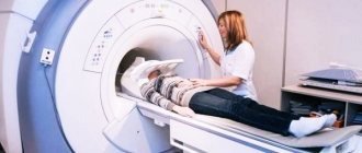
- CT scan of the brain, tomographic, axial - with and without contrast enhancement.
CT helps to see the full picture and determine:
- Severity of injury;
- Fabric density;
- Degree of swelling;
- Petechial hemorrhages;
- Concussion focus;
- Detritus zone, etc.
This is necessary for the doctor to determine the severity and further course of therapy.
For example, a bruise of moderate severity gives a tomogram of lesions with a reduced density, or an increased density in the case of a hemorrhagic type. This allows you to determine the doctor’s further actions.
Also, CT is the main diagnostic method during treatment - to analyze the dynamics of lesion reduction and determine the most effective treatment methods.
Differences from concussion
When diagnosing, it is important for the doctor to rule out a concussion - after all, the symptoms are often very similar, especially with mild symptoms. Both diseases have the same symptoms, only the forms of their manifestation are different. Table of differences:
| Symptoms | Bruises | Shake |
| Headache | Sharp, strong, long lasting | Dull, background |
| Swelling, hemorrhages | Strongly manifested | No |
| Loss of consciousness | Long lasting, up to several days | Short-term, up to several minutes |
| Consciousness | No clarity | Conscious |
| Amnesia | Long lasting | Short term |
| Temperature | Increases to 40 degrees | No |
| Cardiac activity | Problems | No |
| Involuntary eye movement | Intensive | Maybe, but not pronounced |
A professional will never confuse these two completely different diseases.
Possible symptoms of a concussion
· Brief confusion or loss of consciousness. With a strong blow, the moment of injury disappears from memory.
· Dizziness even at rest, and when turning, bending or other changes in body position, the symptom intensifies.
· Severe headache, nausea and vomiting.
· Double vision, inability to concentrate on one point.
· Increased sensitivity to light and sounds.
· Impaired coordination of movements.
· Reaction inhibition—the victim gives an answer to the question after some time.
· Pale skin, weakness, sweating.
Important! A concussion is not always accompanied by visible head injuries, so the absence of wounds does not exclude brain injury.
Head treatment methods
After diagnosis, the doctor determines which method is most effective to treat a particular patient - on a strictly individual basis, taking into account a number of factors:
- The severity of the injury;
- Causes of occurrence;
- Duration of illness;
- Patient's age;
- Presence of concomitant diseases;
- Patient physiology;
- Genetic predisposition;
- Allergic reactions to medications, etc.
Based on the CT readings and general history, the doctor can determine further types of treatment. Table of main types of therapy:
| Type of treatment | Essence and necessity |
| Conservative | Medicines, physiotherapy, folk remedies, gymnastics (more often in mild to moderate cases) |
| Operating | In severe form, according to indications - in moderate severity |
| Emergency | When the life of the victim is threatened |
| Planned | With mild to moderate severity, when there is no threat to health and life. |
Treatment of concussion
Unlike concussion, mild forms of concussion are treated on an outpatient basis. For moderate and severe concussions, therapy is carried out in a medical facility. Another difference is that concussions are not treated with surgery.
For home therapy, long sleep and rest are enough. Drugs are prescribed depending on the symptoms
During inpatient treatment, in addition to symptomatic medications, the doctor prescribes drugs to normalize the functioning of the vascular system, antioxidants, drugs to restore brain function, sleeping pills, and sedatives. Elderly patients are prescribed medications to prevent the development of sclerosis.
In case of a concussion, full recovery occurs within 2 weeks. If a person's main activity is not physical, he can begin work after 14 days. Unlike a concussion, with mild to moderate bruises, recovery occurs within 60 days. If the injury is severe, recovery takes a year and a half.
If you have no time to hesitate
If a patient is admitted to the hospital with a severe injury, then there is simply no time for additional diagnostic measures - the person must be rescued urgently. Therefore, a CT scan is done and the doctor sees the state of the brain: areas of increased density (blood clots) and decreased density (crushing and swelling). Then emergency surgery may be prescribed.
With extremely severe lesions, the zone of destruction of cerebral tissue goes deep into the subcortical structures . Then the decision about surgery is also made. This method is recommended to eliminate primary damage that was caused by traumatic factors. Surgical therapy is used in 15-20% of cases after moderate and severe brain contusions.
“When treating patients with severe brain contusion, accompanied by suppression of the level of wakefulness to the point of coma, it is necessary to monitor intracranial pressure (ICP)” Research Institute of Emergency Medicine named after. N.V. Sklifosovsky.
Indications for surgery
Surgical intervention is prescribed for a number of factors:
- Brain swelling;
- Increased intracranial pressure;
- Problems with the state of consciousness - being in a soporous or comatose state;
- Slow or impaired functioning of internal organs;
- The volumetric focus of pathology is more than 20 cubic meters. cm.
Types of operations
Before the operation, contraindications are also identified, which can play a decisive role in carrying out surgery or refusing it. But often doctors perform an operation - after all, the patient’s life hangs in the balance. The method of surgical intervention is chosen strictly individually, the main methods are in the table
| View | Description |
| Elimination of the area with crushed tissue and performing osteoplastic craniotomy | Opening of the cranium and maximum accessibility to all brain tissues |
| Removal of the lesion with crushed tissue and decompression craniotomy | A certain area of the brain is opened with excision of the temporal region |
| Decompression trepanation without removing the area with crushed brain tissue | If there is slight crushing of the brain tissue, decompression is performed, which allows the blood pressure to normalize. |
But after any operation, conservative methods of therapy will also be used to correct secondary damage, eliminate complications and speed up recovery.
What is brain contusion and concussion?
Head contusions are often confused with brain contusions. The first is a bruise of the skull bones, in which the brain is not injured. In the second version, he suffered certain mechanical injuries. Brain contusion (contusion) is a mild traumatic brain injury. In case of concussion, the victim is diagnosed with: hemorrhages, swelling, concussion.
A concussion is a common consequence of a bruise. The very definition of “concussion” is vague. There is an opinion that this is tissue damage that occurs under external influences. This pathology occurs after industrial, sports, or automobile injuries.
Bruising is a consequence of road traffic accidents, industrial accidents, and impacts. After bruises, the brain hits the skull bones twice. Often a person experiences combined pathologies - bruise + concussion.
Not all concussions are the result of a contusion. Even a sharp, inaccurate shaking of the head can lead to pathology.
Conservative method of treating brain contusion
Conservative treatment methods described in the table will help eliminate secondary factors (often cerebral ischemia):
| Name of therapy | What's the point |
| Respiratory | With problems of the respiratory system, with a clear decrease in oxygen in the blood; Tracheal intubation is performed using equipment that provides artificial ventilation. |
| Infusion therapy (intravenous infusion) | When the volume of circulating blood decreases, maintenance therapy is required to normalize the pressure within 70 mm Hg. |
| Normalization of intracranial pressure indicators | Basic (removal of factors) and emergency treatment (ICP more than 21 mm Hg) is used - using an intraventricular catheter or a pulmonary hyperventilation procedure; if there is no effect - artificial coma, decompression craniotomy. |
| Neuroprotective treatment | Restoration of brain cells, in particular the gray and white matter of brain tissue, using special drugs with neuroprotective effects. |
Protect brain cells
The basis for the treatment of brain contusions is a neuroprotective method that protects nerve cells from the pathogenic influence of secondary damage, promoting a full recovery and reparative process in the gray and white matter of the brain. For neuroprotective effects, various drugs are prescribed - both in the form of injections and in tablet form:
- Erythropoietin - for moderate and severe bruises, in the form of a solution for intravenous infusion;
- Progesterone - in the form of a solution for intravenous administration;
- Leskol - tablets that eliminate the inflammatory process and improve blood supply to the brain.
Folk remedies - as additional therapy
A brain contusion cannot be cured only with folk remedies; they will help you recover after the main treatment.
Then, after discharge from the hospital, the doctor recommends those traditional medicine methods that will help the patient go through rehabilitation and recovery periods faster and speed up rehabilitation at home. Such therapeutic methods include a number of plants, whose decoctions and teas will help to recover from such a severe injury; there are a number of proven recipes:
- A mixture of motherwort, hawthorn, plantain, elecampane and comfrey in equal quantities is simply poured with boiling water, infused and then drunk according to the schedule and in doses - in accordance with the doctor’s recommendations;
- A decoction of rose hips with the addition of honey and lemon;
- Ginkgo Biloba leaves - simply add to teas or food;
- Mix the fruits of hawthorn and sea buckthorn, add honey - turn into a paste;
- Grind raisins, figs, pistachios, mix - the dose and schedule of administration will be determined by the doctor.
Traditional medicine does not tolerate amateurism, so a doctor’s advice in using this or that recipe is simply necessary.
Brain contusion and other TBIs
At the beginning of the article, it was noted that not all TBIs are concussions, but all concussions are traumatic brain injuries. What does it mean? People often consider all injuries, including bruises, compression of the brain, and intracranial hematoma, to be a “concussion.” Traumatic brain injury is an umbrella term. With a TBI, in addition to a concussion, brain structures, cranial nerves, the pathways along which cerebrospinal fluid moves, as well as vessels that deliver nutrients and oxygen with blood can be damaged.
In addition, it should be borne in mind that not only the blow itself, when the brain is damaged at the site of application, can be dangerous for the victim, but also the counter-blow coming from vibrations of the cerebrospinal fluid or from the impact on the processes of the dura mater. Thus, not only the cerebral hemispheres may be damaged, but also the trunk, in which the centers responsible for the activities of many important organs and systems are localized, and metabolic processes will be disrupted. To help the reader correctly assess the situation and navigate such diagnoses if necessary, we will try to briefly dwell on other TBIs:
- Brain contusion, which, unlike a concussion, in addition to general cerebral symptoms, gives local and focal symptoms, depending on the location of the contusion. Brain contusion has 3 degrees of severity, victims with mild and moderate severity are sent to neurosurgical departments, and with grade 3 they are subject to hospitalization in hospitals with intensive care, resuscitation and neurosurgery departments.
- Compression of the brain, as a rule, occurs against the background of severe contusion of the brain and is usually a consequence of the formation of an intracranial hematoma. It manifests itself as psychomotor agitation, an increase in cerebral symptoms, and the development of convulsive syndrome.
- An intracranial hematoma requires urgent surgical intervention in the neurosurgery department. It can manifest itself some time after the injury, which is why seemingly well-being after a TBI does not actually give grounds for peace of mind. It is this symptom, called a light gap
, that is considered an important and insidious sign of a hematoma, and its underestimation is fraught with the development of life-threatening consequences for the victim.
Of course, the therapeutic approach to conditions of this kind is markedly different from the treatment of concussion:
The victim requires not only emergency hospitalization, but also the immediate start of all measures, including surgery, if an intracranial hematoma is diagnosed, which can “deceive” both those around him and the doctor of the arriving ambulance team.
Often the bright period that occurs immediately after the injury is misleading (the person has come to his senses and claims that he feels normal). The thing is that post-traumatic intracranial hematoma can occur at the initial stage without much suffering to the brain, especially if the source of bleeding is venous (when bleeding from an arterial vessel, the light interval lasts minutes). Intensive increase in symptoms of respiratory and vascular disorders, development of mental disorders,
decrease
in heart rate against the background of an increase in blood pressure increases suspicion of an intracranial hematoma, so the patient should under no circumstances be left without hospitalization.
typical areas of hemorrhage and hematoma formation due to head injuries or hemorrhagic strokes
Traumatic brain injury is a common occurrence in our lives, because there are so many dangers around. Often it is limited to a mild degree - a concussion, which, however, does not allow you to relax. You should always keep in mind the possibility of hidden damage and the development of serious complications. Ignorance and underestimation of the insidiousness of TBI can become a tragic mistake that interrupts someone’s life, therefore, in all cases of head injuries, the patient should not be left without attention and help, even if he confidently claims that everything is fine.
Could there be complications?
The degree of complications and the length of the rehabilitation period depend on the severity of the injury. If the bruise is mild, then full recovery will not take long. And after treatment under the supervision of a doctor, patients get rid of the disease forever and completely.
The situation is more complicated with a moderate cerebral contusion; here, development options can develop according to different patterns:
- Full recovery;
- As a consequence - post-traumatic arachnoiditis, post-traumatic hydrocephalus, post-traumatic epilepsy, vegetative-vascular dystonia syndrome, post-traumatic encephalopathy.
A severe degree always develops according to the worst scenario and leaves other types of illness in the form of complications after treatment:
- Brain atrophy;
- Inflammation of the meninges (arachnoiditis, leptomeningitis, pachymeningitis);
- Post-traumatic epilepsy;
- Hydrocephalus with intracranial hypertension;
- Post-traumatic porencephaly (cavities in the thickness of the brain connecting to the ventricles and subarachnoid space);
- CSF cysts;
- Scars in the area of brain tissue and its membranes;
- Liquorhea (leakage of cerebrospinal fluid outward) in the presence of a fracture of the skull bones.
These complications manifest themselves in humans in the form of disorders:
- Propulsion system;
- Speeches;
- Coordination;
- Mental;
- Intellectual, etc.
Such patients may be accompanied by symptoms throughout their lives:
- Headache;
- Convulsions;
- Seizures.
Then you need to register and get a disability group. After all, with such complications, a person cannot fully care for himself and simply live fully.
But still, for patients with severe brain contusions, this is the best prognosis, because according to statistics, about 30-50% of cases of this injury end in death in the acute phase of the disease. But it is worth remembering that “the prognosis for TBI depends on the location and size of the contusion focus, intracranial hypertension, cerebral edema, symptoms of dislocation, hydrocephalus, seizures, and secondary cerebral ischemia.” Medical portal for doctors uMEDp.
Clinical picture of mild traumatic brain injury
Signs of TBI are not always present all together and give a clear clinical picture. In general, the symptoms of a concussion depend on the severity of the condition and include:
- Lethargy, confusion, stupor, lack of concentration.
- Possible (but not obligatory) loss of consciousness lasting from a few seconds to hours and days. Moreover, according to Western experts, the duration of a comatose state should not exceed 6 hours, only then can one count on a favorable prognosis. Otherwise, it becomes obvious that there was some damage to the brain tissue, and this is a different diagnosis and other consequences.
- Nausea, which is often accompanied by vomiting.
- Dizziness, headaches, tinnitus, impaired coordination of movements.
- Paleness of the skin of the face, followed by hyperemia (“vasomotor play”).
- Brady or tachycardia.
- Pain in the eyes, especially when moving the eyeballs, unpleasant sensations in the temporal areas.
- Amnesia (memory loss), when a person cannot remember the events preceding the blow, the strength of which determines the duration of the period lost from memory. This symptom is not very common, and sometimes requires a long recovery.
Considering that a diagnosis such as a concussion, in itself, is the first and mildest degree of serious pathology, united under the general name “traumatic brain injury,” the modern classification does not provide for dividing this form according to severity separately. However, we can agree that not all blows and bruises occur in the same way, so there are certain varieties that make it possible to determine and convey (rather verbally) the degree of damage, which is sometimes used by doctors and quite often patients:
- A mild concussion is avoided without loss of consciousness and amnesia; signs of trouble in the head (lethargy, nausea, severe headache) usually disappear within a quarter of an hour.
- In grade 2 , loss of consciousness is usually absent, but stupor, memory loss and other symptoms occur.
- A severe concussion may be characterized by memory loss and loss of consciousness in combination with the entire set of objective clinical manifestations of pathology, since the patient can make complaints only upon returning to real life (restoration of consciousness).
The damage to health caused by TBI can be significant and depends on what kind of injury the person received: a mild concussion in an adult, with timely first aid and adequate further treatment, can pass and be forgotten. However, it only seems so. Attacks of headaches after a concussion are a common and understandable phenomenon, but the patient himself rarely connects these events with each other, believing that too much time has passed. As for a brain contusion, depending on the severity, it can leave the most serious consequences.
Recovering correctly
There are certain rules of rehabilitation, which include not only proper nutrition and adherence to a daily routine, but also physical therapy and folk remedies. At the hospital, all this is monitored by the attending physician and staff. But when the patient is discharged, he himself, surrounded by his loved ones, must continue to observe the basics of full and effective rehabilitation:
- Eliminate physical activity, over time - only exercise therapy;
- No sudden movements;
- Fresh air - through ventilation or walking;
- Do not overstrain your eyesight (minimize watching TV or computer monitor);
- Follow a diet - the diet should contain a lot of vegetables and fruits rich in vitamins and beneficial microelements;
- Avoid unhealthy foods – sweet, salty, fried, smoked, fatty, etc.;
- Take vitamin complexes;
- Give up addictions - tobacco, alcohol;
- Work with a speech therapist or psychologist - on the recommendation of a doctor;
- Maintain a daily routine;
- On the recommendation of a doctor - physical therapy;
- Don't worry - more positive emotions.
What physical procedures can be prescribed?
To make recovery after a brain injury more active and progressive, the doctor may prescribe physical therapy:
- Electrophoresis;
- UHF therapy;
- Galvanization of the brain;
- Laser therapy;
- DMV therapy
- Air baths.
The prescription of physiotherapy is strictly individual and only upon the direction of a doctor. After all, for many patients, for example with a severe form of the disease, any physical procedure is simply contraindicated.
If, nevertheless, the procedure is prescribed and after such therapy the condition worsens, then you should inform the doctor, he will change the method or eliminate physical treatment altogether.
Therapeutic gymnastics - to help a recovering person
Doctors often prescribe a set of exercises for patients discharged from the hospital for the intermediate period of TBI, with certain recommendations - for example, doing gymnastics with healthy limbs actively, and passively with affected limbs. Always start doing exercises after massaging your body muscles.
Here are the main complexes for the fastest rehabilitation and recovery:
- In a lying position : arms along the body, inhale – reach out to the shoulder joints with your hands, without lifting your elbows from the horizontal surface, pull your toes down, exhale – stretch out your arms and relax;
- Lying down: inhale - raise your forearms and hands up (arms should be straightened at the wrist joint), foot - towards you, exhale - previous position and relaxation;
- Inhale - hands on the chest, legs bent at the knees, exhale - straighten the limbs;
- We raise our arms one by one - perpendicular to the body;
- Inhale - we pull the bent leg to the chest, clasping it with our hands, exhale - we lie down in the original state - arms along the body, legs extended;
- We move our straightened legs to the side one by one;
- We alternately move our arms to the sides - first at a slight angle, then increase the amplitude;
- Sitting with your legs down: inhale - move your hand to the side, turn your head behind it, exhale - lower your hand, do the same with the other hand;
- While sitting, we spread our arms to the sides, head straight, sit straight for a few seconds, maintaining balance, the time should be slightly increased as we do the exercise;
- Sitting and holding the edge of the bed with your hands, inhale - stretch one leg parallel to the floor, exhale - lower the leg, do the same with the other leg;
- While sitting, place your hands on your shoulder girdles and perform rotational movements in the shoulder joints - with both hands at once.
Usually, all these exercises must be repeated 3-4 times at first, gradually increasing the number - up to 8-10. But it is still better to hear the advice of your doctor. After all, each patient is individual and requires his own level of physical activity.
Even with impaired coordination, the presence of paresis of the limbs, and balance disorders, these exercise therapy complexes bring significant results, but they must be done constantly and correctly.
No matter how severe the injury and no matter the degree of brain contusion, the patient himself needs to know what is important:
- See a doctor promptly;
- Comply with all his requirements and recommendations;
- Don't be lazy and don't get discouraged;
- Believe that recovery is inevitable.
In other words, even with severe forms, the patient himself must believe in a good outcome and help doctors achieve a positive treatment result.
Brain contusion
Mild UGM
accompanied by loss of consciousness for up to tens of minutes. Then there is moderate stupor, drowsiness, and there may be incomplete orientation in time and in the environment. Victims complain of constant cephalalgia (headache), weakness, nausea, and dizziness. There is vomiting that does not provide relief, possibly multiple times. Amnesia is observed: the patient does not remember the events preceding the TBI (retrograde amnesia) and for some time after the injury cannot remember what happens to him (anterograde amnesia). Tachycardia or, conversely, bradycardia often develops, and less commonly, arterial hypertension.
In the neurological status: anisocoria, nystagmus, asymmetry of tendon reflexes, unexpressed meningeal symptom complex, there may be mild hemiparesis. When UHM is accompanied by subarachnoid hemorrhage, the meningeal symptom complex is pronounced. With a mild injury, all of these manifestations regress within a period of 2 to 3 weeks.
UGM of moderate degree
manifests itself in an unconscious state for tens of minutes to 4-5 hours. When consciousness is restored, intense cephalgia, repeated vomiting, con-, antero- and retrograde amnesia are observed. Amnesia, moderate or profound stupor, and disorientation may persist for up to several days. Possible mental disorders. Often there is low-grade fever, bradycardia or tachycardia, arterial hypertension, and rapid breathing. The neurological status reveals focal symptoms, varying depending on the location of the bruise area. As a rule, hemiparesis and hemihypesthesia, speech disorders (motor aphasia), anisocoria and oculomotor disorders are observed of varying severity. Typically, these symptoms gradually disappear 4-6 weeks after TBI.
Severe UGM
characterized by a longer duration of unconsciousness (up to several weeks). Motor agitation often occurs. Severe brain contusion occurs with dysfunction of vital systems: arterial hypotension or hypertension, tachy- or bradyarrhythmia, respiratory rhythm disturbance due to tachypnea. In the initial period after TBI, brainstem symptoms dominate: tonic nystagmus, bilateral ptosis and mydriasis, decerebrate rigidity, dysphagia, bilateral foot pathological reflexes, symmetrical hypo- or hyperreflexia. Against this background, signs of damage to the hemispheres are revealed: hemiparesis, hemihypesthesia, oral automatism, etc. Hyperthermia up to 41°C and convulsive paroxysms are possible. Neurological symptoms have a long course and do not fully regress. Mental and/or neurological changes of varying severity remain as persistent residual consequences of TBI.


