What is neurosonography?
Neurosonography of the brain of newborns is a procedure associated with the examination of the brain. This examination replaced the MRI, which was carried out earlier.
This is due to a huge number of contraindications for MRI:
- Having allergies.
- Pituitary gland diseases.
- Early pregnancy.
- Presence of pacemakers and other implants.
That is why doctors tried not to prescribe MRI for babies and older children. Over time, an alternative to MRI was found: neurosonography.
Thanks to this procedure you can explore:
- Spine.
- Spinal cord.
- Skull bones.
- Scalp.
Thanks to neurosonography, it is possible to identify blood supply disorders, tumors or hernias, spinal injuries, as well as head injuries, and inflammation.
Neurosonography is of 3 types:
- Transcranial. This type of neurosonography helps to examine the brain not only in children, but also in adults. How well the state of the brain can be viewed even through the human cranial bones.
- Transfontanelle. The most common way to conduct research. The examination is carried out using a special sensor through the child’s fontanel. The disadvantage of this study is that after the birth of a child, the fontanel overgrows over time, and examination by the transfontanelle method soon becomes impossible.
- Transcranial-transfontanelle. The most informative of these types is transcranial-transfontanelle. This type of study is used for children under 1 year of age.
Preparation and indications
Reliable information about the state of the brain is transmitted to a special device through sensors that are installed on certain anatomical structures found only in children - the fontanelles. They are a dense membrane between the bone bases, which allows the baby's head to shrink somewhat as it passes through the natural tract during childbirth. By the age of one year, the membrane is completely ossified and becomes inseparable from the entire bone skull.
The presence of fontanelles makes it possible to perform an ultrasound of the brain in an infant with a maximum assessment of brain activity. If the pressure in the cranial cavity increases, the structures help to kill some of the excess fluid through sweating.
But most fontanels close almost immediately after performing their main physiological function, and each full-term baby has only one large bony cleft. Somewhat less often, there remains another small fontanel at a short distance from the large one. They can be felt by palpation if you run your hand from the crown of the baby to the forehead. They are usually soft and may throb or appear as small bruises when the child examines them.
Ultrasound of the NSG performed through the fontanelles does not require a preparatory stage in infants. The preparatory nuances are only as follows:
- This procedure is performed on children in any state: during sleep or active wakefulness, when the child falls asleep or even cries. This does not distort the research results or make it difficult to decipher them.
- It is recommended to conduct an ultrasound examination an hour and a half after feeding. There is no need to deviate from the diet, and there is no need to feed the baby or the mother to feed herself with certain products or milk substitutes.
An ultrasound of the child's head is performed according to strict indications. The study is prescribed by a pediatrician or other pediatric specialist for certain conditions of infants to exclude or confirm a particular diagnosis.
The following categories of children undergo ultrasound diagnostics of the brain:
- Infants born during premature birth, namely before the 36th week of gestation.
- Infants born at term or earlier, for some reason receiving an Apgar score of less than 7/7. Preventive ultrasound is also performed in children who cry after some time from birth.
- Full-term babies whose birth weight has not reached 2800 grams.
- Ultrasound of the newborn's head is indicated in cases where there are signs of damage to the central nervous system or peripheral ganglia acquired during childbirth or during fetal development. It is also possible to clarify the cause of cerebral hernias or protrusions of the skull and meninges using an ultrasound wave.
- With pronounced defects of the external structure, especially with the presence of an additional finger or several rudimentary coccygeal processes, with an abnormal shape of the ears.
- In case of sudden convulsive syndrome in a baby immediately after childbirth or within 24 hours after it.
- In case of some pathological conditions on the part of the mother: in particular, with rapid or, on the contrary, prolonged labor, a long anhydrous period, with chronic or acute infection during pregnancy, with Rh incompatibility.
These indications recommend performing an ultrasound of the brain of newborns immediately when they are stabilized from the moment of one or another type of delivery. There are also recommendations for carrying out the procedure a little later: for children at 1 month of life and later than 3 months.
Ultrasound in infants who have reached the age of 1 month is performed when:
- Carrying out childbirth artificially (during a caesarean section) or when using any aid to extract the child (using a vacuum extractor, obstetric forceps).
- If a child has a non-standard head shape, parents detect excessive growth of the head relative to the body.
- At 1 month of life, children diagnosed with birth trauma, cerebral palsy, or convulsive syndrome are subject to a repeat ultrasound of the head.
- For other fetal developmental defects: strabismus, paresis of the limbs, underdevelopment of internal organs.
- In case of unidentified tearfulness or restlessness of the child, with frequent regurgitation and the inability to choose the type of food.
- It is recommended to repeat an ultrasound in the first month of life for children born premature at birth, as well as for babies who have gained 2800 grams by the time of birth.
An ultrasound scan of the head is possible up to one year of the child’s life, until the fontanelles have completely closed. After 3 months the procedure is performed:
- To monitor various neurological abnormalities, establish the effect of treatment of birth trauma, intrauterine infection.
- After a child has had meningitis or other inflammation of the meninges.
- Children with an inferior set of chromosomes or other genetic deficiency.
- In cases of head injuries, intracranial hemorrhages, cysts and abscesses in the brain that occurred before the age of one year.
If ultrasound in newborns and infants under one year of age does not make it possible to establish a diagnosis, MRI under general anesthesia is recommended.
Why is ultrasound performed on newborns and infants?
As a rule, childbirth is a rather complex and traumatic process. It is not uncommon for a child to suffer a brain injury during childbirth. It is impossible to determine this injury without special equipment.
These injuries can subsequently seriously affect the mental and physiological development of the child. Early examination and identification of injury will help to begin treatment on time and avoid serious consequences.
Ultrasound can detect the following pathologies:
- tumors;
- hemorrhage;
- inflammatory processes;
- hydrocephalus.
How often can neurosonography be done on an infant?
Neurosonography of the brain of newborns is a safe procedure. The examination is carried out painlessly, within a few minutes. At the same time, it gives a complete picture of the baby’s brain structures.
As a result, the examination can be carried out without restrictions on the number of procedures. Whether the test should be used and how often it should be done is decided by either the attending physician or the pediatrician. In practice, to determine the health status of a child, once a week is enough.
What does neurosonography of a baby show?
Using neurosonography, you can obtain comprehensive information about the state of the child’s brain.
This examination can show:
- Infant brain damage.
- Presence of a cyst or tumor.
- Clear reflection of convolutions.
- Density of the ventricles of the brain and their size.
- The content of fluid that is located between the cerebral hemispheres.
- A general picture of brain development.
What does the NSG show?
Neurosonography shows the condition of brain tissue and the circulatory system and gives the specialist the opportunity to assess the size and shape of parts of the brain. Using ultrasound, a specialist determines the condition of the membranes of the child’s brain, the amount of cerebrospinal fluid, the state of the vascular system, the degree of formation of brain matter, etc. In addition, ultrasound can track how symmetrical the brain structures are in children, the size of the ventricles of the brain, and assess the development of the cerebellum , detect ischemic areas.
Indications for ultrasound for infants of different ages
Ultrasound is recommended for absolutely all children. Parents have the right to refuse an examination, but it is best not to do this, because it is impossible to confidently say without a full examination that the child does not have any pathologies and is completely healthy.
There are a number of “direct” indications for performing an ultrasound of a child’s brain.
Neurosonography is required if:
- The child was born premature.
- There is a possibility of chromosomal abnormalities.
- Prolonged oxygen starvation of the child.
- The birth occurred with complications (for example: incorrect position of the fetus, trauma during childbirth).
- A score of 7 or less according to the Algar scale (the Algar scale is a rapid assessment system for the general condition of an infant).
- The fontanel either sinks or, on the contrary, protrudes.
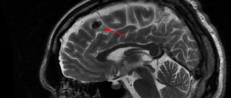
Is the cerebellum the main part of the brain?
Scientists have found that the cerebellum plays a critical role in all higher functions of the brain - movement, attention, thinking, planning and decision-making, writes sciencedaily.com with reference to Neuron.
Scientists usually don't pay attention to the cerebellum. The cerebellum, inconveniently located at the bottom of the brain, was long thought to control only movement. Typically, researchers are interested in higher brain functions.
But scientists at Washington University School of Medicine in St. Louis show that ignoring the cerebellum is a mistake. The results obtained show that the cerebellum is also involved in higher brain functions.
“The biggest surprise for me was the discovery that 80 percent of the cerebellum is dedicated to smart things,” said senior author Nico Dosenbach, MD, assistant professor of neurology and internal medicine and pediatrics. “Everyone thought that the cerebellum was responsible for movement. If your cerebellum is damaged, you can't move smoothly—your hand jerks violently when you try to reach for something. Our research convincingly proves that the cerebellum is responsible not only for the quality of movement, but also for the state of our thoughts, structuring them, correcting them, improving them.”
Dosenbach is one of the founders of the Midnight Scan Club, a group of neuroscientists at the University of Washington who took turns studying their own brains in an MRI scanner at night to generate massive amounts of high-quality data for research. Previous analysis of Midnight Scan Club data showed that measuring functional brain connectivity using functional magnetic resonance imaging can reliably reveal fundamental differences in individual brain function.
Postdoctoral fellow and first author Scott Marek decided to apply a similar analysis to the cerebellum. The problem is that MRIs tend to have a hard time showing the lower floor of the brain, so the images of the cerebellum are of poor quality. But Midnight Scan Club member Marek was able to scan the brains of each of 10 people for more than 10 hours, which was more than enough time to take a serious look at the cerebellum.
For the well-studied cerebral cortex, a map has been built showing how distant areas of the brain are connected into networks that control vision, attention, speech, and movement. Using cortical networks as a template, Marek could identify similar networks in the cerebellum. It is noteworthy that sensory networks - vision, hearing, touch - are absent, and only 20 percent of the cerebellum is devoted to movement - about the same part as in the cerebral cortex. The remaining 80 percent is occupied by networks involved in higher-order cognition: the attention network; a network associated with dreams, memories, idle thoughts, and two networks that control executive functions such as decision making and planning.
“There are a lot of executive function networks in the cerebellum,” Marek said. “We need to completely change our understanding of the cerebellum as controlling movement and see it actively controlling higher levels of cognition.”
The researchers measured the timing of brain activity and found that the cerebellum was always the last step in the neurological chain. Before signals are sent to the cerebellum, they are received through sensory systems and processed in the intermediary networks of the cerebral cortex. According to the researchers, signals undergo final quality checks in the cerebellum and are then sent back to the cerebral cortex for implementation.
“If you imagine an assembly line, the cerebellum is the person who checks the car at the end and says, 'This is good, we'll sell it,' or 'This has a dent, we should go back and fix it,'” Dosenbach said. “This is where everything you think and do becomes more refined and quality controlled.”
People with cerebellar damage are known to experience uncoordinated actions, unsteady gait, slurred speech, and difficulty with fine motor skills such as eating. The cerebellum is also quite sensitive to alcohol, which is one of the causes of unstable gait in people who have consumed too many drinks. But now new evidence may help explain why drunk people have difficulty thinking - the cerebellum fails to control not only motor function, but also executive function.
Marek also conducted individual network analyzes on 10 people. He found that although brain functions were arranged in roughly the same pattern in each cerebellum, there were enough individual differences to distinguish brain scans from two participants from each other. Researchers are now studying how these individual differences in cerebellar networks relate to intelligence, behavior, personality traits such as adaptability, or psychiatric disorders.
“Many people who study the relationship between brain function and behavior simply ignore the cerebellum,” Dosenbach said. “They tear off this data and throw it away because they don’t know what to do with it. But the cerebellum has four times more neurons than the cerebral cortex, so if you refuse to study it, you've already shot yourself in the foot before you start. Imaging the entire human brain should help us understand how it all works. You can’t see how the whole chain works together if you’re missing a major part of it.”
[Photo: sciencedaily.com, friedreichsataxinews.com]
From birth to 2 months
For babies under 2 months of age, neurosonography is performed in the following cases:
- Hyper-reactivity.
- Signs of cerebral palsy.
- The birth was protracted.
- Sepsis.
- Obvious defects of the brain box.
- Cerebral ischemia.
- Childbirth using cesarean section.
- Apert syndrome.
- Developmental anomaly associated with genetics.
- Suspicion of increased intracranial pressure.
In these cases, it is also possible to hospitalize the child and mother during the examination.
From 2 months to six months
There are cases when no pathologies were detected during neurosonography. And after 2-3 months the picture changes dramatically. This is due to the fact that not all pathologies manifest themselves in the first 2 months after the birth of a child.
For older children and up to six months, examination may be prescribed for the following reasons:
- Cramps.
- High nervous excitability.
- The size of the head increases rapidly.
- The infection was able to penetrate the child’s brain in the very first months of his life.
- Muscle weakness.
- Violation of the symmetry of the brain structure.
- There is a suspicion of strabismus.
- Suspicion of high intracranial pressure.
- Suspicion of Cerebral Palsy.
At the end of the examination, the doctor prescribes special measures to improve the child’s condition, and measures are also taken to bring the child’s brain function to the required state.
Main indications for brain NSG
An ultrasound of the child’s brain is prescribed according to the following recommendations:
- birth before 32 weeks of gestation and weight less than 1500 g;
- low Apgar score;
- neurological disorders;
- convulsions;
- suspected periventricular leukomalacia or hemorrhage.
Indications for examination of newborns may include rapid or prolonged labor, hypoxia and anhydrous periods, birth injuries and cesarean section, abnormal skull shape, the use of obstetric manipulations during childbirth, infections and hemolytic disease. It is imperative to do a brain examination in children with encephalopathies, congenital defects and genetic disorders. Some anomalies appear only after a year.
Indications for children up to 2 months
During the first months of life, the child is seen by a neurologist who checks the integration of primitive reflexes. The main one is stepping, when the baby moves his legs while bending his body forward. The reflex normally disappears by 2 months. A neurologist checks the child’s development for age. Suspicion of pathology of the nervous system arises in the following cases:
- lack of physiological flexion of arms and legs (depression of muscle tone);
- increased reflexes (extensor, head tilt);
- weakly expressed neck-tonic reflexes, crawling, grasping.
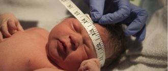
The main symptom of hydrocephalus is a significant increase in head circumference by more than 3 cm from the norm of 39 cm in 2 months. The doctor determines the tension of the fontanel and the condition of the cranial sutures. Often, a baby's spitting up after eating is considered a deviation, but this is only relevant if the sucking reflex is impaired. After examination and suspected pathology, sonography may be prescribed.
Indications for children up to six months
During the first six months of development, the child learns to roll over, raise his head, reach out to a toy, and sit with support. The absence of one of the stages is sometimes regarded as an indication for examination, but in the presence of other factors:
- decreased muscle tone;
- the presence of pathological reflexes for 6 months (automatic walking, crawling, Moro and Galant reflexes, grasping reflex);
- change in the shape of the skull, strabismus;
- lack of response to parents, external stimuli.
Neurosonography of the infant is usually planned in advance if changes are detected in the early neonatal period.
Indications for adults
Neurosonographs help identify various brain diseases at an early stage:
- strokes;
- aneurysms;
- Alzheimer's disease;
- brain tumors;
- epilepsy.
Diagnostics recognizes spinal canal stenosis, myelitis, paralysis, multiple sclerosis.
Are there any contraindications to the study?
There are no contraindications to the study as such. This is explained by the fact that no dangerous drugs are used during the examination. The procedure is painless and without any harm to the child’s health.
With rare exceptions, doctors may refuse to perform neurosonography. For example, a child does not behave calmly during the procedure or a large area of the scalp or neck is damaged.
This procedure can be carried out repeatedly until the fontanel is completely overgrown.
Neurosonography is carried out until the child’s fontanelle closes. As a rule, this period is from birth to 12 months. Next, the procedure is carried out using the transcranial ultrasound method.
Brain defects in infants available with neurosonography
Neurosonography of the brain of newborns is a method through which it is possible to identify almost all pathological changes in the brain, in its vascular bed and in the ventricular system.
Most often, during examination, the following defects are found in infants:
- Subependymal cysts - These are liquid formations. As a rule, they arise near the ventricles of the brain. The child does not have any anxiety because of them. But this pathology should not be ignored. Cysts appear as a result of hemorrhage. Over time, the tumors may begin to grow.
Neurosonography of the brain of newborns with a cyst - Hemorrhage. Hemorrhage can be of two types - parenchymal and intraventricular. Both types of pathology are dangerous for a child. Most often, hemorrhage occurs in premature babies. What awaits the child in the future depends on the degree of hemorrhage. With minor hemorrhage, no complications should be expected in the future. A completely different picture with severe hemorrhage. In the future, the child may develop neurological disorders.
- Choroid plexus cysts. The found pathology does not pose any danger. Absolutely all babies have such cysts. The only difference is that in some children the disease occurs in the womb, while in others it occurs only after birth. Treatment for choroid plexus cysts is not prescribed. They go away gradually on their own, without causing further complications.
- Hydrocephalus. This disease is also called “dropsy of the brain.” With this pathology, a large amount of cerebrospinal fluid appears in the subarachnoid space, as well as in the cerebral ventricles. Liquor is a fluid that constantly circulates in the ventricles and in the subarachnoid brain space. Because of this, the liquor spaces begin to expand greatly. This disease is treated mainly with surgery.
- Porenciphaly. This disease is characterized by the appearance of cysts in the middle parts of the hemispheres. They are associated with the subarachnoid space and the ventricles of the brain. They are round in shape, and their walls are clearly defined.
- Arachnoid cysts. Initially, the size of the cysts is no more than 3 cm. Neoplasms of this size may not bother the child, but over time, such cysts may begin to grow. The larger the size of the cyst, the more it compresses the structure of the brain. In the future, this can lead to serious neurological diseases. That is why the child is under strict supervision of specialists. Treatment for this pathology is mandatory.
- When examining a child in the first days of his life, tumors are often discovered. As a rule, they are located in the midline of the brain (cerebellar belly, 3rd ventricle and pineal gland). Tumors can lead to quite serious consequences: hemorrhage, ventricular asymmetry and cystic brain damage. It is worth noting that neurosonography can only detect a tumor, and not determine its type.
- Hypoxic-ischemic brain damage. This pathology occurs due to a lack of oxygen. For some, it occurs during childbirth, for others, in the womb. Hypoxic-ischemic damage, which occurs in a mild form, does not require anything other than monitoring the child. Over time, the pathology goes away on its own without leaving any serious consequences. The picture is completely different in severe forms of the disease. In these cases, brain cells die. Further, zones of leukomalacia appear in the affected areas.
In addition to neurosonography, additional examinations may be prescribed (for example, an examination by a neurologist).
What does a brain ultrasound show?
Using neurosonography, such pathologies are diagnosed as follows:
- increased intracranial pressure and hydrocephalus;
- congenital malformations (Dandy-Walker anomaly, Arnold-Chiari anomaly, porencephaly and others);
- tumors and cysts;
- complications from infections and injuries (cerebral abscess, subdural hygroma);
- hemorrhages, aneurysms;
- cerebral ischemia, and others.
Ultrasound examination of the brain will help identify intracranial changes, which in the future can lead to the development of very dangerous diseases, for example, epilepsy or cerebral palsy. Enlargement of the ventricles of the brain detected by ultrasound does not always indicate the presence of abnormalities. Gradually, by the age of 1 year, the situation may return to normal. However, if this symptom is detected, constant monitoring by a neurologist is necessary.
Ultrasound of the brain at the Scandinavia clinic
Neurosonography can be done today in many children's medical centers. However, it is obvious that without consulting highly professional ultrasound diagnostic specialists and neurologists, this study is ineffective. Correct interpretation of ultrasound results will save you from unnecessary worries and eliminate the possibility of making an incorrect diagnosis. At the Scandinavia clinic, experienced doctors working in close cooperation with each other are ready to help your child. Modern, high-tech equipment is used to conduct ultrasound examinations. You can sign up for the procedure by calling: +7
or leaving a message on the site.
Preparing the baby for the procedure
There are no special measures when preparing a baby for neurosonography.
The most important thing is that the child eats and is not thirsty before the procedure - this will avoid whims during the examination.
If the child falls asleep, there is no need to wake him up. The baby will move less and the procedure will go much faster.
For the procedure, you should take a diaper on which you can lay the baby. Do not apply ointments or creams before neurosonography. This will worsen the contact of the device’s sensor with the surface of the head, which will not allow obtaining the most accurate results.
Ultrasound process
Neurosonography of the brain of newborns is most often done through the baby’s fontanel. This area is located between the frontal and also the parietal bone. In addition to the anterior fontanel, ultrasound can be done through a special occipital foramen of the child’s head and small lateral temporal fontanelles.
The neurosonography device is no different from a conventional ultrasound machine. It includes:
- Sensor _ With its help, all manipulations during the examination are carried out. There are two types of sensors:
- Sensor with purity up to 6 MHz. This type of sensor is used for children under 2 months of age.
- Sensor with 2 MHz purity. This type of sensor is used for older children.
- Monitor.
Before the procedure, the child is placed on the couch, having first laid a diaper on it. During the procedure, the mother has the right to be present and hold the child’s head if necessary. A special gel is applied to the sensor, after which the doctor moves it over the child’s head. At this time, the impulses that the device receives are displayed on the screen in the form of a moving picture.
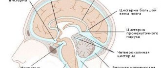
During an ultrasound, the doctor turns his attention to the cerebral ventricles, the cavities of the transparent septum, and the cisterns. Examination of the tank is very important for a doctor. This is due to the fact that her condition shows disturbances in the development of the posterior fossa of the skull.
The study lasts no more than 10 minutes. At the end of the examination, the child’s head should be gently wiped with a cloth to remove any remaining cream. The results of neurosonography will be ready after a few minutes.
Indications for ultrasound
Ultrasound of the newborn’s brain, or neurosonography, is a reliable and safe way to obtain informative data about the structure and functioning of both the brain itself and adjacent structures located in the cranium. The basic methodology for holding the event has been regularly used for the past two decades.
According to the official recommendations of the WHO (World Health Organization), a routine ultrasound of the brain in an infant is carried out at the age of one to one and a half months as part of a comprehensive examination of his health.
However, in some cases, ultrasound diagnostics can be performed repeatedly or more often than once a year, taking into account the following circumstances:
- Any form of prematurity. Regardless of the period of prematurity, newborn children undergo a mandatory procedure for ultrasound examination of the brain;
- Intrauterine hypotrophy. If the baby was born with low body weight, he is prescribed an ultrasound;
- Suspicion of intrauterine infection. Neurosonography is performed in the first weeks after birth;
- Pathologies of the structure of the head or entire structures, the presence of birth injuries. In this case, ultrasound of the brain helps to more objectively assess the condition of individual parts of the brain and develop the necessary principles for comprehensively overcoming the problem;
- Newborns with symptoms of neurological pathologies, as well as those who have experienced hypoxia during fetal development. If there are secondary signs of neurology in the form of seizures, delayed psychomotor development, weakness in the limbs and other problems, as well as taking into account previous oxygen starvation, the neurosonography procedure can be performed on a regular basis, on average once a month, to monitor the dynamics of potential pathological changes.
Explanation of research indicators, table of norms
Neurosonography of the brain has certain data that is deciphered based on specific indicators and parameters. Standard indicators that should be obtained after examining healthy newborns and older children are prescribed in a special table of norms.
It is this that the sonologist will focus on when preparing the results of the study.
Externally, this table looks like this:
| The object in question. | Normal for a newborn. | The norm for a child aged 1 to 3 months. | Normal for a child aged 6 months. |
| Large tank | 44,9 (+/-4,5) | No more than 6 mm. | 82,1 (+/-12,7) |
| Lateral ventricles | Front: 1.5 mm (+/-0.5 mm). Occipital: maximum 1.5 cm. | Front: no more than 2 mm. Occipital: no more than 1.5 cm. | 64,7 (+/-12,8) |
| 3rd stomach | 4.5 mm (+/-0.5 mm) | No more than 5 mm. | 4,8 (+/-1,2) |
| Subarachnoid space | No more than 3 mm. | No more than 2 mm | |
| Brain Cloak | 29,4 (+/-5,7) | 40,1 (+/-2,5) | 46,2 (+/-6,4) |
Small deviations from the indicators in this table should not frighten the child’s parents. Often, small deviations are associated with device errors.
In addition to the above data, the doctor must indicate the symmetrical or asymmetrical shape of the brain tissue.
If there are no deviations from the norms, then, as a rule, the furrows and gyri will be shown very clearly on the screen of the device.
In cases where everything is fine with the ventricles of the brain and there are no abnormalities, the sonologist must make a note that the ventricles are homogeneous and do not have any inclusions. If in the results, when describing the ventricles, the term “flakes” is written, this means that an area of hemorrhage has been found.
In good condition, the space between the hemispheres should not be filled with any liquid. And the blood vessels of the brain must have a homogeneous structure.
The shape of the tentorium of the cerebellum in the absence of any abnormalities can be of two types:
- symmetrical;
- trapezoidal.
In addition, the conclusion of neurosonography uses the following norm for a healthy child:
- The size of the body of the cerebral ventricle should be from 2 mm to 4 mm.
- The size of the gap between the cerebral hemispheres is 2 mm.
When drawing up a conclusion, the doctor pays attention to all aspects of the birth of the child’s mother, namely:
- Baby's weight after birth.
- Duration of labor.
- What injuries were sustained during childbirth?
- How was the birth, were there any complications?
- Did the child have oxygen deprivation?
Interpretation of brain ultrasound results in newborns
As part of an ultrasound examination of the brain in newborns and children up to 3 months, an experienced diagnostician describes the visible structures of the organ and its components.
As modern practice shows, normally, in a child of any age, ultrasound parameters of the brain should be symmetrical, homogeneous and correspond to the dynamics of age-related development.
The standard protocol includes the condition, location and shape of the cerebellar structures , the presence of asymmetry of the above-mentioned elements (if any), the presence or absence of fluid in the interhemispheric fissure, and features of the structure of the cerebellar tent.
The condition of the medullary falx, grooves and convolutions of the organ , the symmetry of the ventricles, as well as the potential presence of various formations, from fluid, hematomas of tumors and cysts to general developmental anomalies and organic changes in the structure of the medulla and vascular bundles are also assessed.
Standard norms for indicators in neurosonography of children under 3 months include the following parameters:
- Lateral ventricles of the brain. The anterior horns are up to 4 millimeters, the body is up to 4 millimeters, and the occipital horns are up to 15 millimeters;
- Third ventricle. On average from 3 to 5 millimeters;
- Fourth ventricle. Up to 4 millimeters;
- Interhemispheric fissure. From 3 to 4 millimeters;
- Large tank. Up to 10 millimeters in newborns and up to 6 millimeters in children about 3 months old;
- Subarachnoid space. Regardless of age – up to 3 millimeters.
In general, the structure under study should not contain any inclusions, including fluid, cysts and tumors. In addition, there are normally no hematomas, developmental anomalies, organic lesions or ischemic foci.
Based on the results of complex neurosonography, a specialized specialist filling out the appropriate standard protocol can add the following potential problems, often associated with various pathological processes:
- Violations of the structure of the arachnoid membrane. These are mainly arachnoid cysts. They can be located anywhere in the designated location and grow linearly. It is a cavity structure containing liquid. Most often, this pathology does not resolve on its own: it requires treatment, control, and observation by other specialists;
- Choroid plexus cysts. They are visualized as small or small bubble-type formations, inside of which there is cerebrospinal fluid. In most cases, they are externally asymptomatic, can resolve on their own and do not require any intervention;
- Subependymal formation. They are formed as a result of various birth and postpartum injuries, hemorrhages. They can increase in size, sometimes resolve on their own, but in any case requires control and observation, and in some cases, qualified complex treatment, both conservative and surgical;
- Hydrocephalus. Represents a significant expansion of the ventricles of the brain due to the accumulation of fluid. It is a fairly acute condition that requires treatment;
- Ischemic lesions of vascular structures. In the described context, it represents a decrease in blood supply to a separate area of the brain or tissue due to a weakening or complete cessation of arterial blood flow. The pathology does not disappear on its own and requires proper therapy from specialized specialists;
- Hematomas. Pathology in the general sense is a limited accumulation of blood due to damage to brain tissue with partial rupture of blood vessels. Liquid or coagulated blood forms in the cavities of hematomas. The pathological condition poses a danger to the health and sometimes the life of a small patient and often requires immediate hospitalization and qualified treatment;
- Increased intracranial pressure. In medical practice, the term “hypertension syndrome” is more often used. It can be caused by excessive accumulation of cerebrospinal fluid in the space of the brain or by a displacement of the position of one of the hemispheres due to the influence of foreign formations, from hematomas and tumors to cysts. If such a pathology is detected, it is necessary to contact specialists and carry out comprehensive qualified treatment.
What to do if deviations are identified?
If any abnormalities were identified during the examination, you must immediately contact a neurologist. It will be very good if this doctor is the one who performs the procedure.
Neurosonography of the brain allows the neurologist to determine whether the child needs treatment right now or can be limited to observation. Situations arise when newborns are prescribed a repeat examination in order to eliminate all possible errors.
Author: Vladimir Kharlamov
Article design: Mila Friedan