Diseases of the nervous system occur when the spinal cord or brain, as well as peripheral nerve trunks and endings, are damaged. The nervous system is a very complex mechanism that connects all organs. That is, as a rule, diseases of the nervous system have a colossal negative impact on the entire body. Neurologists diagnose and treat diseases of the nervous system.
Diseases of the nervous system, based on their origin, are divided into:
- vascular;
- infectious;
- chronic;
- progressive;
- hereditary;
- traumatic.
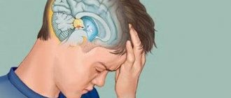
The most common and dangerous are vascular diseases of the nervous system. They are the ones that most often lead to disability or even death. This group includes, among others, such a dangerous disease as stroke. Symptoms of vascular diseases of the nervous system are headache, nausea, vomiting, decreased sensitivity, and changes in movement patterns.
The second common group of diseases of the nervous system are problems that arise after traumatic damage to the nervous system, brain, and nerve endings. This occurs either due to injury or due to compression of nerve endings, such as herniated intervertebral discs. Symptoms include pain in the back and neck, shooting pain in the arm or leg, headaches, and numbness in the limbs.
Diseases of the nervous and vascular systems
Most often, patients come to neurology centers with the following diagnoses:
- Headache
- Migraine
- Dizziness
- Cerebrovascular disorders
- Vegetovascular dystonia (VSD)
- "Manager Syndrome"
- Sciatica (inflammation of the sciatic nerve)
- Encephalopathy and its consequences
- Vertebrobasilar insufficiency
- Inflammation of the occipital nerve
- Neuritis and neuralgia
- Vertebral artery syndrome
- Cognitive disorders (impaired memory and attention)
Important information!
If you have already been diagnosed with diseases such as epilepsy, parkinsonism, stroke in the acute phase, multiple sclerosis and various problems of a similar nature (namely the central nervous system), then you should be recommended TREATMENT IN A HOSPITAL!
Symptoms of nervous system diseases
The body will inform you about disorders in your nervous system with the following “bells”, after which you urgently need to consult a neurologist:
- Back pain;
- Neck pain;
- Lower back pain;
- Headaches (in the occipital, temporal, parietal and other areas);
- "Shots";
- Dizziness;
- Pain or heaviness between the shoulder blades;
- Weakness in arms, legs;
- Chest pain that gets worse with breathing;
- Insomnia;
- Numbness of the arms, legs, fingers;
- Weather dependence (intensification or resumption of symptoms when the weather changes - headache, a feeling of aching in the spine).
Topic 7. METHODS FOR STUDYING THE NERVOUS SYSTEM
Read:- Modern methods of treating uterine fibroids
- I. Immunology. Definition, tasks, methods. History of the development of immunology.
- I. Activities aimed at creating an epidemiological surveillance system
- I. METHODS, APPROACHES AND PROCEDURES FOR DIAGNOSIS AND TREATMENT
- I. Neurogenic tumors from the nervous tissue itself.
- I. Basic examination methods.
- I. Opposing Philosophical Systems
- II) Research methods and symptoms of lesions of III, IV, VI pairs of CN
- II. Additional Methods
- II. Instrumental diagnostic methods
Establishing a neurological diagnosis is based on the results of a patient’s examination, which is carried out according to a certain scheme: - anamnestic data; — clinical and neurological data; — instrumental and laboratory methods.
ANAMNESTIC DATA includes the passport part, complaints, medical history and life history.
Passport part. Last name, first name, patronymic, age, place of residence and study are recorded.
Complaints. Determine the nature of the disorders, their localization, intensity, duration, frequency; factors that increase or decrease the disorder.
History of illness (from Latin anamnesis
- story). The time of appearance of the first symptoms, the nature of their development (sudden, gradual, recurrent), leading signs (pain, paralysis, convulsions), the connection of the disease with previous factors (infections, injuries, hypothermia), the sequence of occurrence and development of symptoms are indicated. Data on treatment and its effectiveness. Hereditary predisposition to the disease is determined.
Anamnesis of life. Find out the peculiarities of pregnancy and childbirth in the mother, type of feeding, development at an early age; At what age did he start walking and talking? Past illnesses, the presence of epileptic seizures, urinary incontinence, sleepwalking, traumatic brain injuries. School performance.
DATA OF CLINICAL-NEUROLOGICAL STUDY
General state. Severity of condition: satisfactory, moderate, severe, extremely severe. Patient position: active (free or forced), passive, can stand, sit independently, with support.
Physical development and condition of internal organs. Body build, height, body weight. Changes in the skeleton, joints. Description of the subcutaneous fat layer, skin and visible mucous membranes (color, presence of wounds, rashes, pigmentation). Lymph nodes and tonsils. Thyroid. Chest shape. Rhythm and frequency of breathing. Heart rate, blood pressure. Condition of the abdominal organs. Body temperature.
State of higher cortical functions. Contact with the patient. State of consciousness: clear, stunned, stupor, coma, delirium, amentia, delirium, hallucinations, agitation. Orientation in time, place. Mental development (age appropriate). Attention and memory (for immediate and distant events). Amnesia. Emotional background: increased irritability, apathy, depression, euphoria, weakness. Suspiciousness, obsessive fears, thoughts, actions.
Attitude to your illness. Assessment of the severity of the condition and life prospects associated with the disease (critical, unreasonably exaggerated fears).
Behavior during research: facial expressions, gestures, manner of presentation (consistent, tendentious, demonstrative).
The patient’s speech (note dysarthria, chanting, monotony, bradylalia, stuttering). Spontaneous speech and speech understanding. Preservation of purposeful actions (praxis), understanding the meaning of visual and auditory stimuli, spatial orientation and topography of parts of one’s body. In the presence of aphasia, apraxia, and agnosia, an in-depth study is carried out according to special schemes.
The functions of the cranial nerves are examined in the following sequence:
I pair – smell;
II pair – visual acuity, color perception, fundus examination;
III, IV, VI pairs - the shape and size of the pupils, their reaction to light; width of palpebral fissures; movements of the eyeballs; the presence of strabismus and double vision;
V pair – facial skin sensitivity; soreness of the nerve exit points on the face; corneal and mandibular reflexes; function of masticatory muscles;
VII pair – movements of facial muscles (raise eyebrows, wrinkle forehead, close eyes, bare teeth, puff out cheeks, whistle), taste;
VIII pair – hearing acuity, air and bone conduction of sound. Determine the presence of tinnitus, dizziness, tolerance to vestibular stress (when traveling in a car, on a ship);
IX, X pair – phonation, swallowing, mobility of the soft palate; sensitivity of the mucous membrane of the soft palate, pharynx; palatal and pharyngeal reflexes;
XI pair – “shrug” of shoulders, turn of head;
XII pair - atrophy and fibrillary twitching of the muscles of the tongue; deviation of the tongue when protruding.
Motor functions. The presence of atrophies, hypertrophies, fibrillary muscle twitching. Volume of active and passive movements, muscle strength and tone. If hyperkinesis is present, indicate its location and nature (amplitude, tempo, rhythm, stereotypy). Precision and smoothness of movements, the presence of clonus, pathological reflexes. Deep and superficial reflexes. Handwriting, walking, coordination tests.
Sensitivity. Presence of pain and paresthesia. They explore superficial, deep and complex types of sensitivity.
Vegetative functions. Local changes in skin temperature and color, trophic changes in the skin (trophic ulcers, hyperkeratosis). Sweating, dermographism, pilomotor, oculocardiac, solar, clinostatic, orthostatic and pupillary reflexes are examined. Find out the presence of fainting, dizziness, urticaria, and bouts of drowsiness.
INSTRUMENTAL AND LABORATORY STUDIES
X-ray of the spine (spondylography) is used to diagnose fractures, tumor displacements, vertebral malformations, osteochondrosis, damage to the spinal cord and roots. Spondylography is performed in direct and lateral projections.
Myelography is a method that involves the introduction of a radiopaque substance into the spinal canal, followed by the production of spondylograms. Against the background of the injected substance, spinal cord tumors, adhesions of the spinal cord membranes (arachnoiditis), and herniated intervertebral discs are well contoured.
X-ray of the skull (craniography) is performed in two projections - frontal and profile. Pay attention to the size and contours of the skull, cranial sutures, and the condition of the fontanelles. Using a craniogram, congenital bone defects, brain malformations, hydrocephalus, fractures, tumors, and signs of increased intracranial pressure are detected. According to indications, targeted photographs of skull fragments are taken, for example, the sella turcica for tumors of the pituitary region.
Pneumoencephalography is a method of X-ray examination of the brain based on the introduction of air into the spinal canal. The air rises to the brain, fills the subarachnoid space and the ventricles of the brain; as a result, they become visible on radiographs. The method is used to diagnose the consequences of inflammatory diseases of the meninges, hydrocephalus, and epilepsy.
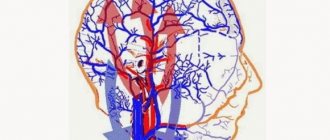
Angiography is an x-ray method for visualizing the vascular bed of the brain. A radiopaque contrast agent is injected into the carotid artery and serial craniograms are taken at short intervals. A clear image of the distribution of blood through the vessels of the brain is obtained. The method is used to diagnose hematoma, aneurysm (pathological dilatation of a vessel with a sharp thinning of its wall), tumor, abscess, cyst.
Rheoencephalography is a method for studying indicators of cerebral hemodynamics, based on measuring the electrical resistance of the brain to high-frequency alternating current. Provides information about the elasticity and degree of blood filling of the cerebral vessels. Used to diagnose migraine, dystonia, atherosclerosis, hypertension.
Doppler ultrasound of cerebral vessels is a method for studying cerebral blood flow based on the Doppler effect - a change in ultrasound parameters when reflected from a moving fluid (blood). Allows you to measure the linear speed of blood flow; used to diagnose vascular diseases of the brain.
Echo-encephalography is a method for studying the brain, based on the ability of ultrasound to be reflected from the interfaces of media with different acoustic densities. The ultrasound beam is delivered from the temporoparietal region, passes through the brain, reflected from the lateral ventricles and midline structures, and is then received by a sensor on the opposite side of the head. The signal is recorded on the device screen in the form of symmetrical peaks. This method reveals displacement of the midline structures of the brain due to a tumor, abscess, hematoma, as well as expansion of the ventricles of the brain due to increased intracranial pressure.
Electroencephalography is a method of recording electrical potentials of the brain from multiple electrodes applied to the surface of the head. This is a summary characteristic of the electrical activity of the brain. Normally, rhythmic oscillations of the correct form with a frequency of 10 Hz are recorded from the occipital-parietal leads (alpha waves) and 20 Hz from the frontotemporal leads (beta waves). With brain pathology, these waves change in frequency, amplitude, shape, and slow waves with a frequency of 2 Hz (delta waves) and 5 Hz (theta waves) appear. To identify hidden pathological activity, functional loads are used in the form of flashes of light, forced breathing, and the introduction of chemicals. Electroencephalography is the most informative for diagnosing epilepsy, tumors and other focal brain lesions.
Electromyography is a method for assessing the condition of muscles and nerves based on recording and analysis of muscle biopotentials. Allows for differential diagnosis of diseases of the nerve (neuritis), muscle (myopathy), disorders of neuromuscular transmission (myasthenia), as well as various levels of damage to the pyramidal tract (conducting tracts, anterior horn, root, peripheral nerve).
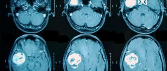
CT scan. A thin X-ray beam scans the brain or spinal cord from different angles in 3mm increments. The part of the beam not absorbed by tissues is recorded by sensors. After processing the results with a computer, the spatial relationship of tissues according to their density is recreated, the epidural space, the substance of the brain, the ventricles, as well as various pathological formations inside the skull are clearly visualized. Computed tomography is used to diagnose tumors, hemorrhages, multiple sclerosis, and herniated intervertebral discs.
Magnetic resonance imaging. The method is based on the fact that when irradiated with an electromagnetic field, water molecules take the direction of the field. After removing the external magnetic field, the molecules return to their original state, and a magnetic signal is generated, which is captured by special sensors, processed by a computer and graphically displayed on the monitor. A special feature of the method is the ability to obtain not only anatomical, but also physicochemical data about the brain. This allows you to more clearly distinguish healthy tissue from damaged tissue. The method is used to diagnose the early stages of brain tumors, multiple sclerosis, and also to analyze cerebral blood flow.
The study of cerebrospinal fluid (CSF) is widely used in neuropathology. Liquor is obtained by puncture: a puncture is made between the third and fourth lumbar vertebrae and 3 ml of liquid is taken from the spinal canal for examination. Normally it is colorless and transparent. With meningitis, the cerebrospinal fluid flows out under increased pressure; with purulent meningitis, it is cloudy. When there is a hemorrhage in the brain or under its membranes, the cerebrospinal fluid contains an admixture of blood. In the laboratory, the cerebrospinal fluid is centrifuged, and the sediment is examined under a microscope. The content of protein and cells in the sediment is determined. The number of cells is increased in meningitis, the amount of protein is increased in tumors. Characteristic changes in the cerebrospinal fluid are observed in tuberculous meningitis.
QUESTIONS AND SITUATIONAL TASKS
· What is the algorithm for establishing a neurological diagnosis?
· What does the anamnestic part of the study include?
· What details are clarified when talking with a patient about his complaints?
· What questions are asked to the patient when taking a medical history?
· What methods of studying the nervous system are considered x-ray?
· To diagnose what diseases of the nervous system are skull radiography used?
· What is the essence of echo-encephalography?
· For the diagnosis of which diseases of the nervous system is electroencephalography most informative?
· What is the difference between computed tomography and magnetic resonance imaging?
· How is cerebrospinal fluid obtained for testing?
· Based on complaints, medical history and neurological examination, the patient is suspected of having a cerebellar tumor. What instrumental studies will help clarify the diagnosis?
· A young man had a grand mal seizure for the first time in his life. What instrumental methods are indicated?
Date added: 2015-05-19 | Views: 1544 | Copyright infringement
| | | | | | | | | | | | | | | | | | | | | | | | 25 | |
Diagnosis of diseases of the nervous system
To correctly diagnose diseases of the nervous system, you need to consult a neurologist. Comprehensive diagnostics includes:
- collection of anamnesis of the disease (conversation);
- special neurological tests;
- reflex diagnostics;
- local palpation examination of the spine and musculoskeletal system.
In addition to the main examination, if necessary, the doctor may prescribe:
- MRI;
- Ultrasound;
- X-ray;
- Lab tests;
- EEG;
Additional examination methods in neurology
Neurological methods for studying the functions of cranial nerves Read more: The cerebellum, its connections with the spinal cord and brain. Symptoms of the lesion
20 Additional examination methods in neurology
The study of cerebrospinal fluid in neurology is of great importance, since many inflammatory, tumor, degenerative and other diseases change its nature and properties. In order to examine the cerebrospinal fluid, a lumbar puncture of the spinal canal is made with a needle. Cerebrospinal fluid is examined for meningitis (inflammation of the meninges), encephalitis (inflammation of the brain substance), tumors of the brain and spinal cord, intracranial hemorrhage, convulsions, dropsy of the brain, etc.
Skull transillumination is a valuable auxiliary research method used to diagnose intracranial diseases in newborns and infants. The principle of the method is to propagate high-intensity light rays in a liquid-filled space. It's like a transillumination of the skull.
Indications for transillumination are an increase in the size of the head in newborns and infants, edema of the brain (hydrocephalus), small sizes of the skull (microcephaly), and suspicion of intracranial hemorrhages.
Currently, X-ray examination is widely used for intracranial diseases, head injuries, diseases of the bones of the skull, spine, joints between vertebrae, etc. On radiographs of the skull, attention is paid to the size and contours of the skull, cranial sutures, the condition of the fontanelles (early or late closure), etc. Using an X-ray of the skull (craniogram), congenital defects of the skull bones, malformations of the brain, hydrocephalus, microcephaly, fractures of the skull bones, and dystrophic changes in the bones of the skull are detected. . X-rays of the spine record congenital malformations of the spine, changes in the vertebral bodies due to tuberculosis, traumatic changes, etc.
Ventriculography is a method of injecting a contrast agent directly into the ventricles of the brain, followed by radiography. X-rays produce images of the ventricles of the brain or the contours of the spinal cord.
Angiography is a valuable method that provides a radiographic image of the cerebral vessels after the injection of a radiocontrast agent into them. Angiography is carried out to clarify the localization of the pathological focus and clarify its nature and character.
Contrast myelography is a radiopaque method for diagnosing diseases of the spinal cord and its membranes.
Computed tomography is a research method that allows you to obtain accurate and detailed images of the slightest changes in the density of brain tissue. The brain is examined using a scanning device containing crystal or gas detectors that are highly sensitive to X-rays. Computed tomography of the brain can detect most congenital malformations, the degree of expansion of the ventricles, the brain and the nature of hydrocephalus, general or local cerebral edema. The method makes it possible to differentiate cerebral vascular disorders, such as infarctions of brain tissue, hemorrhages into the brain substance.
Electroencephalography is a method of recording brain biocurrents. In brain tissue, when nerve cells are excited, a potential difference arises between negatively charged areas of the brain. The potential difference is very small, but with the help of an electroencephalograph they are amplified and recorded.
Electromyography (EMR) is a method of recording muscle biocurrents. It is widely used to diagnose neuromuscular diseases. The electromyogram reflects the electrical activity of muscle fibers. Electromyography is a valuable research method that allows differentiating different levels of damage to the nervous system.
Biochemical research method. In order to determine the nature of the disease of the nervous system, they resort to the study of amino acids, fats, carbohydrates, enzymes (catalysts of biochemical reactions), breakdown products of hemoglobin, red blood cells, proteins, macro- and microelements (potassium, sodium, calcium, phosphorus, magnesium, copper, zinc and etc.).
21. General ideas about the pathology of the nervous system
Disturbances in the functions of the nervous system, which may be the result of some diseases, pathological conditions that developed after illness, trauma to the nervous system, congenital developmental disorders, manifest themselves in the form of any deviations from the normal functioning of one or another functional system or one or another department nervous system. These deviations from normal functioning are a sign, or symptom, of a pathological condition. Often, damage to any part of the nervous system manifests itself in the form of a set of symptoms. For example, damage to the cerebellum is manifested by decreased muscle tone, impaired coordination of movements, impaired balance, etc. This pathological condition, characterized by a persistent combination of several characteristic symptoms, is called a syndrome or symptom complex. As a rule, damage to a certain part of the nervous system corresponds to a certain characteristic syndrome.
Neurological methods for studying the functions of cranial nerves Read more: The cerebellum, its connections with the spinal cord and brain. Symptoms of the lesion
Information about the work "Neuropathy"
Section: Medicine, health Number of characters with spaces: 108824 Number of tables: 0 Number of images: 0
Similar works
Literature - Pediatrics (Book Ultrasound Methods in Neuropathology and Neurosurgery
239764
0
0
... symptoms and prophets of the development of other organs and systems. Sometimes the detection of pathology in NSG is an accidental finding. III. Systematics of B-scanning methods of the brain from the perspective of pediatric neuropathology and neurosurgery Depending on the sensors used, linear scanning or sectoral scanning is performed. Depending on the ultrasonic window used, there are ...
Emergency care in neuropathology
524428
0
0
... laryngospasm. The pain radiates to the ear and is provoked by eating and swallowing. The pain point is determined on the lateral surface of the neck, slightly above the thyroid cartilage. Giving help. Emergency care is similar to that provided to patients with trigeminal neuralgia. Glossalgia. Clinic. Glossalgia is caused by damage to the peripheral somatic formations of the oral cavity, but mainly...
Cerebral palsy as a problem of neuropathology and special pedagogy
42843
4
3
... activity and sound pronunciation side of speech. Such children have a quiet, poorly modulated voice with a nasal tint. Study of the cervical-tonic reflex in cerebral palsy with torticollis symptoms. Depending on the severity and prevalence, the following forms of cerebral palsy are distinguished: spastic diplegia, spastic hemiplegia, double hemiplegia, ...
Bibliography on hypnosis
175737
0
0
... U. M., Belova L. V. “Some issues of psychotherapy in dermatology” - “Bulletin of Dermatology and Venereology” 1982, 11, 62-66. 605. Mirzamukhamedov M. A., Suleymanov A. S., Pak S. T., Shamirzaeva M. Kh. “The effectiveness of hypnosis and acupuncture in some functional diseases in children” - “Medical Journal of Uzbekistan” 1987, 1, 52-54 . 606. Mirzoyan A. S. “Step-by-step psychotherapy of sexual...