CSF analysis is an analysis of cerebrospinal fluid. Cerebrospinal fluid is called cerebrospinal fluid or cerebrospinal fluid. Liquor is created by the filtration of blood through the choroid plexuses of the ventricles of the brain, circulates in the ventricles of the brain and the subarachnoid membrane of the brain and spinal cord. Cerebrospinal fluid serves to soften shocks during mechanical effects on the brain, maintains constant intracranial pressure, protects the brain from infection, maintains normal concentrations of nutrients, helps saturate brain neurons with oxygen and remove carbon dioxide, decay products, and ensures the transport of hormones and other necessary substances. With the help of this biological fluid, the balance of the internal environment of the spinal cord and brain is ensured.
The Neurology Clinic of the Yusupov Hospital uses modern and effective methods for diagnosing diseases of the nervous system; studies are carried out using modern equipment from leading manufacturers from the USA, Europe and other countries. At the Yusupov Hospital you can have your cerebrospinal fluid tested for oligoclonal antibodies and receive effective treatment for multiple sclerosis.
What is cerebrospinal fluid, definition of cerebrospinal fluid
Cerebrospinal fluid (CSF, or CSF) is a liquid medium that fills the space in the ventricles of the brain, flows along the liquor pathway, and circulates in the subarachnoid segment. An alternative name is cerebrospinal fluid .
The synthesis and release of the substance is due to the process of filtration of plasma (the liquid part of the blood) through the capillary wall and the subsequent secretion of substances into exudate from ependymal and secretory cellular structures.
If there is any pathological condition with a violation of the integrity and structure of the bone and soft tissue of the skull, then liquorrhea - the release of cerebrospinal fluid from the ears, nose or defective, damaged areas of the skull and spine. Probable reasons:
- traumatic brain injury;
- hernial tumors or tumors;
- carelessness of medical manipulations;
- postoperative suture weakness.
Any deviation from the norm in the functioning of the organ system affects the density, transparency and quantity of the secreted substance, therefore some pathologies can be determined by its condition.
Indications for cerebrospinal fluid analysis
Not every person needs to know the condition of his spinal shock absorber. Certain indications are required to take a cerebrospinal fluid test. Among the most popular situations in the practice of doctors, when among laboratory tests there is an assessment of the composition of the cerebrospinal fluid, one can indicate:
- suspicion of hemorrhage in the subarochnoid space;
- infectious inflammatory processes in the membrane of the brain - confirmation of meningitis or encephalitis;
- differential diagnosis for brain tumors;
- injuries with penetration through solid structures - open wounds in the area of the spine or skull;
- fever of unknown etiology;
- systemic lesions of nerve endings;
- autoimmune diseases;
- Headaches of unknown etiology – exclude increased cerebrospinal fluid pressure.
In a number of situations, cerebrospinal fluid is analyzed after surgery on brain structures to assess the effectiveness of therapy. Less often, a puncture of cerebrospinal fluid is not diagnostic, but therapeutic in nature - the medication is injected directly into the brain canal. The doctor considers the indications for such a procedure especially carefully to prevent complications.
Functions of cerebrospinal fluid
Like every substance in the human body, CSF performs many vital functions:
- Mechanical protection. Providing a shock-absorbing effect during sudden movements or head impacts - by equalizing intracranial pressure, cerebrospinal fluid protects the brain from damage, ensuring its integrity and normal functioning even in traumatic situations.
- Excretion of metabolites. Some substances can accumulate in the brain space, which will negatively affect its functioning - the cerebrospinal fluid is responsible for their release (excretion) and outflow.
- Transport of necessary connections. Hormones, biologically active substances and metabolites that are responsible for central performance are transported to the gray matter using the cerebrospinal substance.
- Breathing (performing respiratory function). Neuronal clusters, which are responsible for the respiratory function of the body, are located at the very bottom of the fourth ventricle of the brain and are washed by cerebrospinal fluid. If you slightly change the component ratio (for example, increase the concentration of potassium or sodium ions), a change in the amplitude and frequency of inhalations/exhalations will follow.
- Acting as a regulator, stabilizing structure for the central nervous system. It is the CSF that maintains a certain acidity, salt and cation-anion composition, and constant osmotic pressure in the tissues.
- Maintaining a stable brain environment. This barrier must be practically insensitive to changes in the chemical composition of the blood, so that the brain continues to work even when a person is sick or struggling with pathology.
- The work of natural immunoregulators. It is possible to assess the state of the nervous system and trace the course of diseases only with the help of a detailed analysis of punctate, the study of which will help clarify the diagnosis or predict the patient’s health status.
How is the analysis done?
Before taking the test, the patient undergoes an examination. Then the doctor questions the patient about medications taken and recent illnesses. 12 hours before the cerebrospinal fluid analysis, you should stop eating and drinking. A long hollow needle is used to collect cerebrospinal fluid; the doctor’s actions are monitored using a fluoroscope, which converts X-rays into an image on the monitor. Additionally, other devices are used.
The patient is positioned on the treatment table, face down. The procedure is anesthetized. The skin at the site of the future puncture is cleaned and disinfected. Under the control of an X-ray machine, a needle is inserted into the spinal canal between two lumbar vertebrae, then the pressure of the cerebrospinal fluid is measured. After collecting the cerebrospinal fluid, an anesthetic is injected into the spinal canal. The needle is removed and a pressure bandage is applied. After the procedure, the patient must lie on his side for several hours.
Neurologists at the Yusupov Hospital conduct examinations of cerebrospinal fluid, diagnose multiple sclerosis, and treat diseases of the nervous system. The accuracy of diagnosis, experience and knowledge of doctors help in prescribing effective treatment for brain diseases. You can make an appointment with a doctor by phone.
Composition of cerebrospinal fluid
Cerebrospinal substance is produced, on average, at a rate of about 0.40-0.45 ml per minute (in an adult). The volume, rate of production, and most importantly, the component composition of CSF directly depends on the metabolic activity and age of the body. Typically, tests show that the older a person is, the more reduced production is.
This substance is synthesized from the plasma part of the blood, however, both the substrate and the producer differ significantly in ionic and cellular content. Main components:
- Protein.
- Glucose.
- Cations: sodium, potassium, calcium and magnesium ions.
- Anions: chlorine ions.
- Cytosis (presence of cells in the cerebrospinal fluid).
An increased content of protein and cell aggregates indicates a deviation from the norm, which means it is a condition that requires further tests and mandatory consultation with the attending physician.
Analysis and research of cerebrospinal fluid
The study of cerebral spinal puncture is a method that is used to identify and diagnose various disorders of the brain structures and membranes, the central nervous system. Such pathologies include:
- meningitis, tuberculous meningitis;
- inflammatory processes in the membrane;
- tumor formations;
- encephalitis;
- syphilis.
Carrying out the procedure for analyzing and studying SM fluid requires taking a sample as a punctate from the lumbar spinal cord. The collection is made through a small puncture in the desired area of the spine.
A complete analysis of CSF includes macroscopic and microscopic examination, as well as cytology, biochemistry, bacterioscopy and bacterial culture on a nutrient medium.
The spinal tap will be examined according to several parameters:
- Transparency.
The liquor of a healthy person is absolutely transparent, like pure water, so during macroscopic analysis it is compared with a standard - distilled, highly purified water in good lighting. If the sample taken is not clear enough or there is a strong, obvious cloudiness, then there is a reason to look for the disease. After detecting a discrepancy with the standard, the test tube is sent to a centrifuge - the procedure will determine the nature of the turbidity:
- If the sample is still cloudy after centrifugation, this indicates bacterial contamination.
- If the sediment sank to the bottom of the flask, then the turbidity was caused by blood cells or other cells.
- Color.
Liquor produced by a healthy body should be absolutely colorless. The change indicates the presence of any compounds in it that normally should not be there - many pathological conditions of the body are provoked by xanthochromia of the CSF, that is, its coloring in shades of red and orange. Xanthochromia is caused by the presence of hemoglobin and its species in the sample, for example:
- yellowishness - the presence of bilirubin fraction, released during the breakdown of hemoglobin;
- light pink, red-pink shading indicates oxyhemoglobin (hemoglobin saturated with oxygen) in the cerebrospinal fluid;
- orange shades – the sample contains bilirubin compounds that appear as a result of the breakdown of oxyhemoglobin;
- brown colors - reflect the presence of methemoglobin (an oxidized form of hemoglobin) - this condition is observed in tumor phenomena, strokes;
- cloudy green, olive - the presence of pus during purulent meningitis or after opening an abscess.
- redness reflects the presence of blood.
If a little ichor gets into the sample during punctate collection, then such a mixture is considered a “travel” and does not affect the result of macroscopic analysis. Such an admixture is not observed throughout the entire volume of the punctate, but only on top. Impurities can be pale pink, cloudy pink or grayish pink.
The xtanochromic intensity of the sample is assessed according to the “pluses” set by the laboratory assistant during visual assessment:
- first degree (weak).
- second degree (moderate).
- third degree (strong).
- fourth degree (excessive).
Blood fractions or strong saturation of the punctate suggest one of the diagnoses: rupture of aneurysm vessels and subsequent intracranial hemorrhage, hemorrhagic encephalitis or stroke, moderate and severe TBI, hemorrhage in the brain tissue.
- Cytology.
The state of the cerebrospinal fluid of a healthy person allows for a slight content of cells, but within the established values.
Leukocytes in one cubic mm:
- up to 6 units (in adults);
- up to 8-10 units (in children);
- up to 20 units (in infants and toddlers up to 10 months).
There should be no plasma cells. The presence indicates infectious diseases of the central nervous system: multiple sclerosis, encephalitis, meningitis or recovery after surgery with a wound that did not heal for a long time.
Monocytes are observed in numbers up to 2 per cubic mm. If the number increases, then this is a reason to suspect a chronic pathology of the central nervous system: ischemia, neurosyphilis, tuberculosis.
The neutrophil component is present only during inflammatory processes, altered forms are present during recovery from inflammation.
Granular macrophage cells can only be present in the CSF when the body's brain tissue is disintegrating, as in the case of a tumor. Epithelial cells enter the punctate only if a tumor of the central nervous system develops.
Research for meningitis
If cerebrospinal fluid examination does not reveal inflammatory changes, the diagnosis of meningitis is excluded. An important component of the analysis is pleocytosis. Turbid cerebrospinal fluid with a discolored liquid (from milky to greenish) indicates the development of purulent meningitis. Neutrophils predominate in the fluid, which suggests the severity of the inflammatory process. Increased protein, decreased glucose levels. With a decrease in the number of neutrophils and an increase in lymphocytes, and an increase in glucose levels, we can talk about a favorable outcome of the disease.
When testing for tuberculous meningitis, a bacterioscopy test may be negative. This form of meningitis is indicated by the formation of sediment in the cerebrospinal fluid when it settles. In most cases, Mycobacterium tuberculosis is found in sediment, which is a cobweb-like delicate mesh. The cerebrospinal fluid in tuberculous meningitis has no color, is transparent, there is a high level of lymphocytes in pleocytosis, in some cases the level of neutrophils and lymphocytes increases. Poor prognosis with an increase in the number of macrophages and monocytes. With tuberculous meningitis, the protein level is increased, the glucose level is decreased, and in some patients the chloride level decreases.
Meningococcal meningitis is characterized by increased intracranial pressure; when examining the cerebrospinal fluid, mild neutrophilic cytosis is detected, then changes occur as in purulent meningitis.
Serous meningitis is characterized by clear cerebrospinal fluid, slight pleocytosis with an increase in the level of lymphocytes. The protein level is within normal limits or slightly increases; in some patients, the protein level may fall due to overproduction of cerebrospinal fluid. If in the initial stage of the disease there is pleocytosis with the growth of neutrophils, then the prognosis of the disease is considered unfavorable and the course of the disease is severe.
Normal values of cerebrospinal fluid in a healthy person
In addition to the constituent components, transparency and color characteristics, normal cerebrospinal fluid must correspond to other indicators: reaction of the environment, number of cells, chlorides, glucose, protein, maximum cytosis, absence of antibodies, etc.
Deviation from the given indicators can serve as an identifier of the disease - for example, immunoglobulins and oligoclonal antibodies
- Protein in the cerebrospinal fluid : lumbar - 0.21-0.33 g/liter, ventricular - 0.1-0.2 g/liter.
- Pressure in the range of 100-200 mm water column. (sometimes they indicate values of 70-250 mm - in countries outside the post-Soviet space).
- Glucose : 2.70–3.90 mmol per liter (some sources indicate: two thirds of total plasma glucose).
- CSF chloride: 116 to 132 mmol per liter.
- Optimal indicators of the reaction of the environment are considered to be values in the range of 7.310 – 7.330 pH. Changes in acidity have an extremely negative impact on the performance of biological functions, the quality of CSF and the speed of its flow through the liquor-excreting pathways.
- Cytosis in the cerebrospinal fluid : lumbar - up to three units. per µl, ventricular - up to one per µl.
What should NOT be in the puncture of a healthy person?
- Antibodies and immunoglobulins.
- Tumor, epithelial, plasma cells.
- Fibrinogens, fibrinogen film.
The density of the sample is also determined. Norm:
- The total density should not exceed 1.008 grams per liter.
- Lumbar fragment – 1.006-1.009 g/l.
- Ventricular fragment – 1.002-1.004 g/l.
- Suboccipital fragment – 1.002-1.007 g/l.
The value may decrease with uremia, diabetes mellitus or meningitis, and increase with hydrocephalic syndrome (an increase in the size of the head due to the accumulation of fluid and its difficult removal).
Total protein in liquor
Measuring the concentration of total protein in the cerebrospinal fluid (cerebrospinal fluid), used for diagnosis and monitoring of treatment of infectious-inflammatory, oncological, autoimmune and other diseases of the central nervous system.
Synonyms Russian
Total protein in cerebrospinal fluid, CSF; analysis of cerebrospinal fluid for total protein; analysis of CSF for total protein.
English synonyms
Cerebrospinal fluid, Total Protein; Total Protein CSF.
Research method
Colorimetric photometric method.
Units
G/L (grams per liter).
What biomaterial can be used for research?
Liquor.
How to properly prepare for research?
No preparation required.
General information about the study
Liquor (cerebrospinal fluid) is the only biological medium, the study of which in clinical practice allows one to obtain accurate information about the state of the central nervous system. Many diseases of the brain and spinal cord are accompanied by changes in the normal composition of the cerebrospinal fluid. One of the components of the cerebrospinal fluid, the concentration of which changes in diseases of the central nervous system, is total protein. Measuring the concentration of total protein in the cerebrospinal fluid is a mandatory analysis, which is carried out if infectious-inflammatory, oncological, autoimmune and other diseases of the central nervous system are suspected.
Total protein in the cerebrospinal fluid is one of the most sensitive indicators of CNS pathology. Normally, the cerebrospinal fluid of an adult contains 150-500 mg/l of protein. The majority (80%) comes from the blood when it is filtered in the choroid plexus of the ventricles of the brain. A smaller portion is produced directly intrathecally (by choroid plexus cells, glia, neurons and immune cells). The protein composition of the cerebrospinal fluid is very dependent on the protein composition of the blood. As in blood, normally the main protein of the cerebrospinal fluid is albumin (it makes up 35-80% of the total protein of the cerebrospinal fluid), and the main immunoglobulins of the cerebrospinal fluid belong to class G immunoglobulins. It should be noted that normally the main part of IgG enters the cerebrospinal fluid from the blood , and is not synthesized intrathecally.
On the other hand, the protein composition of the cerebrospinal fluid is very different from the protein composition of the blood. This difference is due to the presence of the blood-brain barrier (BBB), a mechanical filter that prevents the entry of blood proteins into the cerebrospinal fluid. Small proteins (albumin) easily pass through the BBB, while the entry of large protein molecules (immunoglobulins) is difficult. Thus, the diffusion of albumin from the blood into the cerebrospinal fluid takes about 1 day, and the diffusion of IgM immunoglobulins takes several days. As a result, the total protein in the cerebrospinal fluid is only 0.1-0.2% of the amount of blood protein. In addition, CNS-specific proteins can be detected in the cerebrospinal fluid, for example, myelin basic protein, glial fibrillary acidic protein, and tau protein. These specific proteins, however, account for about 1-2% of the total cerebrospinal fluid protein.
Changes in the concentration of total protein in the cerebrospinal fluid are often observed in diseases of the central nervous system. An increase in the level of total protein in the cerebrospinal fluid can be observed in 3 situations: 1) An increase in the permeability of the BBB. This mechanism underlies changes in protein levels in the cerebrospinal fluid in most infectious and inflammatory diseases. In this case, changes in the protein composition of the cerebrospinal fluid reflect changes in the composition of the blood protein. 2) Enhanced protein synthesis intrathecally, as, for example, in multiple sclerosis, sarcoidosis or neurosyphilis. 3) Impaired resorption of cerebrospinal fluid protein through arachnoid granulations (processes in the pia mater).
An increase in the level of total protein in the cerebrospinal fluid is a characteristic sign of damage to the central nervous system. It is observed with bacterial (0.4-4.4 g/l), cryptococcal (0.3-3.1 g/l) and tuberculous (0.2-1.5 g/l) meningitis and neuroborreliosis. A total protein concentration of more than 1.5 g/l is a specific (99%) sign of bacterial meningitis. In viral neuroinfections (encephalitis, meningoencephalitis), the level of total protein is not so much increased (usually less than 0.95 g/l). In half of patients with herpetic encephalitis, the level of total protein in the cerebrospinal fluid remains normal. An increase in total protein levels can also be observed in subarachnoid hemorrhage, vasculitis with central nervous system involvement, and in the presence of malignant neoplasms of the central nervous system. An increase in the concentration of total protein with a normal number of cerebrospinal fluid cells (albumin-cell dissociation) is a characteristic sign of acute and chronic inflammatory demyelinating polyneuropathies. It should be noted that during the first week of diseases from this group, the total protein in the cerebrospinal fluid can remain within normal limits. It is elevated in 80% of patients with metastatic lesions of the pia mater (average concentration is about 1 g/l). A moderate increase in total protein in the cerebrospinal fluid can also be observed in a variety of peripheral neuropathies, such as diabetic neuropathy.
A decrease in the level of total protein in the cerebrospinal fluid can be observed with repeated lumbar punctures or with chronic cerebrospinal fluid leaks - conditions in which cerebrospinal fluid is lost faster than it is produced. Also, lower levels of total protein are detected in some children aged 6 months - 2 years, with water intoxication and in some cases of idiopathic intracranial hypertension.
The level of total protein in the cerebrospinal fluid depends on many factors. Thus, the concentration of total protein in the liquor of infants is much higher than that of an adult. “Adult” levels of total protein are established at approximately 6-12 months. A false increase in the concentration of total protein in the cerebrospinal fluid can be obtained during a traumatic lumbar puncture, when a large number of red blood cells are present in the blood sample.
Reference values for total protein vary between laboratories due to different methods for determining it. Therefore, if repeated (multiple) tests of cerebrospinal fluid for total protein are planned, for example, to monitor the treatment of a disease, it is recommended that all tests be performed in one laboratory.
Due to the fact that the amount and composition of protein in the cerebrospinal fluid strongly depends on the amount and composition of protein in the blood, a test for protein in the cerebrospinal fluid should always be supplemented with a test for protein in the blood.
What is the research used for?
- For diagnosis and control of treatment of infectious-inflammatory, oncological, autoimmune and other diseases of the central nervous system.
When is the study scheduled?
- If you suspect the presence of infectious-inflammatory, oncological, autoimmune and other diseases of the central nervous system;
- during a follow-up examination of a patient undergoing treatment for any of these diseases.
What do the results mean?
Reference values
| Age | Reference values |
| 0-16 years | 0.15 - 1.3 g/l |
| More than 16 years | 0.15 - 0.45 g/l |
Reasons for the increase:
- Brain abscess;
- Malignant brain tumor;
- Spinal cord tumor;
- Diabetic neuropathy;
- Guillain–Barré syndrome;
- Freun's syndrome;
- Heavy metal poisoning;
- Traumatic brain injury;
- Hemorrhage (intracerebral, subarachnoid);
- Bacterial meningitis;
- Viral meningitis;
- Tuberculous meningitis;
- Cryptococcal meningitis;
- Multiple sclerosis;
- Myxedema;
- Sarcoidosis;
- Neurosyphilis;
- Systemic lupus erythematosus;
- Thrombosis of cerebral vessels;
- Alcohol consumption;
- Rupture of intracerebral aneurysm;
- Use of isopropanol and phenytoin
Reasons for the downgrade:
- Successful treatment of the disease;
- Water intoxication;
- Multiple lumbar punctures;
- Chronic liquorrhea;
- Some cases of idiopathic intracranial hypertension;
- Age 6 months – 2 years
What can influence the result?
- Age;
- Presence of erythrocytes in the cerebrospinal fluid sample;
- Taking certain medications (phenytoin) and alcohol
Important Notes
- A test for protein in the cerebrospinal fluid should always be supplemented with a test for protein in the blood;
- It is recommended that all tests for total protein in cerebrospinal fluid be performed in one laboratory.
Also recommended
- Glucose in cerebrospinal fluid
- Diagnosis of multiple sclerosis (isoelectric focusing of oligoclonal IgG in cerebrospinal fluid and serum)
- Myelin basic protein in cerebrospinal fluid (CSF)
- Herpes Simplex Virus 1/2, DNA [real-time PCR]
- Treponema pallidum, IgG in cerebrospinal fluid
- Toxoplasma gondii, DNA [real-time PCR]
- Cytomegalovirus, DNA [real-time PCR]
- Borrelia burgdorferi sl, DNA [PCR]
Who orders the study?
Neurologist, neurosurgeon, general practitioner.
Literature
- Dean A. Seehusen, Mark M. Reeves, Demitri A. Fomin. Cerebrospinal Fluid Analysis. Am Fam Physician. 2003 Sep 15;68(6):1103-1109.
- Walker HK, Hall WD, Hurst JW. Clinical Methods: The History, Physical, and Laboratory Examinations. 3rd edition. Boston: Butterworths; 1990.
- Hühmer AF, Biringer RG, Amato H, Fonteh AN, Harrington MG. Protein analysis in human cerebrospinal fluid: Physiological aspects, current progress and future challenges. Dis Markers. 2006;22(1-2):3-26.
- Deisenhammer F, Bartos A, Egg R, Gilhus NE, Giovannoni G, Rauer S, Sellebjerg F; EFNS Task Force.
- Guidelines on routine cerebrospinal fluid analysis. Report from an EFNS task force. Eur J Neurol. 2006 Sep;13(9):913-22.
Violation of cerebrospinal fluid. Causes and symptoms
The main disease states associated with CSF include liquorrhea, liquorodynamic imbalance, cerebral hydrocele, and increased intracranial pressure. Their development mechanism differs, as does the symptom complex.
Liquororrhea
It is the most pathogenetically simple disease, because its mechanism is clear: the integrity of the bones of the base of the skull or meninges is disrupted, which provokes the release of the spinal substance.
Depending on the symptoms and visual manifestations, liquorrhea is called:
- Hidden - liquor flows through the nasal passages, which is not visually noticeable due to aspiration or accidental ingestion.
- Obvious - a clear liquid or mixed with ichor is intensely released from the ears, fracture sites, which is noticeable by the leakage of the bandage headband.
Also distinguished:
- The primary nature of the disease - the discharge manifests itself immediately after injury, after surgery.
- Secondary, or cerebrospinal fluid fistulas - leakage is observed in the later stages of severe complications of infectious diseases.
If the primary pathology is not treated for a long period of time, and then inflammation (meningitis or encephalitis) develops, then this is fraught with the development of a fistula.
Common causes of CSF leak:
- severe bruises with traumatic brain injury;
- injuries and serious injuries of the spine;
- complicated hydrocephalus;
- hernial neoplasms and tumors in dangerous proximity or directly in the brain tissue;
- inaccuracy of medical procedures - washing or draining the ENT profile;
- weakness of the sutures of the dura mater after neurosurgical operations;
- Spontaneous liquorrhea is very rare.
Liquorodynamic disorders
CSF dynamics are disrupted if there is difficulty or improper circulation of cerebrospinal fluid. The course of the disease can be hypertensive (associated with high blood pressure) or hypotensive (on the contrary, with low blood pressure).
The hypertensive form occurs when:
- excessive secretion - due to the strong excitability of the choroid plexuses, which are responsible for the production of CSF;
- insufficient absorption and excretion.
Liquor is produced in large quantities or is simply not absorbed, which provokes the following symptoms:
- severe headaches, especially intense in the morning;
- nausea, frequent gagging, periodic vomiting;
- dizzy;
- slow heartbeat – bradycardia;
- sometimes nystagmus - frequent involuntary eye movements, “trembling” of the pupils;
- symptoms characteristic of meningitis.
The hypotensive form occurs less frequently, with hypofunction or weak activity of the choroid plexuses, the consequence is reduced production of the cerebrospinal fluid substance. Symptoms:
- severe headache in the occipital and parietal regions;
- discomfort, increased pain during sudden movements, excessive physical activity;
- hypotension.
In addition to the listed reasons for the appearance of an imbalance in liquor dynamics, experts also identify inflammatory diseases of the central nervous system, parasitic invasion of the central nervous system, spinal injuries/wounds, as well as congenital genetic characteristics.
Impaired cerebrospinal fluid outflow and resorption
from the brain may be disrupted - due to this, deviations develop that manifest themselves differently in adults and children.
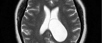
An adult will respond to a deviation by increasing intracranial pressure due to a strong, “overgrown” cranium. The bones of the child’s skull are immature and have not yet fused together, so excessive accumulation of spinal substance provokes hydrocephalus (dropsy) and other unpleasant manifestations.
CSF accumulation in the brain – increased ICP in adults
The cranium contains not only brain tissue and a great many neurons - a significant part of the volume is occupied by CSF. Its larger share is located in the ventricles, and the smaller one washes the GM and moves between its arachnoid and soft membranes.
Intracranial pressure directly depends on the volume of the skull and the amount of fluid circulating in it. Whether the production of a substance increases or its resorption decreases, the body immediately responds to this by increasing ICP.
This indicator reflects how much the pressure inside the skull exceeds atmospheric pressure - the norm is a value from 3 to 15 mm of mercury. Minor fluctuations lead to a deterioration in well-being, but an increase in ICP to 30 mm Hg. Art. already threatens to be fatal.
Manifestations of increased ICP:
- constantly sleepy, low performance;
- severe headaches;
- deterioration of visual acuity;
- forgetfulness, absent-mindedness, low concentration;
- “jumps” in pressure are noticeable - hypertension is regularly replaced by hypotension;
- poor appetite, nausea, vomiting;
- emotional instability: mood swings, depression, apathy, severe irritability;
- spinal pain;
- chills;
- increased sweating;
- failures of respiratory activity, shortness of breath;
- skin is more sensitive;
- muscle paresis.
The presence of 2-3 symptoms is not a reason to suspect increased ICP, but an almost complete complex is a good reason to consult a specialist.
The clearest sign of the disease is a girdling headache that is not expressed in any particular area. Coughing, sneezing and sudden movements only provoke increased pain, which is not relieved even by analgesics.
The second important sign of increased ICP is vision problems. The patient suffers from double vision (diplopia), notices deterioration of vision in the dark and in bright light, sees as if in a fog and suffers from attacks of blindness.
Pressure can increase in a healthy body, but it immediately returns to normal - for example, during physical and emotional stress, stress, coughing or sneezing.
Accumulation of cerebrospinal fluid in the brain - pediatric hydrops GM
Young children cannot report how they are feeling, so parents must be able to identify a violation of the cerebrospinal fluid outflow by the external signs and behavior of the baby. These include:
- noticeable vascular network on the skin of the forehead and back of the head;
- restlessness at night, poor sleep;
- frequent crying;
- vomit;
- protrusion of the fontanel, its pulsation;
- convulsions;
- increase in head size;
- uneven muscle tone - some are tense and some are relaxed.
The most serious sign of increased ICP in a child is hydrocephalus, which occurs with a frequency of up to one case in a couple of thousand newborns. Male babies suffer from hydrocele more often, and the defect itself is usually diagnosed by doctors during the first 3 months of life.
“Cerebral hydrocephalus”, as an independent disease, should not be confused with the diagnosis of “hypertensive-hydrocephalic syndrome”. It reflects that the newborn has a slightly increased ICP, but this does not require therapy or surgery, since it resolves itself.
The childhood form of the disease can be congenital or acquired, depending on the cause of development, of which, according to medical experts, there can be up to 170. The congenital disease is provoked by:
- injury to the child during childbirth;
- hypoxia during childbirth (insufficient oxygen supply);
- genetic failures;
- infectious diseases transmitted by the fetus while in the womb (cytomegalopathy, acute respiratory viral infections, mycoplasma and toxoplasma infections, syphilis, rubella, mumps and herpesvirus).
Genetic abnormalities causing the congenital form:
- underdeveloped cerebrospinal fluid ducts;
- Chiari syndrome - the child’s skull is larger in volume than his brain;
- narrowed liquor duct;
- other chromosomal pathologies.
The acquired form occurs as a result of toxic poisoning, the development of tumors, cerebral hemorrhages, and infectious diseases outside the mother's womb - these include otitis media, meningitis and encephalitis.
Speaking about hydrocephalus in newborns, it is worth considering that normally the head circumference of babies increases quite quickly (one and a half centimeters per month), but if the growth exceeds the indicators, then this is a good reason to examine the child.
The baby’s skull is soft, not yet ossified, and excess cerebrospinal fluid slows down the overgrowth of the fontanel, “pushes” the bones apart and prevents the normal development of the skull - because of this, the head grows disproportionately. Accumulating in the subarachnoid space , which separates the meninges, the cerebrospinal fluid compresses some parts of the brain. Despite the pliability of children's cranial bones, this manifestation of the disease is dangerous and requires immediate treatment. An increase in head size is not the only sign of obstructed liquor outflow in children. Characteristic is:
- the specific sound of a “broken pot”, heard when lightly tapping the skull;
- difficulty raising and holding the head in one position;
- trembling of the chin, hands.
It is important to pay attention to the baby’s eyes, because some signs are indicative:
- involuntary, chaotic eye movements;
- occasional eye rolling;
- eyes “squint”;
- “setting sun” syndrome - when blinking, a thin white stripe is noticeable between the pupil and the upper eyelid.
Hydrocephalus up to 2 years of age is manifested by this symptom complex, and later it is combined with vomiting, nausea, problems with coordination, irritability, diplopia or even blindness.
Sometimes hydrocephalic syndrome develops in adults as a consequence of previous infections, but this is a rare occurrence.
How to improve the outflow of cerebrospinal fluid
The pathology of liquor outflow in a baby is usually learned from a neurologist, whose examination takes place in the first month after birth. An initial examination and identification of signs requires medical correction, since this disease will interfere with the normal development of the child.
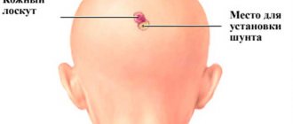
If the condition of a small patient is complex, then specialists use surgical intervention to create “bypass paths” for the CSF and eliminate the poor outflow artificially. If the situation does not threaten the baby’s life, treatment can also take place at home with drug therapy. In order to prescribe the optimal medications for a child, it is necessary to understand what can interfere with the outflow of cerebrospinal fluid in hydrocephalus . The cause, origin and complications will all play a role in the selection of treatment.
Pharmacological correction of outflow disorders in children includes:
- drugs that improve and stimulate blood flow (Actovegin, Pantogam, Cinnarizine);
- medications that help remove excess fluid (Triampur or Diakarb);
- neuroprotective drugs (Ceraxon).
Circulation and resorption of cerebrospinal fluid - history of study and modern presentation
There are references to cerebrospinal fluid in ancient medicine. In the writings of the ancient Greeks and Romans there is already evidence of the presence of fluid in the brain. Thus, F. Rose writes that in the treatises of Hippocrates (about 460-370 BC) there are references to the presence of fluid under the dura mater of the brain. In addition, Hippocrates gave a description of the dura mater of the brain, and suggested the presence and circulation of fluid in the brain (he wrote that the brain “like glands, secretes moisture”), A.P. Friedman points out that Hippocrates also believed that brain fluid plays an important role in diseases of the nervous system.
Aristotle (384-322 BC), who also studied the brain, suggested the existence of a “protective barrier” of the brain. The role of the blood-brain barrier is now generally recognized. According to the conclusion of N.N. Merritt, Fremont-Smith, JB Ayer, Herophilus of Alexandria (335-280 BC) first gave a detailed description of the meninges. He also described the network of blood vessels on the surface of the brain, the sinuses of the dura mater, and proposed the names dura and pia mater. Herophilus gave the name “choroid plexus gland” to the choroid plexuses located inside the cerebral ventricles. The above terms are rooted in anatomy and medicine.
Ringstad, SAS Vatnehol, R.K. Eide note that Anaxagoras (5th century BC), while examining the brain, first drew attention to and gave a description of the lateral ventricles. Erasistratus (3rd century BC) also described the lateral ventricles of the brain. He made an important discovery - a description of the opening that connects the lateral ventricles with the third ventricle of the brain. Subsequently, 2000 years after Erasistratus, these holes were rediscovered by Monroi and named after him.
A.P. Friedman points out that Claudius Galen (131-201) described in detail the meninges and ventricles of the brain, however, like Andrei Vesalius later, Galen did not discover fluid in the ventricles of the brain. Probably, when working with cadaveric material, it flowed out of the brain cavities and therefore disappeared from the field of view of the researcher. In memory of Galen's merits, anatomists named after him the great cerebral vein and the suboccipital liquor tank at the border of the brain and spinal cord. Julian Oribasius (IV century), after Galen, again described the two lateral ventricles of the brain and drew attention to the choroid plexuses.
During the Renaissance, more reliable information about the cerebrospinal fluid itself appeared. A. Vesalius (1514-1565) again described in detail the membranes of the brain, as well as the choroid plexuses in the cerebral ventricles in humans. Vesalius's merit was the discovery of the third meninges - the arachnoid. Like Galen, Vesalius also did not discover cerebrospinal fluid in the ventricles.
JB Ayer et al. indicate that T. Willis (1621-1675) in his work “Cerebri anatome” described the choroid plexuses and the pineal gland, and also described the arachnoid villi. R. Humphrey (1653-1708) in his monograph “Anatomy of the Brain, Including Its Mechanics and Physiology” (1695) wrote about the identity of the choroid plexuses of the third ventricle with the plexuses in other ventricles.
Magendie et al. reported that the presence of this cerebrospinal fluid is a normal rather than pathological phenomenon. He later confirmed the existence of communications between the ventricles of the brain and the arachnoid spaces, as well as the continuity of these spaces in the brain and spinal cord. In his book “Recherches physiologiques et cliniques sur le liquide c phalo-racchidien ou сerebro-spinal”, the author for the first time in science gave the exact designation “cerebrospinal fluid”, described the structure of the ventricles of the brain, arachnoid spaces of the brain and spinal cord. He first noted the movement of cerebrospinal fluid, which is in connection with breathing.
RS Tubbs et al., studying the life and scientific research of N. Luschka, indicate that the German anatomist described the arachnoid membrane, and also studied in detail the histology of the choroid plexuses. This allowed him to consider this formation to be a gland that produces brain fluid. He was the first to study the innervation of the dura mater of the brain and described the presence of an opening between the fourth ventricle and the subarachnoid space of the spinal cord. This hole was subsequently named after him.
In C. Bernard’s work “Lectures on the physiology and pathology of the nervous system,” a separate chapter is devoted to cerebrospinal fluid, in which the author often refers to the works of his teacher F. Magendie, with whom he carried out work on obtaining and studying cerebrospinal fluid using laboratory animals. However, in the medical literature of the 19th century there were still doubts that cerebrospinal fluid was a normal physiological fluid. In the above-mentioned book, C. Bernard wrote: “The fluid found in the ventricles is not serous, it is craniospinal fluid - a completely physiological product, which is completely mistakenly attributed to a pathological origin” and “... one should not trust some anatomical atlases, on which very many, even the newest ones, show the spinal canal filled with the spinal cord. This is incorrect, because the spinal column is separated from the walls of the canal by a fairly significant space, all the more significant since part of the spinal column is considered to have more extensive movements.”
An important event in the domestic school of studying cerebrospinal fluid is the opening at the Military Medical Academy on September 19, 1897 of a clinic for nervous diseases, in which a special operating room was equipped for the first time to perform operations on the nervous system. It should be noted that a graduate (1899) of the Imperial Military Medical Academy N.K. Rosenberg was 12 years ahead of the German researcher Goldman (1913), known in the literature for his “classical” experiments testing the permeability of the blood-brain barrier to dyes.
In 2012, under the leadership of the Danish neurophysiologist Maiken Nedergaard, the glymphatic system was discovered, the discovery of which radically changed the understanding of the pathways of cerebrospinal fluid resorption.
Due to the complexity and significant diversity of the structure, topographical features of the structural elements of the liquor circulation system, A.P. Friedman proposed dividing them into three types of structures: middle, deep and superficial. The median structures include the III and IV ventricles with their choroid plexuses, the cerebral aqueduct; deep - lateral ventricles and their choroid plexuses; superficial - the membranes of the brain (soft, arachnoid, hard) and interthecal spaces (subarachnoid, subdural, epidural).
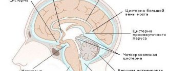
The subarachnoid space of the cerebral hemispheres is differentiated into three types of cavities filled with cerebrospinal fluid, which in a lifetime state is in constant motion: a system of subarachnoid cisterns, a system of cerebrospinal fluid channels and a system of subarachnoid cells.
The functional parts of the cerebrospinal fluid circulation system include:
- liquor production carried out by the choroid plexuses;
- cerebrospinal fluid circulation, including subsections: “ventricular cerebrospinal fluid circulation” (circulation of cerebrospinal fluid within the ventricles and aqueduct of the brain) and “extraventricular cerebrospinal fluid circulation” (circulation of cerebrospinal fluid in the cavities of the subarachnoid space of the brain, cerebellum, spinal cord);
- the outflow of cerebrospinal fluid, which ultimately becomes a component of the venous blood of the dural sinuses.
According to modern concepts, the liquor circulation system has three main links: 1 – liquor production; 2 – liquor circulation; 3 – liquor resorption.
There is a close relationship between the cerebrospinal fluid and cerebral circulation systems. Arterial vascularization of the choroid plexuses (sites of liquor production) is carried out due to the branches of five pairs of arteries belonging to the carotid (anterior villous arteries) and vertebrobasilar (lateral and medial posterior villous arteries, anterior and posterior inferior cerebellar arteries) systems. Resorption of cerebrospinal fluid occurs mainly in the pool of sinuses of the dura mater of the brain, from where, through the venous system, cerebrospinal fluid in the composition of venous blood reaches the right atrium. Thus, the relationship and interdependence of the cerebrospinal fluid circulation system with the circulatory system can be traced.
The amount of cerebrospinal fluid in an adult is from 130 to 150 ml: in the lateral ventricles - 20-30 ml, in the III and IV - 5 ml, in the cranial subarachnoid space - 30 ml, in the spinal ventricle - 75-90 ml. The main way of formation of cerebrospinal fluid is double filtration of blood: first through the basement membrane with access to the interstitial tissue, then through choroidal cells into the ventricles of the brain. The formation of the composition of cerebrospinal fluid occurs with the active participation of the structures of the blood-cerebrospinal fluid barrier. A person produces about 500 ml of cerebrospinal fluid per day, that is, the rate of cerebrospinal fluid circulation is 0.36 ml per minute. Thus, a complete four-fold renewal of cerebrospinal fluid occurs per day.
The circulation paths are determined by the site of cerebrospinal fluid production, the structure of the cerebrospinal fluid pathways and the site of cerebrospinal fluid resorption. Formed in the choroid plexuses of the lateral ventricles, the cerebrospinal fluid enters the third ventricle through the foramina of Monro, mixes with the cerebrospinal fluid produced by the choroid plexus of the third ventricle, enters the fourth ventricle through the cerebral aqueduct, and mixes with the cerebrospinal fluid produced by the choroid plexuses of this ventricle. Diffusion of fluid from the brain substance through the ependyma is also possible into the ventricular system. Through the paired lateral apertures of the fourth ventricle, cerebrospinal fluid enters from the ventricular system into the subarachnoid space of the brain, where it sequentially passes through systems of cisterns communicating with each other, liquor-carrying canals and subarachnoid cells. Some of the cerebrospinal fluid enters the subarachnoid space of the spinal cord.
The forward movement of cerebrospinal fluid in the subarachnoid space of the brain is carried out through the cerebrospinal fluid channels. Research by M.A. Barona, N.A. Mayorova showed that the subarachnoid space of the brain is a system of cerebrospinal fluid channels, which are the main routes of circulation of cerebrospinal fluid, and subarachnoid cells. These microcavities communicate with each other through holes in the walls of the channels and cells. In their atlas “Functional stereomorphology of the meninges,” the authors clearly depicted the structure of the leptomeninges.
The ways of outflow of cerebrospinal fluid outside the subarachnoid space are still being studied, and the views of different researchers differ. The prevailing opinion is that resorption of cerebrospinal fluid from the subarachnoid space of the brain occurs primarily through Pachionian granulations.
A.P. Friedman notes that Pachionian granulations, or arachnoid villi, were first described by T. Willis in 1662. But A. Pacchioni in 1705 described these formations in more detail and called them glands, which are the growth of the arachnoid membrane near the large sinuses of the dura mater and veins of the brain. The largest number of granulations is present in the parietal part of the superior sagittal sinus, a smaller number is in the transverse sinus, single granulations are in the sinus drainage, and in the occipital sinus they are practically absent.
The villus (Pachionian granulation) does not protrude the walls of the sinus, but pierces the dura mater and comes into direct contact with the endothelium of the venous sinus. The surface of the villus is covered with mesothelial cells, which are located in several rows at the top of the pachion granulation. Resorption of cerebrospinal fluid can also occur in the area of the subarachnoid space of the spinal cord through its arachnoid membrane and the blood capillaries of the dura mater of the spinal cord. CSF resorption also partially occurs in the brain parenchyma (mainly in the periventricular region), in the veins of the choroid plexuses and perineural clefts.
The basis for the possibility of cerebrospinal fluid outflow was already given in Schwalbe's classic studies: the German scientist Gustav Schwalbe, in an experiment on rabbits in 1869, injected Prussian blue into the subarachnoid space and obtained staining of the lymphatic vessels of the nasal mucosa, as well as the cervical lymph nodes.
Later, A. Key and G. Retzius conducted similar experiments on animal and human corpses: they injected ink into the subarachnoid space, and then detected it in the pachyonic granulations and venous sinuses.
Today, the most modern theory seems to be the existence of the glymphatic system, the function of which is the outflow of cerebrospinal fluid and the elimination of metabolic products of the central nervous system.
According to this theory, one of the pathways for transport of cerebrospinal fluid is the perivascular spaces of the brain, also known as the Robin-Virchow spaces. They are small (about 100-200 microns), filled with cerebrospinal fluid, channels along the intracerebral blood vessels. Some authors believe that the space is located between the vessel wall and the nervous tissue; others - that the vessel, penetrating from the subarachnoid space into the substance of the brain, involves the arachnoid and pia mater, between which spatia perivascularia is located.
The perivascular space communicates with the brain parenchyma through aquaporins AQP4, through which fluid transport directly occurs. Under the influence of the force of the pulse wave, cerebrospinal fluid flows from the periarterial spaces into the brain parenchyma. With the waste products of neurons (beta-amyloid, tau protein, gliofilaments, etc.), cerebrospinal fluid enters the perivenous spaces or directly into the veins (scientists argue about the existence of perivenous Robin-Virchow spaces). This cerebrospinal fluid pathway for eliminating brain metabolic products was described by a group of researchers from the University of Rochester in 2012 and was called the “glymphatic system.”
The amount of cerebrospinal fluid resorption depends on its production, pressure in the cerebrospinal fluid system and other factors. It undergoes significant changes in conditions of pathology of the nervous system.
Yildiz et al. published the results of a detailed study of the dependence of the speed of cerebrospinal fluid on the functioning of the respiratory and circulatory systems, in particular on the pulsation of the heart, breathing and cough. As a result of their experiment, it was found that the rate of cerebrospinal fluid changes during various physiological processes, namely, it increases slightly during ventricular systole, during forced breathing (breathing in normal mode does not affect the rate of resorption of cerebrospinal fluid) and increases 3 times from the initial value when coughing.
It is obvious that any brain surgery is accompanied by a violation of the integrity of blood vessels, accompanied by hemorrhage into the intrathecal spaces. The issue of the influence of hemorrhage on the rate of cerebrospinal fluid resorption is covered in the work of R. Blasberg et al., R. Blasberg, D. Johnson, J. Fenstermacher measured the rate of cerebrospinal fluid resorption in monkeys before introducing blood into the subarachnoid space, then divided the animals into 2 groups: in the first group (6 observations) were injected with non-heparinized blood, in the second group (4 observations) heparinized blood was injected. After subarachnoid blood injection, CSF resorption was re-checked after 30 minutes, 6 and 12 weeks. In the first group of monkeys, cerebrospinal fluid resorption decreased by 3 times after 30 minutes and by 1.5 times after 12 weeks in comparison with the initial resorption rates. In the second group, after 30 minutes, resorption decreased by 2 times, and after 6 weeks it normalized to the original level. Repeated administration of non-heparinized blood to this group resulted in the same indicators as in the first group.
Also F. Gao et al. The question was raised about the effect of thrombin on the development of hydrocephalus after blood enters the subarachnoid space. It was found that the entry of thrombin into the cerebrospinal fluid contributes to a decrease in the resorption capacity of the meninges and, as a consequence, the development of hydrocephalus as a result of its interaction with the thrombin receptor PAR-1.
Other work by these authors highlights the influence of blood components (iron and thrombin) on the rate of cerebrospinal fluid resorption. This work once again substantiates the statement about the inhibitory effect of thrombin on cerebrospinal fluid resorption, and also proves that ferritin, released during the breakdown of red blood cells after hemorrhage, has a similar effect.
Thus, the currently available literature data indicates a very complex structure of the liquor circulation system, the most controversial part of which is liquor resorption. A number of researchers report a decrease in the rate of resorption of cerebrospinal fluid when blood enters the cerebrospinal fluid. Consequently, after neurosurgical interventions, it is possible to predict hyporesorption of cerebrospinal fluid, which in turn can increase cerebrospinal fluid pressure, increase the risk of postoperative liquorrhea, and contribute to the development of hydrocephalus. However, due to the small number and only experimental nature of published studies, this hypothesis requires further testing and confirmation, which determines the relevance of further experimental and clinical studies.
S.N. Valchuk, D.E. Alekseev, G.V. Gavrilov, A.V. Stanishevsky, D.V. Whistles
2018
Treatment of spinal fluid disorders
Children's diseases of cerebrospinal fluid dynamics are most often corrected by pharmacotherapy, but adults need to be prescribed physiological procedures:
- A course of electrophoresis with aminophylline (ten visits) - drug “recharge” will activate the delivery of oxygen to brain tissue suffering from hypoxia with increased ICP. The condition of the vessels returns to normal, which will ensure normal resorption.
- 15 sessions of massage of the collar area - the procedure is simple, so over time the patient can carry out a similar manipulation himself. With its help, muscle hypertonicity is reduced, spasm is relieved and outflow is improved.
- Magnetic effect on the collar area - reduction of swelling and vascular spasm, improvement of innervation.
- Therapeutic swimming or supportive exercise. charger.


