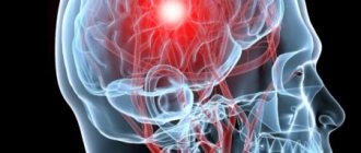
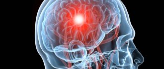
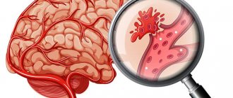
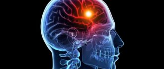
Hemorrhagic stroke is a condition characterized by the effusion of blood from the vessels located in the cranial cavity. Such a diagnosis is valid if the disease did not occur as a result of a traumatic brain injury, but happened spontaneously, most often under the influence of increased pressure in the cranial arteries or weakness of their walls.
Hemorrhagic stroke is an extremely serious disease, often leading to death. This is due to the peculiarity of the cerebral vessels - they collapse poorly, and bleeding when damaged is very difficult to stop. Conventional hemostatic agents do not penetrate the vessels of the brain; only hematomas are removed surgically, and they are not used to clamp a bleeding vessel.
This type of stroke accounts for 8-15%, the remaining 85-92% are ischemic strokes. It can develop at any age (even in children under one year old) and in people of any gender, but most often it is observed in men 50-70 years old.
A brief excursion into the anatomy of the brain
The brain is quite well protected from ischemia, that is, from the condition that can occur when more than 70% of the lumen of one of the arteries of medium or small diameter is blocked. This is due to two circles on its base:
- The first circle, called the circle of Willis, is formed by the branches of the carotid arteries and vertebral arteries that enter the cranial cavity from the thoracic cavity. This is how the cerebral hemispheres and their main sections-lobes are supplied with blood: frontal, temporal, parietal and occipital. The main endocrine organs, the pituitary gland and the hypothalamus, receive blood supply from here.
- The brain stem, a section of it consisting of several parts that differ in structure and function, is supplied with blood from the vertebral and spinal arteries, which also form a vicious circle. The brain stem is the part that seems to “grow” from the middle of the brain. It consists of several parts, one of which is the medulla oblongata, and, emerging from the cranial cavity, without visible anatomical transitions it becomes the spinal cord. And if in the hemispheres there are centers responsible for speech (recognition and reproduction), movements of the limbs, sensitivity of different parts of the body, the brain stem consists of sections responsible for vital functions - breathing, heartbeat, swallowing.
Smaller branches extend from the medium-sized arteries that form the two circles described above, and it turns out that each part of the brain receives nutrition from several sources. This is designed so that if one branch stops functioning, the area it feeds does not die off. But if bleeding occurs from a branch, that section (large - if the branch was large or small, if it had a small diameter) becomes saturated with blood and “switches off”, ceasing to function. Then his nutrition, provided by the “safety” artery, allows him not to die until the blood that has soaked the brain tissue is disposed of by natural methods.
The brain has ventricles. These are cavities lined from the inside with special cells that can produce cerebrospinal fluid and serve as conductors for it inside the brain. For their normal functioning, the ventricles of the brain are richly supplied with blood.
The brain matter has a fairly elastic consistency. If bleeding occurs and quite a lot of blood enters the brain, the areas move away from each other, and an area filled with blood forms in the middle - a hematoma. It compresses the zones along the perimeter and pinches the vessels feeding them, causing disruption of the blood supply. The larger the hematoma, the more the brain regions around it suffer. They, perhaps, would move in order to suffer less, but the hard bones of the skull “do not allow them.”
Also, if a brain bleed occurs, the compressed areas swell, just like skin cells around a scratch. This swelling further disrupts blood circulation in the cranial cavity. It is the reason why coma develops during a stroke.
The brain itself is covered on the outside with several membranes that protect it from traumatic influences. The upper and middle ones are quite close to each other, but between them, when additional liquid appears, there are gaps. Cerebrospinal fluid (CSF) circulates between the middle and lower ones. If a vessel is damaged and blood comes out between the membranes, the following will happen:
- the blood mixes with the cerebrospinal fluid, causing the volume of fluid circulating in the cranial cavity to increase. This leads to increased intracranial pressure;
- the blood in the cerebrospinal fluid coagulates, forming clots. The latter can block those ducts through which cerebrospinal fluid circulates, as a result, the cerebrospinal fluid tracts “swell” and swell;
- Blood clots, when dissolved, become inflamed and can cause meningitis (inflammation of the meninges) or even meningoencephalitis (inflammation of both the membranes and substance of the brain).
In the cranial cavity there are many holes of various diameters through which vessels, nerves, and parts of the brain pass. The largest of these is the foramen magnum, through which the medulla oblongata emerges. Swelling of the brain or a hematoma arising on it can “shift” the brain towards one of these “wells”, surrounded by a dense (bone or connective tissue) ring. If the brain structures reach such a hole, they will be retracted inside it. This is called brain dislocation. The hard ring compresses the part of the brain that gets inside, the blood circulation of the latter is disrupted, and this condition ends in death.
Causes of this condition
The most common cause of hemorrhagic stroke is an increase in blood pressure, which occurs very often in humans and leads to thinning of the wall of the arteries supplying the brain. It is this condition, the cause of which, in most cases remains unclear (it is called hypertension), that causes 85% of all hemorrhagic strokes .
There are other causes of this serious illness. This:
- Arteriovenous malformations are arteries and veins that are improperly connected to each other, when blood from an artery, bypassing the capillary system, enters directly into a vein. The pressure in the vein, which is not designed for that in the artery, increases. Under its influence, the venous wall gradually becomes thinner, and at some unfavorable moment (for example, during stress, sneezing or coughing, when the pressure rises), such a vascular connection is torn. Arteriovenous malformations are the main cause of hemorrhagic strokes in young people and even children.
- Atherosclerosis. A dense plaque formed from lipids leads to damage to the arterial wall. As a result of exercise, smoking, overheating, stress or drinking alcohol, at some point the artery below the plaque becomes damaged and a hemorrhagic stroke occurs.
- Changes in the walls of brain vessels due to: their inflammation (vasculitis), which most often accompanies systemic connective tissue diseases (for example, lupus erythematosus);
- chronic intoxication (manufactured products, alcohol, nicotine);
- avitaminosis C or mixed.
In this case, there is a disruption in the functioning of the inner layer of the arteries, and with a periodic increase in blood pressure, an aneurysm (saccular expansion) gradually forms here. At some point, arterial hypertension caused by stress, physical activity, kidney or adrenal disease, heavy drinking or smoking leads to the rupture of such an aneurysm, which is why a hemorrhagic stroke develops.
- Congenital aneurysms or those that arise during life due to increased pressure (this often occurs especially in places where brain vessels depart from each other at right angles). In these cases, the mechanism of stroke is described above.
- Amyloid angiopathy. In this case, a special protein, amyloid, is deposited in the artery wall. Because of it, the artery along a certain length becomes as if “glazed” and does not show the usual “flexibility” during increases and decreases in blood pressure. At a certain moment, which cannot be predicted, when the pressure is once again greatly increased, the wall of the vessel affected by amyloid deposits “breaks”, blood pours into the cranial cavity.
- Blood diseases: erythremia (increased number of red blood cells), thrombophilia (high level of platelets). Such pathologies lead to an increase in blood viscosity. As a result, in order for blood to be delivered to the brain, the heart has to push it under increased pressure.
- Overdose of blood thinning drugs (anticoagulants): Warfarin, low molecular weight heparin and others.
- Taking drugs that increase blood viscosity: oral contraceptives, narcotic drugs (cocaine, amphetamines).
- Rupture of the vessels feeding the tumor. The arteries leading to the intracranial tumor are tortuous and multiple. They are prone to spontaneous rupture, but are most often damaged due to increased blood pressure.
- In 15% of cases the cause cannot be determined.
Whatever the cause of a hemorrhagic stroke, a provoking factor is needed for its occurrence. Such triggering moments are conditions that lead to an increase in pressure. This:
- overheating in the sun or indoors;
- stress;
- inadequate physical activity;
- hypertensive crisis;
- smoking a large number of cigarettes in a short period of time;
- drinking large amounts of alcohol.
These factors are especially dangerous if:
- pressure is often noted and does not decrease above 160/90 mmHg. (the risk is 4 times higher than for those whose blood pressure is below 140/89 mmHg);
- the person smokes for a long time and/or smokes many cigarettes a day;
- there is atrial fibrillation (the risk of stroke is 3-4 times higher than in the absence of arrhythmias);
- a person suffers from coronary heart disease (the risk doubles);
- there is left ventricular hypertrophy, determined by ECG or ultrasound of the heart (the risk increases 3 times);
- there is heart failure, manifested by swelling in the legs, shortness of breath during exercise or even at rest (the risk increases by 3-4 times);
- the person suffers from diabetes;
- the structure or relative position of the vessels carrying blood to the head is disrupted.
Risk markers also include factors that cannot be corrected. These are: male gender, age over 60 years, heredity in the form of hemorrhagic strokes that have already occurred in close relatives.
When an artery, microaneurysm, or malformation ruptures, bleeding begins, which lasts from several minutes to several days until a blood clot forms in the deformed vessel.
Types of hemorrhagic strokes
Depending on the location, they can be:
- Intracerebral or parenchymal, when the spilled blood saturates an area of the brain. In turn, they also have their division into:
- hemispheric, when an area of the hemispheres is affected;
- subcortical, located under the cortical (gray) matter of the brain;
- to the cerebellum, where the balance centers are located;
- in the brain stem, where the respiratory center and the area that is responsible for the functioning of the heart and maintaining the pressure in the vessels necessary to ensure life are located.
- Subarachnoid (in the space between the meninges). They can be:
- basal - at the base of the brain;
- convexital - located on the convex surface of the brain.
- Ventricular (into the ventricle).
- Mixed: ventricular-parenchymatous, subarachnoid-parenchymatous, parenchymal-ventricular-subarachnoid and so on.
Hemorrhagic strokes also include the development of hematomas - accumulations of blood. They can be:
- Intracerebral, which have their own classification, divided into:
- a) lobar: blood accumulates within the lobe (lobes) of the brain, and can involve both white and gray matter;
- b) lateral – affects the subcortical nuclei;
- c) medial – these are hemorrhages in the thalamic area;
- d) mixed, when blood occupies several of the above zones at once.
- Subdural, which are localized under the dura mater.
Hematomas can vary in volume (it is determined by computed tomography or magnetic resonance imaging) and range from 1-2 ml to 100 ml or more. Small hematomas are those that contain up to 20 ml of blood, medium - 20-50 ml, large - containing more than 50 ml. They occur with equal frequency.
The location and type of stroke depend in part on its cause. Thus, hemorrhage that develops as a result of hypertension is usually localized in areas of white matter on the lower surface of the brain (nuclei), in the region of the pons and cerebellum. The volume of the hematoma depends on the diameter of the damaged vessel, the pressure in it, and the state of the coagulation system.
How does pathology manifest itself?
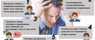
Symptoms such as:
- severe pain in the eyeballs;
- loss of balance;
- tingling or numbness in the legs, arms, or body parts;
- difficulty understanding speech or slurred speech of the person himself.
Such signs are observed in only half of patients with hemorrhagic stroke; the same manifestations may indicate a developed ischemic stroke or transient ischemic attack (popularly called a “micro-stroke”).
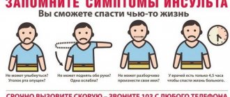
The symptoms of the onset of a hemorrhagic stroke are the following, and they usually occur during the day, after physical or emotional stress (maybe on the beach or in a hot shop):
- a feeling of a strong blow to the head, after which loss of consciousness usually occurs;
- if a person remains conscious, he feels a strong and intensifying pulsating headache, a feeling of strong heartbeat, nausea, vomiting, pain in the eyes when looking at the light;
- the face turns red;
- increased sweating is noted;
- after a short time, motor excitation may develop;
- there may be seizures;
- in most cases, impairment of consciousness gradually increases. At first the person always wants to sleep, but you can wake him up, talk to him, and he will answer questions. Over time, it is possible to wake a person only for a short amount of time, after which he answers in monosyllables and not always on topic. Then a coma develops, during which it is impossible to wake the patient.
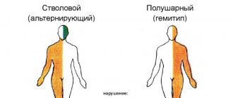
If during a hemorrhagic stroke the right side is lost and at the same time:
- it is impossible to retract the left eyeball, puff out the cheeks (the left cheek “sags”), the left nasolabial fold is lowered, the probable diagnosis is hemorrhage in the pons of the brain on the left;
- movements in the right half of the face are impaired (you can’t puff out your cheeks or bare your teeth), and on the left, pain and temperature sensitivity is reduced, then the parietal lobe of the cortex on the left is soaked in blood or has become a “shelter” for a hematoma;
- the upper eyelid is drooping on the left, the pupil in this eye is dilated, there is difficulty moving the eye to the nose, in addition, on the right it is difficult to bare your teeth or puff out your cheeks, we are talking about a left-sided lesion of the midbrain.
If the right arm is paralyzed, it is bent in all joints, while the leg on the left is paralyzed, which is in the extension position, and the lower parts of the medulla oblongata on the right are affected.
When hemorrhage or hematoma is reflected on the left side of the body:
- if weakness is noted, pain, tactile and temperature sensitivity is reduced more in the left leg than in the arm, hemorrhage occurs in the frontotemporal region on the right;
- if neither the arm nor the leg moves on the left, sensitivity there is also reduced, and on the right it is impossible to look away, the right nasolabial fold is smoothed, a stroke has occurred in the pons of the brain on the right;
- if the arm, leg and left half of the face do not move on the left, there are also disturbances in pain and temperature sensitivity, the parietal lobe of the cortex on the right is soaked in blood or compressed by a hematoma;
- if the arm and leg are paralyzed on the left, deep sensitivity is also impaired there, while pain and temperature sensitivity are not affected, and, in addition, there is difficulty in moving the tongue on the right (when opening the mouth, it is turned to the left), the medulla oblongata on the left is affected;
- if there is no movement of the upper and lower extremities on the left, there is no sensitivity there, and on the right side of the face there is a loss of pain and temperature sensitivity with its preservation around the mouth, hemorrhage has occurred in the bridge;
- loss of all types of sensitivity on the left side of the face, left arm and leg indicates right-sided hemorrhage in the thalamus.
The described symptoms only allow the doctor to determine the location of the lesion. It is impossible to distinguish hemorrhage from a hematoma using them.
If there is a hemorrhage in the cerebellum, a headache appears in the back of the head and neck area, the person cannot pronounce words clearly, and the tone of his arms and legs is reduced (they hang “like whips”). Strabismus is also observed, when one eye looks down and inward, the second - up and out.
If hemorrhage occurs in the ventricles, the person’s condition deteriorates quickly, as well as:
- muscle tone of all four limbs decreases;
- breathing becomes difficult;
- a person chokes when swallowing;
- temperature rises;
- seizures may develop;
- consciousness is impaired.
A hemorrhagic stroke in the brain stem is manifested by disturbances in breathing (it becomes irregular and may be superficial) and in the activity of the heart. Strabismus develops, the pupils become wide and may be of unequal size. The eyeballs are not fixed, but “float” and tremble when moving. The swallowing process is disrupted.
Subarachnoid hemorrhage has slightly different symptoms. This:
- severe headache;
- nausea;
- vomit;
- photophobia;
- increased sensitivity of the skin of the whole body;
- there may be seizures;
- impairment of consciousness often increases, which can be reversible with adequate assistance.
Paralysis, disturbances in eye movements, and changes in the pupils are not typical for the initial stages of subarachnoid hemorrhage. They join with the development of cerebral edema.
Subdural hematoma, that is, an accumulation of blood between the membranes of the brain, has its own symptoms:
- initially, against the background of inadequate physical activity, stress or hypertensive crisis, there is a sharp headache and loss of consciousness;
- After a while, the person regains consciousness and nothing bothers him for several hours to several days;
- After this “bright period” of time, the condition sharply worsens, the person loses consciousness, against the background of which convulsions develop, two or one limbs stop moving, strabismus and facial asymmetry appear.
Symptoms
For damage to the left hemisphere of the brain, the development of the following symptoms is typical:
- Speech impairment. It becomes the primary sign indicating a stroke has occurred. The patient exhibits slurred pronunciation of words, explanations in inconsistent/fragmentary phrases, poor articulation, and lack of understanding of speech addressed to him.
- Loss of speech and verbal memory. Accompanied by forgetfulness of words and speech patterns.
- Paralysis of the right side of the body. It is characterized by problems with motor activity against the background of absent muscle atrophy. The patient may lose an arm and/or leg.
- Synkinesia (uncontrolled flexion of paralyzed limbs).
- Psychological disorders - depression, isolation.
Externally, paralysis after a stroke of the left hemisphere of the brain manifests itself in the following:
- right hand bent at the elbow joint, pressed to the body;
- clenching the hand into a fist;
- the leg is extended at the knee, the foot is turned inward;
- the corner of the lips and the corner of the lower eyelid on the right side of the face are lowered down.
Course and prognosis of pathology
The prognosis of hemorrhagic stroke is unfavorable. It depends on the location and extent of the lesion. Hemorrhage into the brainstem is dangerous, which is accompanied by respiratory failure and a sharp, poorly corrected by drugs, decrease in blood pressure to critical levels. Hemorrhage into the ventricles with their breakthrough is severe and often ends in death.
How long do people live with hemorrhagic stroke? This pathology is fatal in 50-90% of cases. Death may occur on the very first day - against the background of generalized convulsions, when breathing is impaired. More often, death occurs later, by 2 weeks. This is due to a cascade of biochemical reactions triggered by the outpouring of blood into the cranial cavity and leading to the death of brain cells. If there is no displacement of the brain, no herniation (entry into a bone hole), no breakthrough of blood into the ventricles, and the compensatory capabilities of the brain are sufficiently large (this is more typical for children and young people), then the person has a great chance of survival.
At 1-2 weeks, in addition to neurological disorders, complications associated with the immobility of the patient, exacerbation of his chronic diseases or connecting him to an artificial respiration apparatus (pneumonia, bedsores, liver, kidney, cardiovascular failure) occur. And if they do not lead to death, then by the end of 2-3 weeks the cerebral edema will stop. By week 3, it becomes clear what the consequences of a hemorrhagic stroke are in this case.
The following symptoms are prognostically unfavorable:
- coma, especially if it developed in less than 3 hours, led to a sharp drop in pressure and breathing problems;
- a combination of paralysis of the arm and leg on one side with violent movements (attempting to cover oneself with a blanket, adjusting clothes, checking the condition of the genitals) of the limbs on the other side;
- chills;
- cold sweat;
- fever that does not respond to antipyretic drugs;
- nasal voice;
- irregular, slower or faster (deep and noisy) breathing;
- swallowing disorder.
Following a special lifestyle after a hemorrhagic stroke, that is:
- constantly monitoring blood pressure;
- excluding salty foods;
- removing coffee, black tea, and alcoholic beverages from the diet;
- quitting smoking;
- having performed an MRI of the brain and magnetic resonance angiography, which will allow one to see pathological vessels and treat them surgically as planned;
- avoiding exposure of the body to harmful substances (varnishes, paints, heavy metals and others);
- doing light physical activity;
- excluding physical inactivity;
- controlling the coagulability and saturation of lipids in one’s own blood,
you can still live a sufficient amount of time, calculated not in years, but in their dozens (if the stroke occurred in youth). Survival will also depend on the health of the heart, liver, kidneys and other internal organs.
Rehabilitation
Based on the fact that with a hemorrhagic cerebral stroke on the right side, there is a decrease or complete absence of motor functions, suppression of the speech, visual and tactile apparatus, the recovery period includes measures aimed at restoring these brain functions.
The basic list of rehabilitation measures for recovery after a stroke on the right side includes the following items:
- Ensuring continued emotional and physical rest for the affected person;
- Compliance with dietary recommendations that include excluding foods rich in low-density lipoproteins from the diet;
- Therapeutic and restorative massage;
- Physical exercises to combat paralysis (physical therapy);
- An important step is compliance with the restrictive regime, which includes abstaining from drinking alcohol and smoking;
- Minimizing psychological and physical stress;
- Treatment of chronic diseases;
- Rehabilitation sessions with a physiotherapist, psychologist and neurologist.
The duration of recovery for a stroke is individual for each individual clinical case. These periods and their duration depend on the severity of damage to the brain structures of the right hemisphere, as well as the individual characteristics of the human body. In some patients, recovery of the right side after a cerebral infarction lasts about 14 days, while in others this figure exceeds 6 months.
First aid and treatment
First aid for a hemorrhagic stroke primarily involves calling an ambulance. Then the patient needs to be placed in bed, ensuring that the head end is raised by 30 degrees. The person must be freed from constrictive elements of clothing: unbutton the collar, buttons, and belt. It is necessary to provide a flow of fresh air. It is impossible to feed and drink until the permission of the neurologist or resuscitator of the hospital, even if the patient is in clear consciousness (if you have a strong desire to drink, you can wet your lips).
Anti-pressure pills should not be given before the ambulance arrives: there is a possibility of a sharp decrease in blood pressure, which is dangerous in this case, since otherwise the blood-damaged brain may not receive enough oxygen, which it now needs to continue functioning. Only specially trained medical personnel know the rules for lowering blood pressure when it rises above 150/100 mmHg.
If seizures develop, it is necessary to prevent additional head injuries to the patient as much as possible. You should also try to remove the lower jaw to prevent the tongue from retracting and blocking the airways. To do this, you need to stand facing the feet of a lying person and place your hands on his jaw so that the little fingers, ring and middle fingers are in the area of the temporomandibular joints, and the index and thumbs are where the jaw meets the chin. With a synchronized movement of both hands, you need to try to push this moving bone so that the lower teeth take place in front of the upper ones.
How is the treatment performed?
Treatment of hemorrhagic stroke begins with emergency doctors, who must slightly, by 20% within an hour, reduce blood pressure, ensure airway patency and sufficient oxygen supply to the patient. Further it occurs only in the hospital.
Therapy depends on the type of stroke: a hematoma or blood soaking of the brain substance, which is determined using computed tomography or magnetic resonance imaging:
- If this is a hematoma, then after a short-term stabilization of the condition (if possible), an operation is performed in the neurosurgery department aimed at eliminating the volume of blood compressing the brain. Based on tomography data indicating the location of the hematoma, the neurosurgeon performs a craniotomy (makes a “window” in the bone), after which he punctures and evacuates the blood, stopping the bleeding. The bone hole is often left open, suturing only the soft tissue above it, so that when the brain edema, the latter is provided with additional space.
- Parenchymal and subarachnoid hemorrhages are treated with medication.
The following medications are used in the treatment of hemorrhagic stroke:
- calcium channel blockers (“Nimotop”) intravenous drip or micro-jet. They, by lowering blood pressure, protect parts of the brain from death;
- osmodiuretics (“Mannitol”). These drugs effectively, but short-term, reduce intracranial pressure;
- neuroprotectors – drugs that protect brain cells from death: “Somazina”; "Neuroxon", "Cerebrolysin";
- antibiotics (Ceftriaxone, Cefepime) to prevent suppuration of blood spilled into the cranial cavity;
- solutions of electrolytes ("Sodium chloride", "Ringer's solution") and gelatin-based ("Gelafusin");
- hemostatic agents (Kontrikal, Etamzilat) - if there are disturbances in the coagulation system. Otherwise they have no effect.
Oxygen therapy, prevention of bedsores, and prevention of thromboembolic complications are also provided. If necessary, medications that increase blood pressure and glucose-lowering medications (insulin) are administered.
What can you do about a hemorrhagic stroke:
- Nutrition. If the person is conscious and has no swallowing problems, solid food is fed: small amounts, excluding fatty, sour, smoked, salty, tea and coffee. In case of problems with consciousness and swallowing, a probe (tube) is installed through the nose, the end of which is located in the stomach. Special mixtures for enteral nutrition are introduced into the tube (their appearance and preparation resemble baby food for infants).
- Mode. Strict bed rest is observed for about 3 weeks, and you cannot even get up to go to the toilet. Feeding is carried out in a position with the head end raised 60 degrees. Physiological functions are also carried out in a supine position: if the patient is conscious, he urinates on the bedpan; if unconscious, a catheter is inserted into the bladder through the urethra.
- You can use anti-bedsore mattresses or sandbags that are placed under those parts of the body that are subject to friction.
- Plaster splints can be used to place paralyzed limbs in the correct physiological position.
Smoking, walking around the ward, eating fresh fruits, vegetables, chocolate, seafood, playing on electronic devices is prohibited. This behavior increases the chance of recurrent hemorrhage.
Life prognosis after stroke and life expectancy
The mortality rate for ischemic stroke is about 15%. The highest risk of death is in the first 30 days after an attack. The death rate at this time is 35%. The second critical period is the first year of rehabilitation. During this time, the risk of high mortality remains up to 50%.
Risks of an unfavorable outcome:
- Speech or motor activity is impaired;
- Elderly patient;
- Muscular-articular sensation is impaired;
- The tone is gone;
- Have depression or cognitive impairment.
Stages of rehabilitation
- The first period takes place in an inpatient department and lasts approximately 1 month.
- The second begins after discharge from the hospital or transfer of the patient to the rehabilitation department. For the second stage, neurological sanatoriums are suitable, where there are conditions for patients to stay immediately after the hospital.
- The third period lasts up to a year from the attack. It includes home rehabilitation and treatment at the medical rehabilitation department of a community clinic.
Recovery
Successful rehabilitation requires an integrated approach. First of all, you should pay attention to blood pressure control. The patient must regularly use the medications recommended by doctors.
Physiotherapy and gymnastics
Physiotherapy can be performed 1 month after the attack, when the patient’s blood pressure and hemodynamics have stabilized. Hydrotherapy procedures (whirlpool baths) for swelling in paralyzed limbs, darsonval and other currents are effective for stimulating the tone of paralyzed and paretic muscles.
Gymnastics can be started from the first weeks of an attack. It is recommended to perform it in bed if the patient's activity is low. Exercises for gymnastics are selected by a physical therapy specialist. As the patient's skills are restored, the complex should become more complex.
Massage
Massage is suitable for restoring blood circulation and normalizing muscle tone. It is carried out by a massage therapist after the first month from the onset of the attack. The course consists of 10-15 sessions. The massage can be repeated after 3-4 months.
Rehabilitation
On average, effective rehabilitation should last about 6 months. The first three are key to skill recovery. It is important to carry out rehabilitation measures under the supervision of experienced specialists and with the permission of the attending physician.
Consequences of hemorrhagic stroke
As a result of the disease, the following may occur:
- paralysis (complete lack of movement) and paresis (partial lack of movement) of the limbs, face or part thereof;
- impairment of speech production or understanding;
- inappropriate behavior;
- blindness;
- loss of sensation in the trunk or limbs;
- deafness;
- memory loss;
- depression;
- severe pain in the limbs;
- lack of proper sleep;
- loss of reading/writing skills;
- inadequacy of perception of the surrounding world, due to which a person becomes unable to care for himself or may even harm himself.
Complications
A certain proportion of patients develop severe complications, in particular paraplegia (paralysis of both arms/legs). The cause is incorrectly chosen or incorrectly carried out treatment, extensive brain damage. Irreversible consequences develop when medical care is not provided in a timely manner.
Possible consequences of a stroke:
- Speech impairment. It is possible that there is either a complete impairment of speech functionality or slow pronunciation.
- Formation of bedsores, pneumonia, blood clots. The cause is prolonged immobility of a person.
- Violation of the processes of defecation and release of the bladder. Develops as a result of damage to large areas of the brain.
- Memory impairment, inability to navigate in space, difficulty comparing facts. Caused by poor brain function.
As a rule, such complications are reversible and gradually disappear with adequate therapy.
Rehabilitation period
Recovery after a hemorrhagic stroke is long. It depends on the lost functions and does not guarantee their complete rehabilitation. Lost abilities are restored most quickly in the first year after a stroke, then this process is slower. The neurological deficit that remains after three years will most likely remain for life.
Neurologists and rehabilitation specialists are ready to help restore lost functions as much as possible (see rehabilitation after a stroke). For this:
- for paresis or paralysis, physiotherapy is carried out (for example, on the Myoton apparatus), massage and exercise therapy are performed with an instructor;
- if speech reproduction is impaired, the person will have to work with a speech therapist;
- classes are held with a psychologist or psychotherapist;
- if reading/writing skills are lost, classes are held to restore them;
- drugs are prescribed that will help restore lost neural connections (“Cerakson”, “Somazina”), reduce high blood pressure (“Enalapril”, “Nifedipine”), antidepressants and sedatives;
- hydrotherapy is carried out (massage in the pool, light exercises in the water);
- classes on special simulators;
- color therapy - treatment with visual images.
Only when the patient and the doctor unite in the fight against the consequences of a hemorrhagic stroke can it be defeated, restoring lost capabilities as much as possible.
Author:
Krivega Maria Salavatovna resuscitator
Restoration of the speech apparatus
Often, a right-sided stroke in men and women has negative consequences in terms of speech loss. Speech may be lost within the first hour after an attack. The function can be restored in a few weeks or in a few years.
Articulation exercises
It is important to conduct speech restoration classes from the first days after the stroke is stopped. Speech is difficult to recover. This may take more than one year, but don't panic. Even if a person does not produce the desired results, at some point he will still speak.
Main recommendations:
- Constantly talk to the patient, even if he cannot produce sounds. While listening to sounds, he will try to repeat them.
- Start by studying individual syllables or sounds. The patient must independently continue the word he started. It is important to separate words into syllables. For example: the word “mo-lo-ko”, “le-to”, etc.
- To speed up speech ability, it is important for a person to turn on audio recordings to listen to other people singing. This method will begin to reproduce individual sounds faster.
- It is important to do exercises to improve articulation. This could be: curling your lips into a tube, sticking out your tongue, hissing, licking your lips, baring your teeth, etc.
When speech is restored, begin to resume and strengthen memory. To do this, continue to learn tongue twisters and short poems with the patient. Board games that will distract a person have a beneficial effect.
It all depends on the severity of the stroke. Of course, the most dangerous option is a massive stroke, but even in such a situation you cannot give up.